

The DOI matches its Crossref record; 49 of the 49 references it cites were found in a registry or an index.
Retinosomes
Open accessAbstract
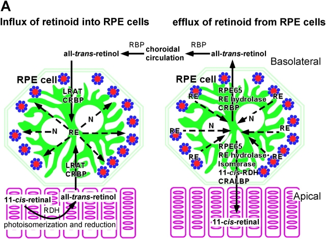
Lipid bodies form autonomous intracellular structures in many model cells and in some cells of specific tissue origin. They contain hydrophobic substances, a set of structural proteins such as perilipin or adipose differentiation-related protein, enzymes implicated in lipid metabolism, and proteins that participate in signaling and membrane trafficking. Retinosomes, particles reminiscent of lipid bodies, have been identified in retinal pigment epithelium as distinct structures compartmentalizing a metabolic intermediate involved in regeneration of the visual chromophore. These observations suggest that lipid bodies, including retinosomes, carry out specific functions that go beyond those of mere lipid storage organelles.
Overview
The claims and statistics of this paper haven’t been checked yet.
Free: it reads the open-access text and checks the first 5 claims that cite a source we can check. Create a free account to check every claim.
Integrity signals
Registry
Cites
Statistics
Figures


Verification
Claims the paper makes
The claims and statistics of this paper haven’t been checked yet.
Free: it reads the open-access text and checks the first 5 claims that cite a source we can check. Create a free account to check every claim.
Claims other papers make about this one
Each is checked against this paper’s own text: does it say what the citing paper says it does?
The source backs part of the claim, not all of it — or backs it for a different population, condition or comparison.
In the citing paper
“Because retinols and retinyl esters (but not retinals) are intrinsically fluorescent, their transformation can be followed by fluorescence induced by two-photon excitation.”
From Chemistry of the Retinoid (Visual) Cycle (2014) · cites this paper as [282]
Experimental — accuracy not yet measuredThe source backs part of the claim, not all of it — or backs it for a different population, condition or comparison.
In the citing paper
“LRAT, through its ability to catalyze the formation of retinyl esters that are readily sequestered by aggregation into lipid droplets called retinosomes, changes the mass action ratio to favor retinoid uptake from both photoreceptors and the choroidal circulation.”
From Chemistry of the Retinoid (Visual) Cycle (2014) · cites this paper as [282]
Experimental — accuracy not yet measuredThe source backs part of the claim, not all of it — or backs it for a different population, condition or comparison.
In the citing paper
“Retinosomes are located close to the RPE plasma membrane and are essential components for 11-cis-retinal production.”
From Chemistry of the Retinoid (Visual) Cycle (2014) · cites this paper as [282]
Experimental — accuracy not yet measured
Self-check
Percentages, sums and reported differences re-computed from the paper’s own numbers.
The self-check needs the paper’s full text.
Citations
Cites
Two-photon laser scanning fluorescence microscopy
Denk, W., Strickler, J. H., & Webb, W. W. (1990) · Science · doi:10.1126/science.2321027 · Cited by 9,488
Allikmets, R., Singh, N., Sun, H., Shroyer, N. F., Hutchinson, A., Chidambaram, A., Gerrard, B., Baird, L., Stauffer, D., Peiffer, A., et al. (1997) · Nature Genetics · doi:10.1038/ng0397-236 · Cited by 1,435 · cited 2×
“The significance of transport across the disk membrane is underscored by the finding that mutations in the ABCR gene in humans (Allikmets et al., 1997) or ABCR gene ablation in mice cause a buildup of bis-retinoids in photoreceptor cells (Mata et al.”
BackgroundThe Surface of Lipid Droplets Is a Phospholipid Monolayer with a Unique Fatty Acid Composition
Tauchi-Sato, K., Ozeki, S., Houjou, T., Taguchi, R., & Fujimoto, T. (2002) · Journal of Biological Chemistry · doi:10.1074/jbc.m207712200 · Cited by 753
Tansey, J. T., Sztalryd, C., Gruia-Gray, J., Roush, D. L., Zee, J. V., Gavrilova, O., Reitman, M. L., Deng, C.-X., Li, C., Kimmel, A. R., et al. (2001) · Proceedings of the National Academy of Sciences · doi:10.1073/pnas.101042998 · Cited by 734
Absence of perilipin results in leanness and reverses obesity in Leprdb/db mice
Martinez-Botas, J., Anderson, J. B., Tessier, D., Lapillonne, A., Chang, B. H.-J., Quast, M. J., Gorenstein, D., Chen, K.-H., & Chan, L. (2000) · Nature Genetics · doi:10.1038/82630 · Cited by 581
Liu, P., Ying, Y., Zhao, Y., Mundy, D. I., Zhu, M., & Anderson, R. G. W. (2004) · Journal of Biological Chemistry · doi:10.1074/jbc.m311945200 · Cited by 527
Mata, N. L., Weng, J., & Travis, G. H. (2000) · Proceedings of the National Academy of Sciences · doi:10.1073/pnas.130110497 · Cited by 456
Londos, C., Brasaemle, D. L., Schultz, C. J., Segrest, J. P., & Kimmel, A. R. (1999) · Seminars in Cell & Developmental Biology · doi:10.1006/scdb.1998.0275 · Cited by 435
Isomerization and Oxidation of Vitamin A in Cone-Dominant Retinas
Mata, N. L., Radu, R. A., Clemmons, R. S., & Travis, G. H. (2002) · Neuron · doi:10.1016/s0896-6273(02)00912-1 · Cited by 382
Miura, S., Gan, J.-W., Brzostowski, J., Parisi, M. J., Schultz, C. J., Londos, C., Oliver, B., & Kimmel, A. R. (2002) · Journal of Biological Chemistry · doi:10.1074/jbc.m204410200 · Cited by 372
Research works printed but not linked: 1
- 2002, Invest. Ophthalmol. Vis. Sci., 43, 1730
Cited by
Lipid droplets: a unified view of a dynamic organelle
Martin, S., & Parton, R. G. (2006) · Nature Reviews Molecular Cell Biology · doi:10.1038/nrm1912 · Cited by 1,169
Leber congenital amaurosis: Genes, proteins and disease mechanisms
den Hollander, A. I., Roepman, R., Koenekoop, R. K., & Cremers, F. P. M. (2008) · Progress in Retinal and Eye Research · doi:10.1016/j.preteyeres.2008.05.003 · Cited by 811
G Protein–Coupled Receptor Rhodopsin
Palczewski, K. (2006) · Annual Review of Biochemistry · doi:10.1146/annurev.biochem.75.103004.142743 · Cited by 764
Self-citationImaging lipid bodies in cells and tissues using third-harmonic generation microscopy
Débarre, D., Supatto, W., Pena, A.-M., Fabre, A., Tordjmann, T., Combettes, L., Schanne-Klein, M.-C., & Beaurepaire, E. (2006) · Nature Methods · doi:10.1038/nmeth813 · Cited by 578
Hepatic stellate cell lipid droplets: A specialized lipid droplet for retinoid storage
Blaner, W. S., O'Byrne, S. M., Wongsiriroj, N., Kluwe, J., D'Ambrosio, D. M., Jiang, H., Schwabe, R. F., Hillman, E. M. C., Piantedosi, R., & Libien, J. (2009) · Biochimica et Biophysica Acta (BBA) - Molecular and Cell Biology of Lipids · doi:10.1016/j.bbalip.2008.11.001 · Cited by 444
Metadata
- Authors
- Imanishi, Y., Gerke, V., & Palczewski, K.
- Affiliations
- University of Washington (US)
- University of Münster (DE)
- Corresponding author
- Palczewski, Krzysztof
- Venue
- The Journal of Cell Biology
- Publisher
- Rockefeller University Press
- Published
- 2004-08-16
- Volume
- 166
- Issue
- 4
- Pages
- 447-453
- ISSN
- 0021-9525 (ISSN-L)
- 1540-8140
- PMC
- PMC1360213
- OpenAlex
- W2006378422
- Cited by
- 102
- References
- 49
- Citations by year
- 2026: 3 · 2025: 1 · 2024: 4 · 2023: 3 · 2022: 4 · 2021: 5 · 2020: 4 · 2019: 1 · 2018: 3 · 2017: 2 · 2016: 8 · 2015: 1 · 2014: 6 · 2013: 4 · 2012: 7
- FWCI
- 0.88
- Type
- journal article
- Language
- en
- Open access
- green
- www.ncbi.nlm.nih.gov
- Licence
- cc-by-nc-sa
- Locations
- The Journal of Cell Biology (publishedVersion)
- Europe PMC (PubMed Central) (publishedVersion, cc-by-nc-sa)
- PubMed Central (publishedVersion, cc-by-nc-sa)
- PubMed (publishedVersion)
- Funders
- National Eye Institute (R01EY008061, EY13385, EY 08061, R01EY013385)
- Topics
- Life Sciences › Biochemistry, Genetics and Molecular Biology › Biochemistry › Lipid metabolism and biosynthesis
- Life Sciences › Biochemistry, Genetics and Molecular Biology › Cell Biology › Cellular transport and secretion
- Health Sciences › Medicine › Ophthalmology › Retinal Diseases and Treatments
- Keywords
- lipid droplets, retinal pigment epithelium, lipid metabolism, membrane trafficking, retinol
- MeSH terms
- Perilipin-1, Perilipin-2, Animals, Carrier Proteins, Cell Membrane, Cytoplasm, Immunohistochemistry, Lipids, Membrane Proteins, Mice, Transgenic, Models, Biological, Phosphoproteins, Retina, Time Factors, Tretinoin, Signal Transduction, Lipid Metabolism, Mice
About these checks
Registry, retraction, Cites and statistics results are computed, not judged, with one exception: a printed reference that no index lists is read once by a model before it counts as a citation error. Claims are extracted by a model and kept only when their quote appears verbatim in the text; each cited source is then checked, with a model’s help, for whether it says what the claim relies on. Those verdicts can be wrong — read the claim’s quote and the cited source before relying on one.
Statistics and arithmetic are recomputed, not judged.
Confidence bands are shown only for fields calibrated on our annotated test set. None is calibrated yet, so no band is shown.
What counts as a claim ›How we measure confidence ›How we check this ›
Are you an author, or do you see a problem with this page? Request a review
How to cite
Cite CoChat for the checks on this page, and the paper for its own findings.
- This page
- CoChat. (2026). Citation check: Retinosomes. https://app.cochat.ai/papers/10.1083/jcb.200405110
- The paper
- Imanishi, Y., Gerke, V., & Palczewski, K. (2004). Retinosomes. The Journal of Cell Biology. https://doi.org/10.1083/jcb.200405110
Data sources
Bibliographic data: Semantic Scholar, OpenAlex, Crossref.
Contains information from the Semantic Scholar Academic Graph Datasets, made available under the ODC Attribution License (ODC-BY 1.0).
Semantic Scholar’s data is described in: Kinney, R., et al. (2023). The Semantic Scholar Open Data Platform. arXiv:2301.10140.
