

The DOI matches its Crossref record; 46 of the 46 references it cites were found in a registry or an index.
Imaging analysis reveals mechanistic differences between first- and second-phase insulin exocytosis
Open accessAbstract
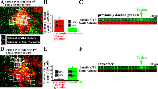
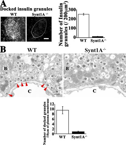
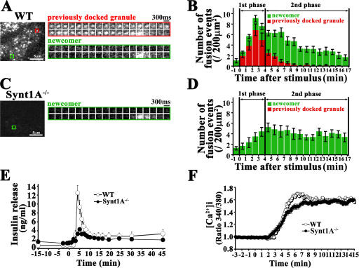
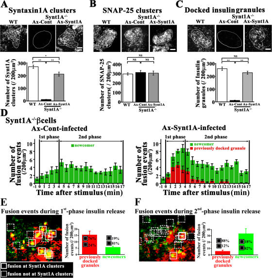
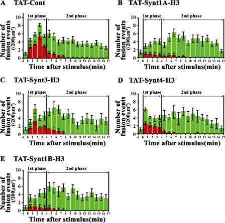
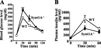
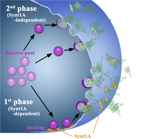
The mechanism of glucose-induced biphasic insulin release is unknown. We used total internal reflection fluorescence (TIRF) imaging analysis to reveal the process of first- and second-phase insulin exocytosis in pancreatic β cells. This analysis showed that previously docked insulin granules fused at the site of syntaxin (Synt)1A clusters during the first phase; however, the newcomers fused during the second phase external to the Synt1A clusters. To reveal the function of Synt1A in phasic insulin exocytosis, we generated Synt1A-knockout (Synt1A−/−) mice. Synt1A−/− β cells showed fewer previously docked granules with no fusion during the first phase; second-phase fusion from newcomers was preserved. Rescue experiments restoring Synt1A expression demonstrated restoration of granule docking status and fusion events. Inhibition of other syntaxins, Synt3 and Synt4, did not affect second-phase insulin exocytosis. We conclude that the first phase is Synt1A dependent but the second phase is not. This indicates that the two phases of insulin exocytosis differ spatially and mechanistically.
Overview
The claims and statistics of this paper haven’t been checked yet.
Run the check — free, no account needed. It reads the open-access text and checks the sources it cites.
Integrity signals
Registry
Cites
Statistics
Figures
Verification
Claims the paper makes
The claims and statistics of this paper haven’t been checked yet.
Run the check — free, no account needed. It reads the open-access text and checks the sources it cites.
Claims other papers make about this one
Each is checked against this paper’s own text: does it say what the citing paper says it does?
The source backs part of the claim, not all of it — or backs it for a different population, condition or comparison.
In the citing paper
“There is general agreement that many docked granules are released in response to membrane depolarization produced by sulfonylureas or high [K+]o (288, 446, 487, 617).”
From Pancreatic β-Cell Electrical Activity and Insulin Secretion: Of Mice and Men (2018) · cites this paper as [487]
Experimental — accuracy not yet measuredThe cited source says what the claim attributes to it.
In the citing paper
“In one study where TIRF microscopy was used to monitor granule exocytosis, Stx1a deficiency resulted in a marked reduction of docked granules and strong suppression of 1st phase insulin secretion (487). However, 2nd phase secretion was unaffected.”
From Pancreatic β-Cell Electrical Activity and Insulin Secretion: Of Mice and Men (2018) · cites this paper as [487]
Self-check
Percentages, sums and reported differences re-computed from the paper’s own numbers.
The self-check needs the paper’s full text.
Citations
Cites
Südhof, T. C. (2004) · Annual Review of Neuroscience · doi:10.1146/annurev.neuro.26.041002.131412 · Cited by 2,491
Wilschut, J. (1990) · Cited by 2,407
Jahn, R., Lang, T., & Südhof, T. C. (2003) · Cell · doi:10.1016/s0092-8674(03)00112-0 · Cited by 1,364
Syntaxin: A Synaptic Protein Implicated in Docking of Synaptic Vesicles at Presynaptic Active Zones
Bennett, M. K., Calakos, N., & Scheller, R. H. (1992) · Science · doi:10.1126/science.1321498 · Cited by 1,263
Membrane fusion and exocytosis.
Jahn, R., & Südhof, T. (1999) · Annual Review of Biochemistry · doi:10.1146/annurev.biochem.68.1.863 · Cited by 1,253
Triggering and amplifying pathways of regulation of insulin secretion by glucose.
Henquin, J. C. (2000) · Diabetes · doi:10.2337/diabetes.49.11.1751 · Cited by 1,201
Hemizygosity at the elastin locus in a developmental disorder, Williams syndrome
Ewart, A. K., Morris, C. A., Atkinson, D., Jin, W., Sternes, K., Spallone, P., Stock, A. D., Leppert, M., & Keating, M. T. (1993) · Nature Genetics · doi:10.1038/ng0993-11 · Cited by 1,138
Insulin granule dynamics in pancreatic beta cells
Rorsman, P., & Renstr�m, E. (2003) · Diabetologia · doi:10.1007/s00125-003-1153-1 · Cited by 844
Natural history of Williams syndrome: Physical characteristics
Morris, C. A., Demsey, S. A., Leonard, C. O., Dilts, C., & Blackburn, B. L. (1988) · The Journal of Pediatrics · doi:10.1016/s0022-3476(88)80272-5 · Cited by 670
SNARE Function Analyzed in Synaptobrevin/VAMP Knockout Mice
Schoch, S., Deák, F., Königstorfer, A., Mozhayeva, M. G., Sara, Y., Südhof, T. C., & Kavalali, E. T. (2001) · Science · doi:10.1126/science.1064335 · Cited by 664
Research works printed but not linked: 4
- 2004, Am. J. Med. Genet. A., 131, 255
- 1986, Lancet., 2, 360
- 1997, Am. J. Hum. Genet., 61, 449
- 2000, News Physiol. Sci., 15, 72
Cited by
Sorted by citations
Items 201–209 of 209
Hypoxia causes pancreatic β-cell dysfunction by activating a transcriptional repressor BHLHE40Preprint
Tsuyama, T., Sato, Y., Yoshizawa, T., Matsuoka, T.-A., & Yamagata, K. (2022) · bioRxiv · doi:10.1101/2022.06.28.498031
Rahman, M. M., Pathak, A., Schueler, K. L., Alsharif, H. A., Michl, A. N., Alexander, J., Kim, J.-A., Chapman, E. R., & Bhatnagar, S. (2022) · bioRxiv (Cold Spring Harbor Laboratory) · doi:10.1101/2022.10.20.513128
Insulin Secretion and the β-Cell 102 Years After the Discovery of the Hormone
Ortis, F., Rebelato, E., Carpinelli, Â. R., & Abdulkader, F. (2024) · Current Molecular Biology Reports · doi:10.1007/s40610-024-00158-9
VAMP8 Is an Endosomal v-SNARE That Supports GLP-1 Receptor Recycling in Pancreatic β-Cells
Liu, L., Marshall, M. R., Chadeuf, E., Saras, J., & Barg, S. (2025) · Diabetes · doi:10.2337/db24-0921
Use of mouse models to establish genotype-phenotype correlations in Williams-Beuren syndrome
Puimedon, S., & Fragaki, M. (2012) · LA Referencia (Red Federada de Repositorios Institucionales de Publicaciones Científicas)
Etude du rôle de la protéine PCSK9 dans la cellule bêta pancréatique
Saitoski, K. (2021) · theses.fr (ABES) · doi:10.70675/58b8c006z57e2z47a2zbb66z1e469544d6a5
Martín-Díaz, L., Patrikainen, M. S., Chongtham, C., Gupta, A., Dotsenko, V., Ojanen, M., Martins, F. T. A., George, J. J., Davidsson, R., Hakanen, S., et al. (2026) · bioRxiv (Cold Spring Harbor Laboratory) · doi:10.64898/2026.05.28.728433
Carter, T. (2026) · Digital Commons - DU (University of Denver) · doi:10.56902/etdcrp.2026.60
Metadata
- Authors
- Ohara-Imaizumi, M., Fujiwara, T., Nakamichi, Y., Okamura, T., Akimoto, Y., Kawai, J., Matsushima, S., Kawakami, H., Watanabe, T., Akagawa, K., & Nagamatsu, S.
- Affiliations
- 1Department of Biochemistry
- 2Department of Cell Physiology
- 5Division of Animal Models, Department of Infectious Diseases, Research Institute, International Medical Center of Japan, Tokyo 162-8655, Japan
- 3Department of Anatomy,
- Juntendo University (JP)
- Kyorin University (JP)
- Corresponding author
- Nagamatsu, Shinya
- Venue
- The Journal of Cell Biology
- Publisher
- Rockefeller University Press
- Published
- 2007-05-14
- Volume
- 177
- Issue
- 4
- Pages
- 695-705
- ISSN
- 0021-9525 (ISSN-L)
- 1540-8140
- PMC
- PMC2064214
- OpenAlex
- W2124167615
- Cited by
- 211
- References
- 46
- Citations by year
- 2026: 2 · 2025: 6 · 2024: 4 · 2023: 9 · 2022: 7 · 2021: 10 · 2020: 6 · 2019: 7 · 2018: 6 · 2017: 13 · 2016: 5 · 2015: 11 · 2014: 16 · 2013: 10 · 2012: 23
- FWCI
- 18.64
- Type
- journal article
- Language
- en
- Open access
- green
- www.ncbi.nlm.nih.gov
- Licence
- cc-by-nc-sa
- Locations
- The Journal of Cell Biology (publishedVersion)
- PubMed Central (publishedVersion, cc-by-nc-sa)
- PubMed (publishedVersion)
- Topics
- Health Sciences › Medicine › Endocrinology, Diabetes and Metabolism › Pancreatic function and diabetes
- Life Sciences › Biochemistry, Genetics and Molecular Biology › Cell Biology › Cellular transport and secretion
- Life Sciences › Biochemistry, Genetics and Molecular Biology › Physiology › Calcium signaling and nucleotide metabolism
- Keywords
- insulin exocytosis, first-phase insulin secretion, second-phase insulin secretion, syntaxin 1A, syntaxin 3, syntaxin 4, granule fusion, pancreatic beta cells, glucose-induced insulin release
- MeSH terms
- Insulin Secretion, Animals, Cells, Cultured, Exocytosis, Insulin, Islets of Langerhans, Male, Microscopy, Fluorescence, Mice, Knockout, Mice
About these checks
Registry, retraction, Cites and statistics results are computed, not judged, with one exception: a printed reference that no index lists is read once by a model before it counts as a citation error. Claims are extracted by a model and kept only when their quote appears verbatim in the text; each cited source is then checked, with a model’s help, for whether it says what the claim relies on. Those verdicts can be wrong — read the claim’s quote and the cited source before relying on one.
Statistics and arithmetic are recomputed, not judged.
Confidence bands are shown only for fields calibrated on our annotated test set. None is calibrated yet, so no band is shown.
What counts as a claim ›How we measure confidence ›How we check this ›
Are you an author, or do you see a problem with this page? Request a review
