Cited by 631Autophagy in Disease and TherapyCellular transport and secretionAmyotrophic Lateral Sclerosis Research

The DOI matches its Crossref record; 62 of the 62 references it cites were found in a registry or an index.

Functional multivesicular bodies are required for autophagic clearance of protein aggregates associated with neurodegenerative disease

Open access

Abstract

The endosomal sorting complexes required for transport (ESCRTs) are required to sort integral membrane proteins into intralumenal vesicles of the multivesicular body (MVB). Mutations in the ESCRT-III subunit CHMP2B were recently associated with frontotemporal dementia and amyotrophic lateral sclerosis (ALS), neurodegenerative diseases characterized by abnormal ubiquitin-positive protein deposits in affected neurons. We show here that autophagic degradation is inhibited in cells depleted of ESCRT subunits and in cells expressing CHMP2B mutants, leading to accumulation of protein aggregates containing ubiquitinated proteins, p62 and Alfy. Moreover, we find that functional MVBs are required for clearance of TDP-43 (identified as the major ubiquitinated protein in ALS and frontotemporal lobar degeneration with ubiquitin deposits), and of expanded polyglutamine aggregates associated with Huntington's disease. Together, our data indicate that efficient autophagic degradation requires functional MVBs and provide a possible explanation to the observed neurodegenerative phenotype seen in patients with CHMP2B mutations.

Overview

The claims and statistics of this paper haven’t been checked yet.

Run the check — free, no account needed. It reads the open-access text and checks the sources it cites.

A few free checks a day · papers already verified open instantly · results are public

Integrity signals

Registry

Crossref · 10.1083/jcb.200702115

Cites

62 of 62 references verified0 citation errors · 0 not checkable

Statistics

Figures

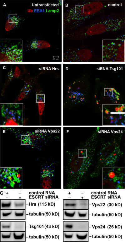
Figure 1. Non-endosomal ubiquitin-positive structures accumulate in ESCRT depleted cells. HeLa cells were left untransfected (A) or transfected with control (B), Hrs (C), Tsg101 (D), Vps22 (E), or Vps24 (F) siRNA for 5 d and processed for immunofluorescence microscopy. Cells were labeled with antibodies against EEA1 (blue), ubiquitin (mono and poly-Ub, red), and Lamp2 (green). Colocalization between EEA1 and Ub is indicated in purple and between Ub and Lamp2 in yellow. Bar, 10 μm. Single channel images of the insets are shown in Fig. S1 (available at http://www.jcb.org/cgi/content/full/jcb.200702115/DC1). (G) Knock-down efficiencies were analyzed by Western blotting. Equal loading was verified by anti-α-tubulin immunoblotting.
Figure 1. Non-endosomal ubiquitin-positive structures accumulate in ESCRT depleted cells. HeLa cells were left untransfected (A) or transfected with control (B), Hrs (C), Tsg101 (D), Vps22 (E), or Vps24 (F) siRNA for 5 d and processed for immunofluorescence microscopy. Cells were labeled with antibodies against EEA1 (blue), ubiquitin (mono and poly-Ub, red), and Lamp2 (green). Colocalization between EEA1 and Ub is indicated in purple and between Ub and Lamp2 in yellow. Bar, 10 μm. Single channel images of the insets are shown in Fig. S1 (available at http://www.jcb.org/cgi/content/full/jcb.200702115/DC1). (G) Knock-down efficiencies were analyzed by Western blotting. Equal loading was verified by anti-α-tubulin immunoblotting.CC BY-NC-SA · PubMed Central
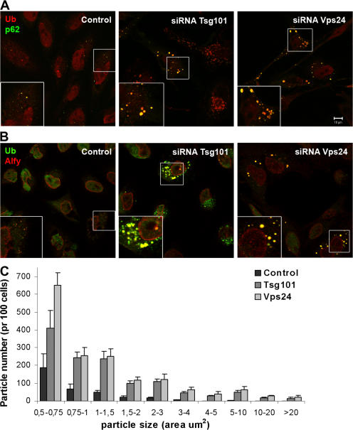
Figure 2. p62- and Alfy-positive structures accumulate in cells depleted of Tsg101 or Vps24. HeLa cells transfected with siRNA against Tsg101 or Vps24 were fixed, permeabilized, and stained with antibodies against ubiquitin (red) and p62 (green) (A) or against ubiquitin (green) and Alfy (red) (B). Colocalization is indicated in yellow. Bar, 10 μm. (C) The number and size (area) of p62-positive particles were quantified using ImageJ software. Approximately 100 cells from three different experiments (n = 334 (control), 309 (Tsg101), and 246 (Vps24)) were used for the quantification and the data is presented as average number per 100 cells. Error bars = SEM.
Figure 2. p62- and Alfy-positive structures accumulate in cells depleted of Tsg101 or Vps24. HeLa cells transfected with siRNA against Tsg101 or Vps24 were fixed, permeabilized, and stained with antibodies against ubiquitin (red) and p62 (green) (A) or against ubiquitin (green) and Alfy (red) (B). Colocalization is indicated in yellow. Bar, 10 μm. (C) The number and size (area) of p62-positive particles were quantified using ImageJ software. Approximately 100 cells from three different experiments (n = 334 (control), 309 (Tsg101), and 246 (Vps24)) were used for the quantification and the data is presented as average number per 100 cells. Error bars = SEM.CC BY-NC-SA · PubMed Central
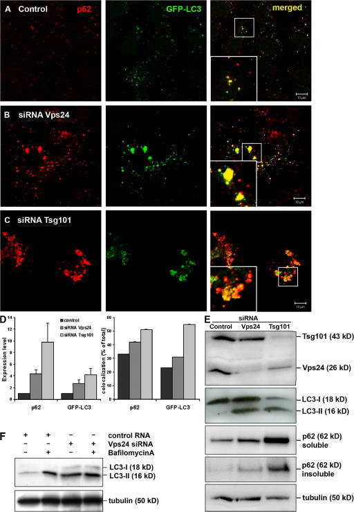
Figure 3. Autophagic degradation is impeded in Tsg101- and Vps24-depleted cells. HeLa cells stably expressing GFP-LC3 (green) were transfected with control (A), Vps24 (B), or Tsg101 (C) siRNA for 5 d and processed for immunofluorescence analysis. Cells were labeled with antibodies against p62 (red) and analyzed by confocal microscopy. Colocalization is indicated in yellow. Bar, 10 μm. (D) The total level of p62 and GFP-LC3 (normalized to control cells) and their degree of colocalization (% of total) in control and Tsg101- and Vps24-depleted cells were quantified using the Zeiss LSM 510 Meta software. 30 cells from three independent experiments were used for quantification. Error bars = SEM. (E) Western blot showing accumulation of LC3-II and p62 in HeLa cells depleted of Vps24 and Tsg101. p62 also accumulates in the insoluble fraction (bottom picture), indicating a shift to a more aggregated form upon depleted of Vps24 and Tsg101. (F) Western blot showing that LC3-II levels were similar in control and Vps24-depleted cells treated with Bafilomycin A.
Figure 3. Autophagic degradation is impeded in Tsg101- and Vps24-depleted cells. HeLa cells stably expressing GFP-LC3 (green) were transfected with control (A), Vps24 (B), or Tsg101 (C) siRNA for 5 d and processed for immunofluorescence analysis. Cells were labeled with antibodies against p62 (red) and analyzed by confocal microscopy. Colocalization is indicated in yellow. Bar, 10 μm. (D) The total level of p62 and GFP-LC3 (normalized to control cells) and their degree of colocalization (% of total) in control and Tsg101- and Vps24-depleted cells were quantified using the Zeiss LSM 510 Meta software. 30 cells from three independent experiments were used for quantification. Error bars = SEM. (E) Western blot showing accumulation of LC3-II and p62 in HeLa cells depleted of Vps24 and Tsg101. p62 also accumulates in the insoluble fraction (bottom picture), indicating a shift to a more aggregated form upon depleted of Vps24 and Tsg101. (F) Western blot showing that LC3-II levels were similar in control and Vps24-depleted cells treated with Bafilomycin A.CC BY-NC-SA · PubMed Central
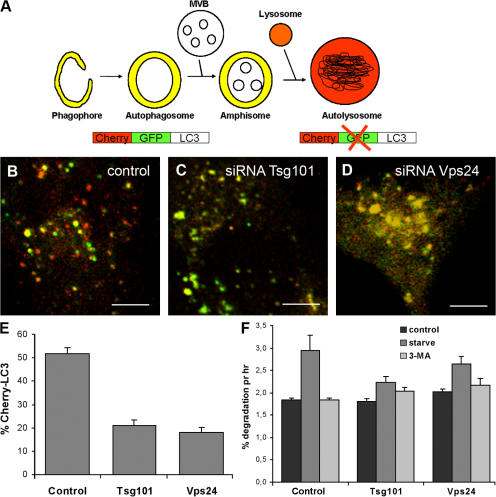
Figure 4. Formation of autolysosomes is inhibited in cells depleted of Tsg101 or Vps24. (A) The double-tagged LC3 protein (mCherry-GFP-LC3) will emit yellow (green merged with red) fluorescence in non-acidic structures and appear as red only in the autolysosomes due to quenching of GFP in these acidic structures. (B–F) HeLa cells were transfected with control (B), Tsg101 (C), or Vps24 (D) siRNA for 4 d and then with mCherry-GFP-LC3 for another 24 h before confocal microscopy analysis. Bar, 5 μm. (E) The mCherry-LC3 (red) signal was quantified in ∼30 cells with similar expression levels from three independent experiments using the Zeiss LSM 510 Meta software and the data are presented as % of total mCherry-GFP-LC3. Error bar = SEM. (F) Degradation of [14C]-valine- labeled proteins in cells incubated in complete medium (control), serum- and amino acid-deficient medium (starve), or starvation media in the presence of 3-methyl adenine (3-MA) to inhibit autophagic degradation. Average degradation per hour from four experiments done in duplicates ±1 SEM are shown.
Figure 4. Formation of autolysosomes is inhibited in cells depleted of Tsg101 or Vps24. (A) The double-tagged LC3 protein (mCherry-GFP-LC3) will emit yellow (green merged with red) fluorescence in non-acidic structures and appear as red only in the autolysosomes due to quenching of GFP in these acidic structures. (B–F) HeLa cells were transfected with control (B), Tsg101 (C), or Vps24 (D) siRNA for 4 d and then with mCherry-GFP-LC3 for another 24 h before confocal microscopy analysis. Bar, 5 μm. (E) The mCherry-LC3 (red) signal was quantified in ∼30 cells with similar expression levels from three independent experiments using the Zeiss LSM 510 Meta software and the data are presented as % of total mCherry-GFP-LC3. Error bar = SEM. (F) Degradation of [14C]-valine- labeled proteins in cells incubated in complete medium (control), serum- and amino acid-deficient medium (starve), or starvation media in the presence of 3-methyl adenine (3-MA) to inhibit autophagic degradation. Average degradation per hour from four experiments done in duplicates ±1 SEM are shown.CC BY-NC-SA · PubMed Central
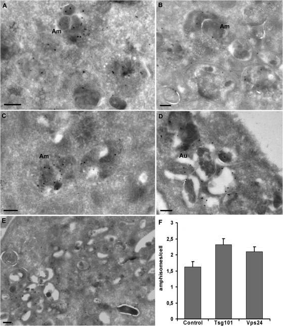
Figure 5. Autophagosomes and amphisomes do form in ESCRT-depleted cells. HeLa cells stably expressing GFP-LC3 were transfected with control (A), Tsg101 (B, D, and E), or Vps24 (C) siRNA and processed for immuno-EM analysis. Cryosections were incubated with antibodies against GFP (15-nm gold) and LBPA (10-nm gold). Amphisomes (Am), characterized by their content of electron-dense LC3-positive areas and intraluminal vesicles, were observed in both control (A) and ESCRT-depleted cells (B and C). (D) In the Tsg101-depleted cells we found large clusters of more typical autophagosomes (Au) and (E) clusters of double-membrane structures, consisting of autophagosomes and tubular structures which might represent phagophores, all labeling strongly for GFP-LC3. We never observed similar clusters in control cells and very rarely in Vps24-depleted cells. Bars, 200 nm. (F) Quantification of amphisomes in control cells and cells depleted for Tsg101 or Vps24. Approximately 20 cells were included per siRNA depletion per experiment (n = 3). Error bars = SEM.
Figure 5. Autophagosomes and amphisomes do form in ESCRT-depleted cells. HeLa cells stably expressing GFP-LC3 were transfected with control (A), Tsg101 (B, D, and E), or Vps24 (C) siRNA and processed for immuno-EM analysis. Cryosections were incubated with antibodies against GFP (15-nm gold) and LBPA (10-nm gold). Amphisomes (Am), characterized by their content of electron-dense LC3-positive areas and intraluminal vesicles, were observed in both control (A) and ESCRT-depleted cells (B and C). (D) In the Tsg101-depleted cells we found large clusters of more typical autophagosomes (Au) and (E) clusters of double-membrane structures, consisting of autophagosomes and tubular structures which might represent phagophores, all labeling strongly for GFP-LC3. We never observed similar clusters in control cells and very rarely in Vps24-depleted cells. Bars, 200 nm. (F) Quantification of amphisomes in control cells and cells depleted for Tsg101 or Vps24. Approximately 20 cells were included per siRNA depletion per experiment (n = 3). Error bars = SEM.CC BY-NC-SA · PubMed Central
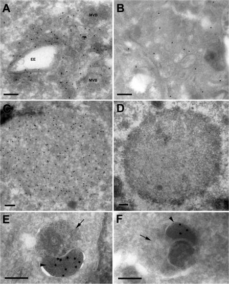
Figure 6. Immuno-EM of p62-positive structures in Vps24- and Tsg101-depleted cells. Cryosections of HeLa cells transfected with Vps24 (A, C, and E) or Tsg101 (B, D, and F) siRNA were incubated with antibodies against p62 (15-nm protein A gold, 10-nm in D). Membrane-free p62-positive aggregates (C and D) or p62-positive structures contained within dense clusters of vesicular-tubular elements (A and B) were detected. Vesicles with early endosomal morphology (EE) or multivesicular appearance (MVB) associated with these clusters. We also observed p62 labeling in electron-dense structures sequestered within amphisomes in cell depleted of Vps24 (E) or Tsg101 (F). The amphisomes typically consist of an electron-dense p62 positive body (E and F, arrowhead), and a multivesicular endosomal structure (E and F, arrow). Bars, 200 nm.
Figure 6. Immuno-EM of p62-positive structures in Vps24- and Tsg101-depleted cells. Cryosections of HeLa cells transfected with Vps24 (A, C, and E) or Tsg101 (B, D, and F) siRNA were incubated with antibodies against p62 (15-nm protein A gold, 10-nm in D). Membrane-free p62-positive aggregates (C and D) or p62-positive structures contained within dense clusters of vesicular-tubular elements (A and B) were detected. Vesicles with early endosomal morphology (EE) or multivesicular appearance (MVB) associated with these clusters. We also observed p62 labeling in electron-dense structures sequestered within amphisomes in cell depleted of Vps24 (E) or Tsg101 (F). The amphisomes typically consist of an electron-dense p62 positive body (E and F, arrowhead), and a multivesicular endosomal structure (E and F, arrow). Bars, 200 nm.CC BY-NC-SA · PubMed Central
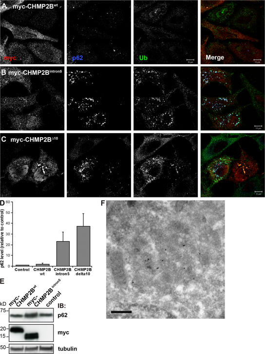
Figure 7. Ubiquitin and p62 positive structures accumulate in cells expressing CHMP2 mutants. HeLa cells transfected with myc-tagged wild-type CHMP2B (A), CHMP2BIntron5 (B), or CHMP2BΔ10 (C) were labeled with antibodies against c-Myc (red), ubiquitin (green), and p62 (blue) and analyzed by confocal microscopy. Single channel images in black and white are shown. Bars,10 μm. (D) Total levels of p62 in these cells were quantified in 20 cells from three independent experiments using the Zeiss LSM 510 Meta software and the average normalized to the average p62 level in control (mock transfected) cells. Error bars = SEM. (E) Western blot analysis showing accumulation of p62 in cells transfected with myc-CHMP2BIntron5 compared with mock-transfected cells (control) and cells transfected with CHMP2Bwt. (F) Immuno-EM showing p62-positive aggregates containing dense clusters of vesicular–tubular structures in cells transfected with myc-CHMP2BIntron5. Bar, 200 nm.
Figure 7. Ubiquitin and p62 positive structures accumulate in cells expressing CHMP2 mutants. HeLa cells transfected with myc-tagged wild-type CHMP2B (A), CHMP2BIntron5 (B), or CHMP2BΔ10 (C) were labeled with antibodies against c-Myc (red), ubiquitin (green), and p62 (blue) and analyzed by confocal microscopy. Single channel images in black and white are shown. Bars,10 μm. (D) Total levels of p62 in these cells were quantified in 20 cells from three independent experiments using the Zeiss LSM 510 Meta software and the average normalized to the average p62 level in control (mock transfected) cells. Error bars = SEM. (E) Western blot analysis showing accumulation of p62 in cells transfected with myc-CHMP2BIntron5 compared with mock-transfected cells (control) and cells transfected with CHMP2Bwt. (F) Immuno-EM showing p62-positive aggregates containing dense clusters of vesicular–tubular structures in cells transfected with myc-CHMP2BIntron5. Bar, 200 nm.CC BY-NC-SA · PubMed Central
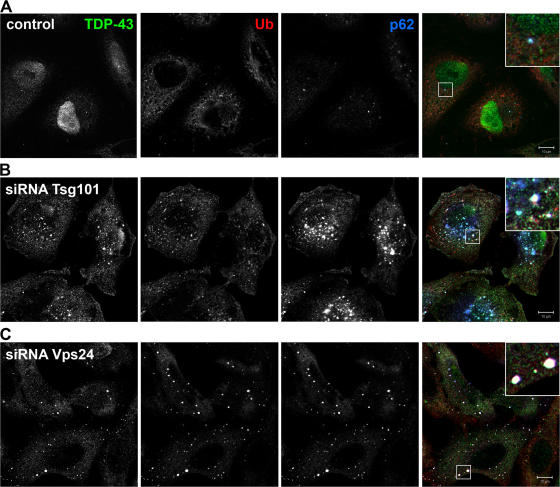
Figure 8. TDP-43 accumulates in cytoplasmic ubiquitin-positive structures in Tsg101- and Vps24-depleted cells. HeLa cells transfected with control (A), Tsg101 (B), or Vps24 (C) siRNA were fixed, permeabilized, and stained with antibodies against TDP-43 (green), ubiquitin (red), and p62 (blue). Single channel images in black and white are shown. Colocalization of all proteins is indicated in white in the merged picture. Bars, 10 μm.
Figure 8. TDP-43 accumulates in cytoplasmic ubiquitin-positive structures in Tsg101- and Vps24-depleted cells. HeLa cells transfected with control (A), Tsg101 (B), or Vps24 (C) siRNA were fixed, permeabilized, and stained with antibodies against TDP-43 (green), ubiquitin (red), and p62 (blue). Single channel images in black and white are shown. Colocalization of all proteins is indicated in white in the merged picture. Bars, 10 μm.CC BY-NC-SA · PubMed Central
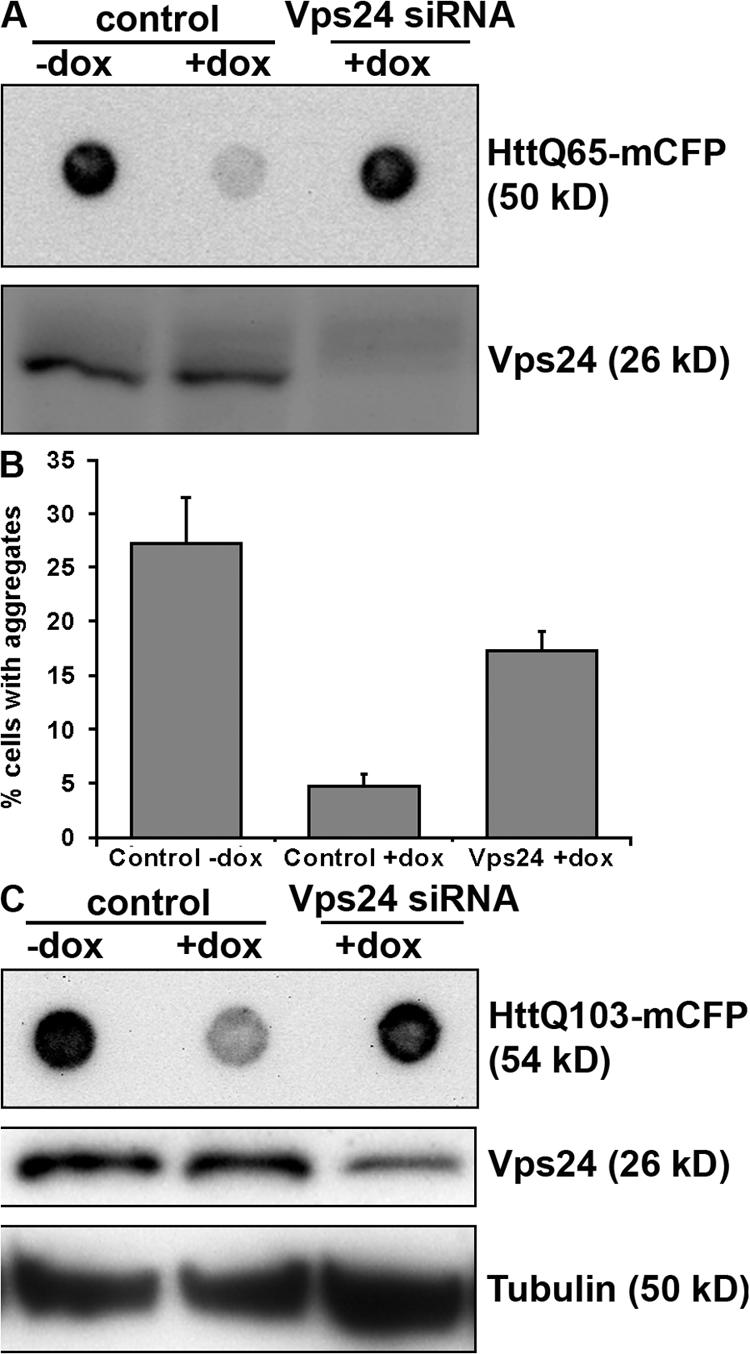
Figure 9. Vps24 is required for efficient clearance of Htt inclusions. HeLa cells expressing HttQ65-mCFP (A) or HttQ103-mCFP (B) and N2a cells expressing HttQ103-mCFP (C) were transfected with control or Vps24 siRNA for 2 d and then incubated in the absence or presence of doxycycline for 3 d to turn off expression of HttQ65/Q103-mCFP. Vps24 depletion was determined by Western blot analysis. (A and C) Clearance of SDS-insoluble Htt inclusions was analyzed by filter-trap assays. (B) Alternatively, aggregate clearance was analyzed by confocal quantification of the number of HeLa HttQ103 cells having visible inclusions after 3 d in the absence or presence of dox. 300 cells from three independent experiments were counted for each condition. Error bars = SD.
Figure 9. Vps24 is required for efficient clearance of Htt inclusions. HeLa cells expressing HttQ65-mCFP (A) or HttQ103-mCFP (B) and N2a cells expressing HttQ103-mCFP (C) were transfected with control or Vps24 siRNA for 2 d and then incubated in the absence or presence of doxycycline for 3 d to turn off expression of HttQ65/Q103-mCFP. Vps24 depletion was determined by Western blot analysis. (A and C) Clearance of SDS-insoluble Htt inclusions was analyzed by filter-trap assays. (B) Alternatively, aggregate clearance was analyzed by confocal quantification of the number of HeLa HttQ103 cells having visible inclusions after 3 d in the absence or presence of dox. 300 cells from three independent experiments were counted for each condition. Error bars = SD.CC BY-NC-SA · PubMed Central
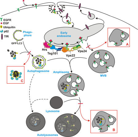
Figure 10. Model for autophagic degradation in control and ESCRT-depleted cells. In control cells, cytoplasmic cargo (proteins and organelles) is sequestered by an isolation membrane/ phagophore, forming double-membrane autophagosomes that can fuse with MVBs, forming amphisomes. Amphisomes, containing both endocytic and autophagic cargo, then fuse with lysosomes, forming autolysosomes, where the content is degraded. Autophagosomes may also fuse directly with lysosomes, although the amphisome pathway seems to be the major pathway in HeLa cells. The ESCRT complexes are required for proper sorting and degradation of ubiquitinated integral membrane proteins (e.g., EGFR) and for proper MVB morphology, and depletion of ESCRT subunits results in the formation of aberrant MVBs (morphology depending of which ESCRT subunit is depleted) (red box A). Degradation of autophagic cargo is also inhibited in ESCRT-depleted cells, proposedly because of inhibited formation of autolysosomes (red box B), although autophagosomes and amphisomes are still formed. In addition, large p62- and ubiquitin-positive membrane-free aggregates accumulate in ESCRT- depleted cells (red box C), indicating that continuous autophagic clearance of cytoplasmic proteins is important to avoid accumulation of ubiquitin-positive aggregates that may cause neurodegeneration.
Figure 10. Model for autophagic degradation in control and ESCRT-depleted cells. In control cells, cytoplasmic cargo (proteins and organelles) is sequestered by an isolation membrane/ phagophore, forming double-membrane autophagosomes that can fuse with MVBs, forming amphisomes. Amphisomes, containing both endocytic and autophagic cargo, then fuse with lysosomes, forming autolysosomes, where the content is degraded. Autophagosomes may also fuse directly with lysosomes, although the amphisome pathway seems to be the major pathway in HeLa cells. The ESCRT complexes are required for proper sorting and degradation of ubiquitinated integral membrane proteins (e.g., EGFR) and for proper MVB morphology, and depletion of ESCRT subunits results in the formation of aberrant MVBs (morphology depending of which ESCRT subunit is depleted) (red box A). Degradation of autophagic cargo is also inhibited in ESCRT-depleted cells, proposedly because of inhibited formation of autolysosomes (red box B), although autophagosomes and amphisomes are still formed. In addition, large p62- and ubiquitin-positive membrane-free aggregates accumulate in ESCRT- depleted cells (red box C), indicating that continuous autophagic clearance of cytoplasmic proteins is important to avoid accumulation of ubiquitin-positive aggregates that may cause neurodegeneration.CC BY-NC-SA · PubMed Central

Verification

Claims the paper makes

The claims and statistics of this paper haven’t been checked yet.

Run the check — free, no account needed. It reads the open-access text and checks the sources it cites.

A few free checks a day · papers already verified open instantly · results are public

Claims other papers make about this one

Each is checked against this paper’s own text: does it say what the citing paper says it does?

Self-check

Percentages, sums and reported differences re-computed from the paper’s own numbers.

The self-check needs the paper’s full text.

Citations

Cites

Show all 62 references

Research works printed but not linked: 3

  1. 1993, Eur. J. Cell Biol., 61, 54
  2. 2006, Am. J. Med. Genet. B. Neuropsychiatr. Genet., 141, 944
  3. 2007, Nat. Rev. Mol. Cell Biol., 8, 355

Cited by

Sorted by citations

See all 631

Metadata

Authors
Filimonenko, M., Stuffers, S., Raiborg, C., Yamamoto, A., Malerød, L., Fisher, E. M. C., Isaacs, A., Brech, A., Stenmark, H., & Simonsen, A.
Affiliations
  • University of Oslo (NO)
  • Columbia University (US)
  • University College London (GB)
  • MRC Prion Unit (GB)
Corresponding author
Simonsen, Anne
Venue
The Journal of Cell Biology
Published
2007-11-05
Volume
179
Issue
3
Pages
485-500
ISSN
  • 0021-9525 (ISSN-L)
  • 1540-8140
OpenAlex
W2103275390
Cited by
626
References
62
Citations by year
2026: 13 · 2025: 26 · 2024: 20 · 2023: 15 · 2022: 18 · 2021: 35 · 2020: 29 · 2019: 27 · 2018: 41 · 2017: 31 · 2016: 46 · 2015: 34 · 2014: 43 · 2013: 36 · 2012: 40
FWCI
29.80
Type
journal article
Language
en
Open access
Locations
  • The Journal of Cell Biology (publishedVersion)
  • PubMed Central (publishedVersion, cc-by-nc-sa)
  • PubMed (publishedVersion)
  • UCL Discovery (University College London) (submittedVersion)
Topics
  • Life Sciences › Biochemistry, Genetics and Molecular Biology › Cell Biology › Autophagy in Disease and Therapy
  • Life Sciences › Biochemistry, Genetics and Molecular Biology › Cell Biology › Cellular transport and secretion
  • Health Sciences › Medicine › Clinical Neurology › Amyotrophic Lateral Sclerosis Research
Keywords
multivesicular bodies, ESCRT, CHMP2B, autophagy, protein aggregates, neurodegenerative diseases, TDP-43, polyglutamine aggregates, Huntington's disease, amyotrophic lateral sclerosis, frontotemporal dementia, p62, ESCRT-III
MeSH terms
Autophagy, Cell Membrane, HeLa Cells, Humans, Huntington Disease, Models, Biological, Mutation, Nerve Tissue Proteins, Neurons, Phenotype, Protein Binding, Endosomes, Neurodegenerative Diseases, Ubiquitin, Endosomal Sorting Complexes Required for Transport

About these checks

Registry, retraction, Cites and statistics results are computed, not judged, with one exception: a printed reference that no index lists is read once by a model before it counts as a citation error. Claims are extracted by a model and kept only when their quote appears verbatim in the text; each cited source is then checked, with a model’s help, for whether it says what the claim relies on. Those verdicts can be wrong — read the claim’s quote and the cited source before relying on one.

Statistics and arithmetic are recomputed, not judged.

Confidence bands are shown only for fields calibrated on our annotated test set. None is calibrated yet, so no band is shown.

What counts as a claim ›How we measure confidence ›How we check this ›

Are you an author, or do you see a problem with this page? Request a review