

The DOI matches its Crossref record; 23 of the 23 references it cites were found in a registry or an index.
USP7 counteracts SCFβTrCP- but not APCCdh1-mediated proteolysis of Claspin
Open accessAbstract
Claspin is an adaptor protein that facilitates the ataxia telangiectasia and Rad3-related (ATR)-mediated phosphorylation and activation of Chk1, a key effector kinase in the DNA damage response. Efficient termination of Chk1 signaling in mitosis and during checkpoint recovery requires SCFβTrCP-dependent destruction of Claspin. Here, we identify the deubiquitylating enzyme ubiquitin-specific protease 7 (USP7) as a novel regulator of Claspin stability. Claspin and USP7 interact in vivo, and USP7 is required to maintain steady-state levels of Claspin. Furthermore, USP7-mediated deubiquitylation markedly prolongs the half-life of Claspin, which in turn increases the magnitude and duration of Chk1 phosphorylation in response to genotoxic stress. Finally, we find that in addition to the M phase–specific, SCFβTrCP-mediated degradation, Claspin is destabilized by the anaphase-promoting complex (APC) and thus remains unstable in G1. Importantly, we demonstrate that USP7 specifically opposes the SCFβTrCP- but not APCCdh1-mediated degradation of Claspin. Thus, Claspin turnover is controlled by multiple ubiquitylation and deubiquitylation activities, which together provide a flexible means to regulate the ATR–Chk1 pathway.
Overview
The claims and statistics of this paper haven’t been checked yet.
Run the check — free, no account needed. It reads the open-access text and checks the sources it cites.
Integrity signals
Registry
Cites
Statistics
Figures
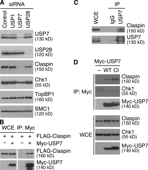
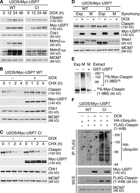
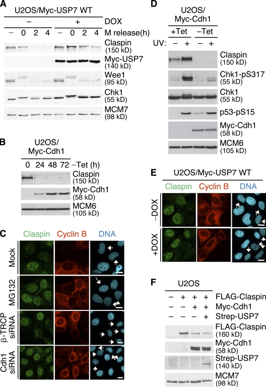
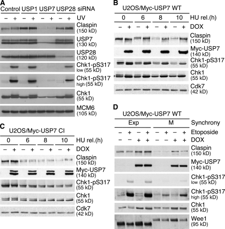
Verification
Claims the paper makes
The claims and statistics of this paper haven’t been checked yet.
Run the check — free, no account needed. It reads the open-access text and checks the sources it cites.
Claims other papers make about this one
Each is checked against this paper’s own text: does it say what the citing paper says it does?
In the citing paper
“USP7 also influences DSB repair by regulating checkpoint kinase 1 (Chk1) (Faustrup et al., 2009; Alonso-de Vega et al., 2014; Zhang P. et al., 2014).”
From USP7 Is a Master Regulator of Genome Stability (2020) · cites this paper as Faustrup et al., 2009
In the citing paper
“USP7 controls Chk1 signaling by deubiquitinating and stabilizing both Chk1 and its activator protein, Claspin (Faustrup et al., 2009; Alonso-de Vega et al., 2014; Zhang P. et al., 2014).”
From USP7 Is a Master Regulator of Genome Stability (2020) · cites this paper as Faustrup et al., 2009
Self-check
Percentages, sums and reported differences re-computed from the paper’s own numbers.
The self-check needs the paper’s full text.
Citations
Cites
The DNA damage response: putting checkpoints in perspective
Zhou, B.-B. S., & Elledge, S. J. (2000) · Nature · doi:10.1038/35044005 · Cited by 3,213
A genomic and functional inventory of deubiquitinating enzymes.
Nijman, S. M. B., Luna-Vargas, M. P. A., Velds, A., Brummelkamp, T. R., Dirac, A. M. G., Sixma, T. K., & Bernards, R. (2005) · Cell · doi:10.1016/j.cell.2005.11.007 · Cited by 1,854
The anaphase promoting complex/cyclosome: a machine designed to destroy
Peters, J.-M. (2006) · Nature Reviews Molecular Cell Biology · doi:10.1038/nrm1988 · Cited by 1,315
RNF8 Ubiquitylates Histones at DNA Double-Strand Breaks and Promotes Assembly of Repair Proteins
Mailand, N., Bekker‐Jensen, S., Kildegaard, H. F., Melander, F., Bártek, J., Lukas, C., & Lukas, J. (2007) · Cell · doi:10.1016/j.cell.2007.09.040 · Cited by 1,193
Self-citationRapid Destruction of Human Cdc25A in Response to DNA Damage
Mailand, N., Falck, J., Lukas, C., Syljuåsen, R. G., Welcker, M., Bartek, J., & Lukas, J. (2000) · Science · doi:10.1126/science.288.5470.1425 · Cited by 787
Self-citationDNA damage checkpoints: from initiation to recovery or adaptation
Bartek, J., & Lukas, J. (2007) · Current Opinion in Cell Biology · doi:10.1016/j.ceb.2007.02.009 · Cited by 784
Self-citationA dynamic role of HAUSP in the p53-Mdm2 pathway
Li, M., Brooks, C. L., Kon, N., & Gu, W. (2004) · Molecular Cell · doi:10.1016/s1097-2765(04)00157-1 · Cited by 679
Spatial organization of the mammalian genome surveillance machinery in response to DNA strand breaks
Bekker‐Jensen, S., Lukas, C., Kitagawa, R., Melander, F., Kastan, M. B., Bártek, J., & Lukas, J. (2006) · The Journal of Cell Biology · doi:10.1083/jcb.200510130 · Cited by 647
Self-citationThe Deubiquitinating Enzyme USP1 Regulates the Fanconi Anemia Pathway
Nijman, S. M. B., Huang, T. T., Dirac, A. M. G., Brummelkamp, T. R., Kerkhoven, R. M., D’Andrea, A. D., & Bernards, R. (2005) · Molecular Cell · doi:10.1016/j.molcel.2005.01.008 · Cited by 601
Regulation of monoubiquitinated PCNA by DUB autocleavage
Huang, T. T., Nijman, S. M. B., Mirchandani, K. D., Galardy, P. J., Cohn, M. A., Haas, W., Gygi, S. P., Ploegh, H. L., Bernards, R., & D’Andrea, A. D. (2006) · Nature Cell Biology · doi:10.1038/ncb1378 · Cited by 597
Research works printed but not linked: 2
- 2006, Nat. Cell Biol., 8, 339
- 2006, Nat. Rev. Mol. Cell Biol., 7, 644
Cited by
Sorted by citations
Breaking the chains: deubiquitylating enzyme specificity begets function
Clague, M. J., Urbé, S., & Komander, D. (2019) · Nature Reviews Molecular Cell Biology · doi:10.1038/s41580-019-0099-1 · Cited by 878
ATM-mediated stabilization of ZEB1 promotes DNA damage response and radioresistance through CHK1
Zhang, P., Wei, Y., Wang, L., Debeb, B. G., Yuan, Y., Zhang, J., Yuan, J., Wang, M., Chen, D., Sun, Y., et al. (2014) · Nature Cell Biology · doi:10.1038/ncb3013 · Cited by 462
Ubiquitylation in apoptosis: a post-translational modification at the edge of life and death
Vucic, D., Dixit, V. M., & Wertz, I. E. (2011) · Nature Reviews Molecular Cell Biology · doi:10.1038/nrm3143 · Cited by 468
Activity-Based Chemical Proteomics Accelerates Inhibitor Development for Deubiquitylating Enzymes
Altun, M., Kramer, H., Willems, L. I., McDermott, J. L., Leach, C. A., Goldenberg, S. J., Kumar, S., Konietzny, R., Fischer, R., Kogan, E., et al. (2011) · Chemistry & Biology · doi:10.1016/j.chembiol.2011.08.018 · Cited by 415
Du, Z., Song, J. K., Wang, Y., Zhao, Y., Guda, K., Yang, S., Kao, H., Xu, Y., Willis, J. E., Markowitz, S. D., et al. (2010) · Science Signaling · doi:10.1126/scisignal.2001462 · Cited by 348
Metadata
- Authors
- Faustrup, H., Bekker-Jensen, S., Bartek, J., Lukas, J., & Mailand, N.
- Affiliations
- Institute of Cancer Biology and Centre for Genotoxic Stress Research, Danish Cancer Society, DK-2100 Copenhagen, Denmark
- Danish Cancer Society (DK)
- ORCID
- Mailand, Niels
- Venue
- The Journal of Cell Biology
- Publisher
- Rockefeller University Press
- Published
- 2009-01-05
- Volume
- 184
- Issue
- 1
- Pages
- 13-19
- ISSN
- 0021-9525 (ISSN-L)
- 1540-8140
- PMC
- PMC2615094
- OpenAlex
- W2087983227
- Cited by
- 113
- References
- 23
- Citations by year
- 2026: 3 · 2025: 3 · 2024: 1 · 2023: 4 · 2022: 5 · 2021: 10 · 2020: 7 · 2019: 8 · 2018: 9 · 2017: 7 · 2016: 12 · 2015: 6 · 2014: 11 · 2013: 9 · 2012: 5
- FWCI
- 2.41
- Type
- journal article
- Language
- en
- Open access
- hybrid
- doi.org
- Licence
- cc-by-nc-sa
- Locations
- The Journal of Cell Biology (publishedVersion, cc-by-nc-sa)
- PubMed Central (publishedVersion, cc-by-nc-sa)
- PubMed (publishedVersion)
- Funders
- National Center for Chronic Disease Prevention and Health Promotion (U58 DP006009)
- Topics
- Life Sciences › Biochemistry, Genetics and Molecular Biology › Molecular Biology › Ubiquitin and proteasome pathways
- Life Sciences › Biochemistry, Genetics and Molecular Biology › Molecular Biology › DNA Repair Mechanisms
- Life Sciences › Biochemistry, Genetics and Molecular Biology › Cell Biology › Microtubule and mitosis dynamics
- Keywords
- Claspin, USP7, deubiquitination, anaphase-promoting complex, CDH1, protein stability, Chk1 phosphorylation, ATR-Chk1 pathway, DNA damage response, spindle assembly checkpoint, ubiquitination, genotoxic stress
- MeSH terms
- Checkpoint Kinase 1, Ubiquitin-Specific Peptidase 7, Cell Line, DNA Damage, Humans, Phosphorylation, Protein Kinases, Substrate Specificity, G1 Phase, Ubiquitin Thiolesterase, Ubiquitin-Protein Ligase Complexes, SKP Cullin F-Box Protein Ligases, Adaptor Proteins, Signal Transducing, Ubiquitination, Anaphase-Promoting Complex-Cyclosome
About these checks
Registry, retraction, Cites and statistics results are computed, not judged, with one exception: a printed reference that no index lists is read once by a model before it counts as a citation error. Claims are extracted by a model and kept only when their quote appears verbatim in the text; each cited source is then checked, with a model’s help, for whether it says what the claim relies on. Those verdicts can be wrong — read the claim’s quote and the cited source before relying on one.
Statistics and arithmetic are recomputed, not judged.
Confidence bands are shown only for fields calibrated on our annotated test set. None is calibrated yet, so no band is shown.
What counts as a claim ›How we measure confidence ›How we check this ›
Are you an author, or do you see a problem with this page? Request a review
