

The DOI matches its Crossref record; 33 of the 33 references it cites were found in a registry or an index.
PINK1 stabilized by mitochondrial depolarization recruits Parkin to damaged mitochondria and activates latent Parkin for mitophagy
Open accessAbstract
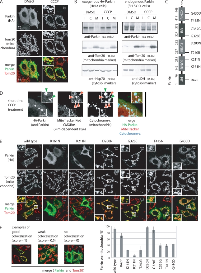
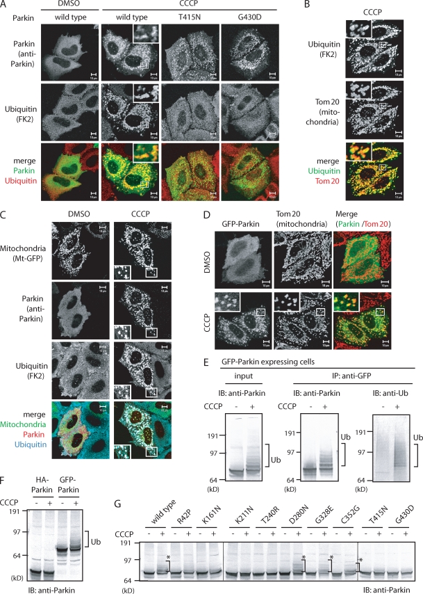
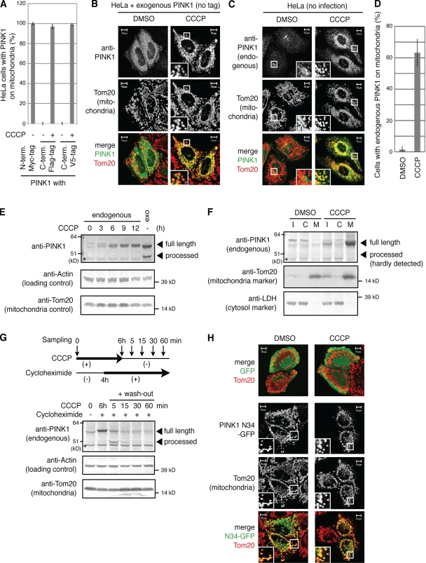
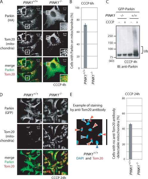
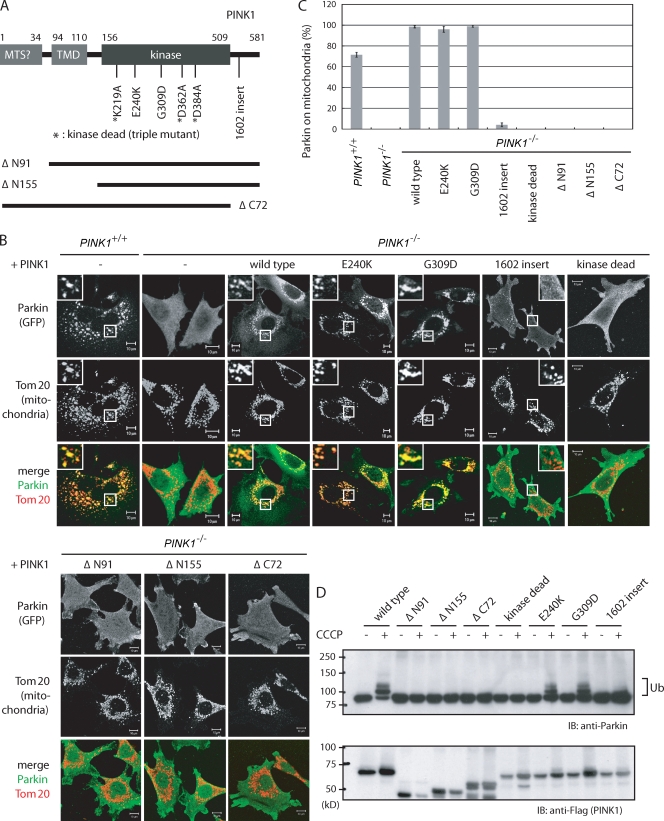
Parkinson's disease (PD) is a prevalent neurodegenerative disorder. Recent identification of genes linked to familial forms of PD such as Parkin and PINK1 (PTEN-induced putative kinase 1) has revealed that ubiquitylation and mitochondrial integrity are key factors in disease pathogenesis. However, the exact mechanism underlying the functional interplay between Parkin-catalyzed ubiquitylation and PINK1-regulated mitochondrial quality control remains an enigma. In this study, we show that PINK1 is rapidly and constitutively degraded under steady-state conditions in a mitochondrial membrane potential-dependent manner and that a loss in mitochondrial membrane potential stabilizes PINK1 mitochondrial accumulation. Furthermore, PINK1 recruits Parkin from the cytoplasm to mitochondria with low membrane potential to initiate the autophagic degradation of damaged mitochondria. Interestingly, the ubiquitin ligase activity of Parkin is repressed in the cytoplasm under steady-state conditions; however, PINK1-dependent mitochondrial localization liberates the latent enzymatic activity of Parkin. Some pathogenic mutations of PINK1 and Parkin interfere with the aforementioned events, suggesting an etiological importance. These results provide crucial insight into the pathogenic mechanisms of PD.
Overview
The claims and statistics of this paper haven’t been checked yet.
Run the check — free, no account needed. It reads the open-access text and checks the sources it cites.
Integrity signals
Registry
Cites
Statistics
Figures
Verification
Claims the paper makes
The claims and statistics of this paper haven’t been checked yet.
Run the check — free, no account needed. It reads the open-access text and checks the sources it cites.
Claims other papers make about this one
Each is checked against this paper’s own text: does it say what the citing paper says it does?
The cited source doesn’t support the claim as written.
In the citing paper
“Concurrently with the changes in parkin levels, the electrophoretic mobility of parkin was transiently shifted upward, consistent with a CCCP-induced activation of parkin and parkin auto-ubiquitination, as reported previously (Matsuda et al, 2010;Lazarou et al, 2013).”
From USP8 regulates mitophagy by removing K6‐linked ubiquitin conjugates from parkin (2014) · cites this paper as Matsuda et al, 2010
Checked by a reasoning model — may be wrong.
Experimental — accuracy not yet measuredThe cited source doesn’t support the claim as written.
In the citing paper
“where it ubiquitinates a number of mitochondrial proteins (Chan et al, 2011;Sarraf et al, 2013), in addition to ubiquitinating itself (Matsuda et al, 2010;Lazarou et al, 2013).”
From USP8 regulates mitophagy by removing K6‐linked ubiquitin conjugates from parkin (2014) · cites this paper as Matsuda et al, 2010
Checked by a reasoning model — may be wrong.
Experimental — accuracy not yet measuredThe source backs part of the claim, not all of it — or backs it for a different population, condition or comparison.
In the citing paper
“Parkin-mediated ubiquitination is critical for the efficient elimination of depolarized dysfunctional mitochondria by autophagy (mitophagy).”
From USP8 regulates mitophagy by removing K6‐linked ubiquitin conjugates from parkin (2014) · cites this paper as Matsuda et al, 2010
Experimental — accuracy not yet measuredThe cited source says what the claim attributes to it.
In the citing paper
“Upon mitochondrial damage, PINK1 is quickly stabilized on damaged mitochondria, which leads to translocation of cytosolic Parkin to damaged mitochondria [11–13].”
From miR-27a and miR-27b regulate autophagic clearance of damaged mitochondria by targeting PTEN-induced putative kinase 1 (PINK1) (2016) · cites this paper as [13]
In the citing paper
“The most extensively studied mitophagy pathway involves the E3 ligase Parkin and PINK1 (Phosphatase and tensin homolog (PTEN)-induced kinase 1) [121,122,123,124,125].”
From Mitochondrial Dynamics and Mitophagy in Skeletal Muscle Health and Aging (2021) · cites this paper as [125]
In the citing paper
“Early-onset Parkinson's disease-associated PINK1-Parkin signaling maintains mitochondrial health.”
From A Cell-Based High-Throughput Screening Identified Two Compounds that Enhance PINK1-Parkin Signaling (2020) · cites this paper as Matsuda et al., 2010
Self-check
Percentages, sums and reported differences re-computed from the paper’s own numbers.
The self-check needs the paper’s full text.
Citations
Cites
Mutations in the parkin gene cause autosomal recessive juvenile parkinsonism
Kitada, T., Asakawa, S., Hattori, N., Matsumine, H., Yamamura, Y., Minoshima, S., Yokochi, M., Mizuno, Y., & Shimizu, N. (1998) · Nature · doi:10.1038/33416 · Cited by 5,227
Self-citationParkin is recruited selectively to impaired mitochondria and promotes their autophagy
Narendra, D., Tanaka, A., Suen, D.-F., & Youle, R. J. (2008) · The Journal of Cell Biology · doi:10.1083/jcb.200809125 · Cited by 4,026
Hereditary Early-Onset Parkinson's Disease Caused by Mutations in PINK1
Valente, E. M., Abou-Sleiman, P. M., Caputo, V., Muqit, M. M. K., Harvey, K., Gispert, S., Ali, Z., Del Turco, D., Bentivoglio, A. R., Healy, D. G., et al. (2004) · Science · doi:10.1126/science.1096284 · Cited by 3,481
PINK1 Is Selectively Stabilized on Impaired Mitochondria to Activate Parkin
Narendra, D. P., Jin, S. M., Tanaka, A., Suen, D.-F., Gautier, C. A., Shen, J., Cookson, M. R., & Youle, R. J. (2010) · PLoS Biology · doi:10.1371/journal.pbio.1000298 · Cited by 2,944
Self-citationPINK1/Parkin-mediated mitophagy is dependent on VDAC1 and p62/SQSTM1
Geisler, S., Holmström, K. M., Skujat, D., Fiesel, F. C., Rothfuss, O. C., Kahle, P. J., & Springer, W. (2010) · Nature Cell Biology · doi:10.1038/ncb2012 · Cited by 2,835
Impairment of starvation-induced and constitutive autophagy in Atg7 -deficient mice
Komatsu, M., Waguri, S., Ueno, T., Iwata, J., Murata, S., Tanida, I., Ezaki, J., Mizushima, N., Ohsumi, Y., Uchiyama, Y., et al. (2005) · The Journal of Cell Biology · doi:10.1083/jcb.200412022 · Cited by 2,331
Self-citationFamilial Parkinson disease gene product, parkin, is a ubiquitin-protein ligase
Shimura, H., Hattori, N., Kubo, S.-I., Mizuno, Y., Asakawa, S., Minoshima, S., Shimizu, N., Iwai, K., Chiba, T., Tanaka, K., et al. (2000) · Nature Genetics · doi:10.1038/77060 · Cited by 2,024 · cited 1×
“In 2000, we and others found that dysfunction of an E3 ubiquitin ligase (Imai et al., 2000; Shimura et al., 2000; Zhang et al., 2000) termed Parkin causes autosomal recessive juvenile Parkinsonism (Kitada et al., 1998).”
BackgroundSelf-citationDrosophila pink1 is required for mitochondrial function and interacts genetically with parkin
Clark, I. E., Dodson, M. W., Jiang, C., Cao, J. H., Huh, J. R., Seol, J. H., Yoo, S. J., Hay, B. A., & Guo, M. (2006) · Nature · doi:10.1038/nature04779 · Cited by 1,788
Mitochondrial dysfunction in Drosophila PINK1 mutants is complemented by parkin
Park, J., Lee, S. B., Lee, S., Kim, Y., Song, S., Kim, S., Bae, E., Kim, J., Shong, M., Kim, J.-M., et al. (2006) · Nature · doi:10.1038/nature04788 · Cited by 1,705
PINK1-dependent recruitment of Parkin to mitochondria in mitophagy
Vives-Bauza, C., Zhou, C., Huang, Y., Cui, M., de Vries, R. L. A., Kim, J., May, J., Tocilescu, M. A., Liu, W., Ko, H. S., et al. (2010) · Proceedings of the National Academy of Sciences · doi:10.1073/pnas.0911187107 · Cited by 1,616
Cited by
Sorted by citations
Items 601–700 of 1,005
Intracellular Dynamics of Synucleins
Surguchov, A. P. (2015) · International review of cytology · doi:10.1016/bs.ircmb.2015.07.007 · Cited by 77
Quansah, E., Peelaerts, W., Langston, J. W., Simon, D. K., Colca, J. R., & Brundin, P. (2018) · Molecular Neurodegeneration · doi:10.1186/s13024-018-0260-x · Cited by 76
Parkinson's disease and mitophagy: an emerging role for LRRK2
Singh, F., & Ganley, I. G. (2021) · Biochemical Society Transactions · doi:10.1042/bst20190236 · Cited by 79
Rahman, F. A., & Quadrilatero, J. (2021) · Cellular and Molecular Life Sciences · doi:10.1007/s00018-021-03807-9 · Cited by 77
Panes-Fernández, J., Wendt, A., Ramírez-Molina, O., Castro, P. A., & Fuentealba, J. (2021) · Neural Regeneration Research · doi:10.4103/1673-5374.317957 · Cited by 77
Yamamoto‐Imoto, H., Minami, S., Shioda, T., Yamashita, Y., Sakai, S., Maeda, S., Yamamoto, T., Oki, S., Takashima, M., Yamamuro, T., et al. (2022) · Cell Reports · doi:10.1016/j.celrep.2022.110444 · Cited by 76
Mitochondrial Fission and Autophagy in the Normal and Diseased Heart
Iglewski, M., Hill, J. A., Lavandero, S., & Rothermel, B. A. (2010) · Current Hypertension Reports · doi:10.1007/s11906-010-0147-x · Cited by 75
Mitochondrial autophagy in cardiomyopathy
Tong, M., & Sadoshima, J. (2016) · Current Opinion in Genetics & Development · doi:10.1016/j.gde.2016.02.006 · Cited by 75
PINK1-Mediated Phosphorylation of Parkin Boosts Parkin Activity in Drosophila
Shiba‐Fukushima, K., Inoshita, T., Hattori, N., & Imai, Y. (2014) · PLoS Genetics · doi:10.1371/journal.pgen.1004391 · Cited by 75
Self-citationStudying Autophagy in Zebrafish
Mathai, B. J., Meijer, A. H., & Simonsen, A. (2017) · Cells · doi:10.3390/cells6030021 · Cited by 75
PTENα regulates mitophagy and maintains mitochondrial quality control
Li, G., Yang, J., Yang, C., Zhu, M., Jin, Y., McNutt, M. A., & Yin, Y. (2018) · Autophagy · doi:10.1080/15548627.2018.1489477 · Cited by 77
Structural basis for specific cleavage of Lys6-linked polyubiquitin chains by USP30
Sato, Y., Okatsu, K., Saeki, Y., Yamano, K., Matsuda, N., Kaiho, A., Yamagata, A., Goto‐Ito, S., Ishikawa, M., Hashimoto, Y., et al. (2017) · Nature Structural & Molecular Biology · doi:10.1038/nsmb.3469 · Cited by 76
Self-citationMitophagy Contributes to the Pathogenesis of Inflammatory Diseases
Zhao, Y., Huang, S., Liu, J., Wu, X., Zhou, S., Dai, K., & Kou, Y. (2018) · Inflammation · doi:10.1007/s10753-018-0835-2 · Cited by 74
Sala, D., Cunningham, T. J., Stec, M. J., Etxaniz, U., Nicoletti, C., Dall’Agnese, A., Puri, P., Duester, G., Latella, L., & Sacco, A. (2019) · Nature Communications · doi:10.1038/s41467-019-09746-1 · Cited by 74
Patergnani, S., Missiroli, S., Morciano, G., Perrone, M., Mantovani, C. M., Anania, G., Fiorica, F., Pinton, P., & Giorgi, C. (2021) · Cancers · doi:10.3390/cancers13225622 · Cited by 77
Parkinson's disease proteins: Novel mitochondrial targets for cardioprotection
Mukherjee, U., Ong, S., Ong, S., & Hausenloy, D. J. (2015) · Pharmacology & Therapeutics · doi:10.1016/j.pharmthera.2015.10.005 · Cited by 72
Neuronal Autophagy: Self-eating or Self-cannibalism in Alzheimer’s Disease
Ułamek-Kozioł, M., Furmaga-Jabłońska, W., Januszewski, S., Brzozowska, J., Ściślewska, M., Jabłoński, M., & Pluta, R. M. (2013) · Neurochemical Research · doi:10.1007/s11064-013-1082-4 · Cited by 75
Chambers, R. P., Call, G. B., Meyer, D., Smith, J., Techau, J. A., Pearman, K., & Buhlman, L. M. (2013) · Behavioural Brain Research · doi:10.1016/j.bbr.2013.07.020 · Cited by 71
Yasuda, T., Hayakawa, H., Nihira, T., Ren, Y., Nakata, Y., Nagai, M., Hattori, N., Miyake, K., Takada, M., Shimada, T., et al. (2011) · Journal of Neuropathology & Experimental Neurology · doi:10.1097/nen.0b013e3182269ecd · Cited by 69
Self-citationSelect E2 enzymes differentially regulate parkin activation and mitophagy
Fiesel, F. C., Moussaud-Lamodière, E. L., Ando, M., & Springer, W. (2014) · Journal of Cell Science · doi:10.1242/jcs.147520 · Cited by 72
Identifying Functional Cysteine Residues in the Mitochondria
Bak, D. W., Pizzagalli, M. D., & Weerapana, E. (2017) · ACS Chemical Biology · doi:10.1021/acschembio.6b01074 · Cited by 70
The Multifaceted Roles of Autophagy in Flavivirus-Host Interactions
Ke, P. (2018) · International Journal of Molecular Sciences · doi:10.3390/ijms19123940 · Cited by 78
PINK1 Content in Mitochondria is Regulated by ER-Associated Degradation
Guardia‐Laguarta, C., Liu, Y., Lauritzen, K. H., Erdjument‐Bromage, H., Martin, B. M. S., Swayne, T. C., Jiang, X., & Przedborski, S. (2019) · Journal of Neuroscience · doi:10.1523/jneurosci.1691-18.2019 · Cited by 70
Regulation of neuronal autophagy and the implications in neurodegenerative diseases
Cai, Q., & Ganesan, D. (2021) · Neurobiology of Disease · doi:10.1016/j.nbd.2021.105582 · Cited by 70
Chakkittukandiyil, A., Sajini, D. V., Karuppaiah, A., & Selvaraj, D. (2022) · Neurochemistry International · doi:10.1016/j.neuint.2022.105325 · Cited by 75
Zhao, X., Wang, Z., Wang, L., Jiang, T., Dong, D., & Sun, M. (2024) · Pharmacological Research · doi:10.1016/j.phrs.2024.107466 · Cited by 76
Qu, D., Hage, A., Don-Carolis, K., Huang, E., Joselin, A. P., Safarpour, F., Marcogliese, P. C., Rousseaux, M. W. C., Hewitt, S. J., Huang, T., et al. (2015) · Journal of Biological Chemistry · doi:10.1074/jbc.m115.677815 · Cited by 73
Zhang, H., Mi, L., Wang, T., Yuan, L., Li, X., Dong, L., Zhao, P., Fu, J., Yao, B., & Zhou, Z. (2016) · Toxicology in Vitro · doi:10.1016/j.tiv.2016.04.006 · Cited by 69
The multi‐faceted role of mitochondria in the pathology of Parkinson’s disease
Trinh, D., Israwi, A. R., Arathoon, L. R., Gleave, J. A., & Nash, J. E. (2020) · Journal of Neurochemistry · doi:10.1111/jnc.15154 · Cited by 74
Role of mitochondria in renal ischemia-reperfusion injury
Huang, R., Zhang, C., Xiang, Z., Lin, T., Ling, J., & Hu, H. (2024) · The FEBS Journal · doi:10.1111/febs.17130 · Cited by 72
Chi, H.-C., Chen, S.-L., Lin, S.-L., Tsai, C.-Y., Chuang, W.-Y., Lin, Y.-H., Huang, Y.-H., Tsai, M.-M., Yeh, C.-H., & Lin, K.-H. (2017) · Oncogene · doi:10.1038/onc.2017.136 · Cited by 70
Yang, Q., Sun, G., Yin, H., Li, H., Cao, Z., Wang, J., Zhou, M., Wang, H., & Li, J. (2018) · Free Radical Biology and Medicine · doi:10.1016/j.freeradbiomed.2018.02.025 · Cited by 70
Regulation of cell death in the cardiovascular system
Patel, P., & Karch, J. M. (2019) · International review of cytology · doi:10.1016/bs.ircmb.2019.11.005 · Cited by 74
Wen, H., Li, L., Zhan, L., Zuo, Y., Li, K., Qiu, M., Li, H., Sun, W., & Xu, E. (2021) · Cell Death and Disease · doi:10.1038/s41419-021-03900-8 · Cited by 69
Mitophagy in depression: Pathophysiology and treatment targets
Tripathi, A., Scaini, G., Barichello, T., Quevedo, J., & Pillai, A. (2021) · Mitochondrion · doi:10.1016/j.mito.2021.08.016 · Cited by 71
Mitophagy Regulation Following Myocardial Infarction
Turkieh, A., Masri, Y. E., Pinet, F., & Dubois‐Deruy, E. (2022) · Cells · doi:10.3390/cells11020199 · Cited by 69
Crosstalk Between ROS and Autophagy in Tumorigenesis: Understanding the Multifaceted Paradox
Hasan, A., Rizvi, S. F., Parveen, S., Pathak, N., Nazir, A., & Mir, S. S. (2022) · Frontiers in Oncology · doi:10.3389/fonc.2022.852424 · Cited by 77
Lacoursiere, R. E., Hadi, D., & Shaw, G. S. (2022) · Biomolecules · doi:10.3390/biom12030467 · Cited by 74
Zhou, Y., Liu, Y., Kang, Z., Yao, H. P., Song, N., Wang, M., Song, C., Zhang, K., Ding, J., Tang, J., et al. (2023) · Autophagy · doi:10.1080/15548627.2023.2196889 · Cited by 72
Fésüs, L., Demény, M. Á., & Petrovski, G. (2010) · Antioxidants and Redox Signaling · doi:10.1089/ars.2010.3485 · Cited by 69
Mitochondria: key players in the neurotoxic effects of amphetamines
Barbosa, D. J., Capela, J. P., Feio‐Azevedo, R., Gomes, A., Bastos, M. D. L., & Carvalho, F. D. (2015) · Archives of Toxicology · doi:10.1007/s00204-015-1478-9 · Cited by 70
Autophagic flux control in neurodegeneration: Progress and precision targeting—Where do we stand?
Lumkwana, D., du Toit, A., Kinnear, C. J., & Loos, B. (2017) · Progress in Neurobiology · doi:10.1016/j.pneurobio.2017.03.006 · Cited by 69
Ren, Y., Li, Y., Yan, J., Ma, M., Zhou, D., Xue, Z., Zhang, Z., Liu, H., Yang, H., Jia, L., et al. (2017) · Scientific Reports · doi:10.1038/s41598-017-03319-2 · Cited by 70
Research progress on the pharmacological effects of berberine targeting mitochondria
Fang, X., Wu, H., Wei, J., Miao, R., Zhang, Y., & Tian, J. (2022) · Frontiers in Endocrinology · doi:10.3389/fendo.2022.982145 · Cited by 75
Wang, H., Luo, W., Chen, H., Cai, Z., & Xu, G. (2024) · Mitochondrion · doi:10.1016/j.mito.2024.101847 · Cited by 70
Programming cancer cells for high expression levels of Mcl1
Ertel, F., Nguyen, M., Roulston, A., & Shore, G. C. (2013) · EMBO Reports · doi:10.1038/embor.2013.20 · Cited by 67
Regulation of Parkin E3 ubiquitin ligase activity
Walden, H., & Martinez-Torres, R. J. (2012) · Cellular and Molecular Life Sciences · doi:10.1007/s00018-012-0978-5 · Cited by 70
Mitochondrial dynamics and mitophagy in Parkinson's disease: A fly point of view
von Stockum, S., Nardin, A., Schrepfer, E., & Ziviani, E. (2015) · Neurobiology of Disease · doi:10.1016/j.nbd.2015.11.002 · Cited by 70
Ubiquitin and Parkinson's disease through the looking glass of genetics
Walden, H., & Muqit, M. M. K. (2017) · Biochemical Journal · doi:10.1042/bcj20160498 · Cited by 69
Goiran, T., Duplan, E., Chami, M., Bourgeois, A., Manaa, W. E., Rouland, L., Dunys, J., Lauritzen, I., You, H., Stambolic, V., et al. (2017) · Biological Psychiatry · doi:10.1016/j.biopsych.2017.04.011 · Cited by 68
Inhibition of apoptotic Bax translocation to the mitochondria is a central function of parkin
Charan, R. A., Johnson, B. N., Zaganelli, S., Nardozzi, J. D., & LaVoie, M. J. (2014) · Cell Death & Disease · doi:10.1038/cddis.2014.278 · Cited by 67
Role of autophagy in alcohol and drug-induced liver injury
Williams, J. A. R., & Ding, W. (2019) · Food and Chemical Toxicology · doi:10.1016/j.fct.2019.111075 · Cited by 66
Role of Selective Autophagy in Spermatogenesis and Male Fertility
Lv, C., Wang, X., Guo, Y., & Yuan, S. (2020) · Cells · doi:10.3390/cells9112523 · Cited by 73
Zhang, L., Yang, F., Li, Y., Cao, H., Huang, A., Zhuang, Y., Zhang, C., Hu, G., Mao, Y., Luo, J., et al. (2021) · Environmental Pollution · doi:10.1016/j.envpol.2021.117301 · Cited by 67
Acute focal brain damage alters mitochondrial dynamics and autophagy in axotomized neurons
Cavallucci, V., Bisicchia, E., Cencioni, M. T., Ferri, A., Latini, L., Nobili, A., Biamonte, F., Nazio, F., Fanelli, F., Moreno, S., et al. (2014) · Cell Death and Disease · doi:10.1038/cddis.2014.511 · Cited by 68
Koh, H., Kim, H., Kim, M. J., Park, J., Lee, H.-J., & Chung, J. (2012) · Journal of Biological Chemistry · doi:10.1074/jbc.m111.337907 · Cited by 68
A Mechanistic Review of Mitophagy and Its Role in Protection against Alcoholic Liver Disease
Williams, J. A. R., & Ding, W. (2015) · Biomolecules · doi:10.3390/biom5042619 · Cited by 68
The Close Interconnection between Mitochondrial Dynamics and Mitophagy in Cancer
Bordi, M., Nazio, F., & Campello, S. (2017) · Frontiers in Oncology · doi:10.3389/fonc.2017.00081 · Cited by 71
Gleave, J. A., Arathoon, L. R., Trinh, D., Lizal, K. E., Giguère, N., Barber, J., Najarali, Z., Khan, M. H., Thiele, S. L., Semmen, M. S., et al. (2017) · Neurobiology of Disease · doi:10.1016/j.nbd.2017.06.009 · Cited by 66
Wu, Z., Wu, A., Dong, J., Sigears, A., & Lu, B. (2018) · Experimental Gerontology · doi:10.1016/j.exger.2018.09.014 · Cited by 67
Even, B., Fayad‐Kobeissi, S., Gagliolo, J., Motterlini, R., Boczkowski, J., Foresti, R., & Dagouassat, M. (2018) · Aging Cell · doi:10.1111/acel.12837 · Cited by 68
Yamaguchi, A., Ishikawa, K., Inoshita, T., Shiba‐Fukushima, K., Saiki, S., Hatano, T., Mori, A., Oji, Y., Okuzumi, A., Li, Y., et al. (2020) · Stem Cell Reports · doi:10.1016/j.stemcr.2020.04.011 · Cited by 66
Self-citationSwerdlow, N., & Wilkins, H. (2020) · International Journal of Molecular Sciences · doi:10.3390/ijms21249661 · Cited by 66
Wang, X., Feng, S., Wang, Y., Yuan, Y., Li, Z., Chen, N., Wang, Z., & Zhang, Y. (2021) · Cellular and Molecular Neurobiology · doi:10.1007/s10571-021-01039-w · Cited by 66
Chemotherapy Resistance: Role of Mitochondrial and Autophagic Components
Bahar, E., Han, S., Kim, J., & Yoon, H. (2022) · Cancers · doi:10.3390/cancers14061462 · Cited by 67
Phospho-ubiquitin: upending the PINK–Parkin–ubiquitin cascade
Matsuda, N. (2016) · The Journal of Biochemistry · doi:10.1093/jb/mvv125 · Cited by 68
Self-citationPaez, H. G., Pitzer, C. R., & Alway, S. E. (2023) · Cells · doi:10.3390/cells12020249 · Cited by 71
Optineurin provides a mitophagy contact site for TBK1 activation
Yamano, K., Sawada, M., Kikuchi, R., Nagataki, K., Kojima, W., Endo, R., Kinefuchi, H., Sugihara, A., Fujino, T., Watanabe, A., et al. (2024) · The EMBO Journal · doi:10.1038/s44318-024-00036-1 · Cited by 68
Self-citationPINK1 signalling in cancer biology
O’Flanagan, C. H., & O’Neill, C. (2014) · Biochimica et Biophysica Acta (BBA) - Reviews on Cancer · doi:10.1016/j.bbcan.2014.10.006 · Cited by 66
Regional variation in mitochondrial DNA copy number in mouse brain
Fuke, S., Kubota‐Sakashita, M., Kasahara, T., Shigeyoshi, Y., & Kato, T. (2010) · Biochimica et Biophysica Acta (BBA) - Bioenergetics · doi:10.1016/j.bbabio.2010.11.016 · Cited by 62
Reggiori, F., Komatsu, M., Finley, K. D., & Simonsen, A. (2012) · International Journal of Cell Biology · doi:10.1155/2012/156272 · Cited by 68
Self-citationBrown, K., Yang, P., Salvador, D., Kulikauskas, R. M., Ruohola‐Baker, H., Robitaille, A. M., Chien, A. J., Moon, R. T., & Sherwood, V. (2017) · Oncogene · doi:10.1038/onc.2016.450 · Cited by 67
Liu, N., Wu, J., Zhang, L., Gao, Z., Sun, Y., Yu, M., Zhao, Y., Dong, S., Lu, F., & Zhang, W. (2017) · Journal of Cellular and Molecular Medicine · doi:10.1111/jcmm.13223 · Cited by 65
Drake, J. C., Laker, R. C., Wilson, R. J., Zhang, M., & Yan, Z. (2018) · Cell Cycle · doi:10.1080/15384101.2018.1559556 · Cited by 68
Müller, M., Kötter, P., Behrendt, C., Walter, E., Scheckhuber, C. Q., Entian, K.-D., & Reichert, A. S. (2015) · Cell Reports · doi:10.1016/j.celrep.2015.01.044 · Cited by 67
Mitochondrial Importance in Alzheimer’s, Huntington’s and Parkinson’s Diseases
Correia, S. C., Santos, R. X., Perry, G. L., Zhu, X., Moreira, P. I., & Smith, M. A. (2012) · Advances in experimental medicine and biology · doi:10.1007/978-1-4614-0653-2_16 · Cited by 65
Bereiter‐Hahn, J., & Jendrach, M. (2010) · International review of cytology · doi:10.1016/s1937-6448(10)84001-8 · Cited by 67
Xu, H. M., Liu, Y., Li, L.-S., & Liu, Y. (2023) · Aging and Disease · doi:10.14336/ad.2022.1123 · Cited by 69
Alpha-synuclein-induced neurodegeneration is exacerbated in PINK1 knockout mice
Oliveras-Salvá, M., Macchi, F., Coessens, V., Deleersnijder, A., Gérard, M., der Perren, A. V., den Haute, C. V., & Baekelandt, V. (2014) · Neurobiology of Aging · doi:10.1016/j.neurobiolaging.2014.04.032 · Cited by 66
Unbreak My Heart: Targeting Mitochondrial Autophagy in Diabetic Cardiomyopathy
Kubli, D. A., & Gustafsson, Å. B. (2015) · Antioxidants and Redox Signaling · doi:10.1089/ars.2015.6322 · Cited by 66
Mitochondrial Reactive Oxygen Species and Kidney Hypoxia in the Development of Diabetic Nephropathy
Schiffer, T. A., & Friederich‐Persson, M. (2017) · Frontiers in Physiology · doi:10.3389/fphys.2017.00211 · Cited by 66
Evidence that phosphorylated ubiquitin signaling is involved in the etiology of Parkinson’s disease
Shiba‐Fukushima, K., Ishikawa, K., Inoshita, T., Izawa, N., Takanashi, M., Sato, S., Onodera, O., Akamatsu, W., Okano, H., Imai, Y., et al. (2017) · Human Molecular Genetics · doi:10.1093/hmg/ddx201 · Cited by 64
Self-citationMyricetin improves endurance capacity and mitochondrial density by activating SIRT1 and PGC-1α
Jung, H., Lee, D.-Y., Ryu, H. G., Choi, B.-H., Go, Y., Lee, N., Lee, D., Son, H. G., Jeon, J., Kim, S., et al. (2017) · Scientific Reports · doi:10.1038/s41598-017-05303-2 · Cited by 67
Onnis, A., Cianfanelli, V., Cassioli, C., Samardžić, D., Pelicci, P. G., Cecconi, F., & Baldari, C. T. (2018) · Autophagy · doi:10.1080/15548627.2018.1505153 · Cited by 61
Loss of HSPA9 induces peroxisomal degradation by increasing pexophagy
Jo, D. S., Park, S. J., Kim, A., Park, N. Y., Kim, J. B., Bae, J.-E., Park, H. J., Shin, J. H., Chang, J. W., Kim, P. K., et al. (2020) · Autophagy · doi:10.1080/15548627.2020.1712812 · Cited by 64
Tokarew, J. M., Kodsi, D. N. E., Lengacher, N. A., Fehr, T. K., Nguyen, A., Shutinoski, B., O’Nuallain, B., Jin, M., Khan, J. M., Ng, A. C., et al. (2021) · Acta Neuropathologica · doi:10.1007/s00401-021-02285-4 · Cited by 64
Self-citationGuan, Z., Chen, J., Wang, L., Hao, M., Dong, X., Luo, T., Jiang, J., Lin, Z., Li, X., Chen, P., et al. (2022) · Phytomedicine · doi:10.1016/j.phymed.2022.154494 · Cited by 63
Potentiation of neurotoxicity in double-mutant mice with Pink1 ablation and A53T-SNCA overexpression
Gispert, S., Brehm, N., Weil, J., Seidel, K., Rüb, U., Kern, B., Walter, M. J., Roeper, J., & Auburger, G. W. J. (2014) · Human Molecular Genetics · doi:10.1093/hmg/ddu520 · Cited by 64
The Aging Heart: Mitophagy at the Center of Rejuvenation
Liang, W., & Gustafsson, Å. B. (2020) · Frontiers in Cardiovascular Medicine · doi:10.3389/fcvm.2020.00018 · Cited by 65
Mitochondria and the culture of the Borg
Braschi, E., & McBride, H. M. (2010) · BioEssays · doi:10.1002/bies.201000073 · Cited by 65
Synergistic recruitment of UbcH7~Ub and phosphorylated Ubl domain triggers parkin activation
Condos, T., Dunkerley, K. M., Freeman, E. A., Barber, K. R., Aguirre, J. D., Chaugule, V. K., Xiao, Y., Konermann, L., Walden, H., & Shaw, G. S. (2018) · The EMBO Journal · doi:10.15252/embj.2018100014 · Cited by 61
Regulation of Iron Homeostasis and Related Diseases
Li, Y., Huang, X., Wang, J., Huang, R., & Wan, D. (2020) · Mediators of Inflammation · doi:10.1155/2020/6062094 · Cited by 66
Oxidative stress and synaptic dysfunction in rodent models of Parkinson's disease
Imbriani, P., Martella, G., Bonsi, P., & Pisani, A. (2022) · Neurobiology of Disease · doi:10.1016/j.nbd.2022.105851 · Cited by 63
Imai, Y., & Lu, B. (2011) · Current Opinion in Neurobiology · doi:10.1016/j.conb.2011.10.016 · Cited by 63
Hara, Y., Yanatori, I., Ikeda, M., Kiyokage, E., Nishina, S., Tomiyama, Y., Toida, K., Kishi, F., Kato, N., Imamura, M., et al. (2014) · American Journal Of Pathology · doi:10.1016/j.ajpath.2014.07.024 · Cited by 62
TSPO is a REDOX regulator of cell mitophagy
Gatliff, J., & Campanella, M. (2015) · Biochemical Society Transactions · doi:10.1042/bst20150037 · Cited by 64
Specificity and disease in the ubiquitin system
Chaugule, V. K., & Walden, H. (2016) · Biochemical Society Transactions · doi:10.1042/bst20150209 · Cited by 65
Ntsapi, C., & Loos, B. (2016) · Experimental Gerontology · doi:10.1016/j.exger.2016.07.014 · Cited by 65
Yamaguchi, O. (2019) · Circulation Journal · doi:10.1253/circj.cj-18-1065 · Cited by 62
OGT Regulates Hematopoietic Stem Cell Maintenance via PINK1-Dependent Mitophagy
Murakami, K., Kurotaki, D., Kawase, W., Soma, S., Fukuchi, Y., Kunimoto, H., Yoshimi, R., Koide, S., Oshima, M., Hishiki, T., et al. (2021) · Cell Reports · doi:10.1016/j.celrep.2020.108579 · Cited by 62
Metadata
- Authors
- Matsuda, N., Sato, S., Shiba, K., Okatsu, K., Saisho, K., Gautier, C. A., Sou, Y.-S., Saiki, S., Kawajiri, S., Sato, F., Kimura, M., Komatsu, M., Hattori, N., & Tanaka, K.
- Affiliations
- Tokyo Metropolitan Institute of Medical Science (JP)
- Juntendo University (JP)
- Center for Neurologic Diseases, Brigham and Women's Hospital, Harvard Medical School, Boston, MA 02115 4
- Japan Science and Technology Agency (JP)
- Corresponding author
- Matsuda, Noriyuki
- Tanaka, Keiji
- ORCID
- Venue
- Journal of Cell Biology
- Publisher
- Rockefeller University Press
- Published
- 2010-04-19
- Volume
- 189
- Issue
- 2
- Pages
- 211-221
- ISSN
- 0021-9525 (ISSN-L)
- 1540-8140
- PMC
- PMC2856912
- OpenAlex
- W2138311396
- Cited by
- 1,969
- References
- 32
- Citations by year
- 2026: 98 · 2025: 115 · 2024: 118 · 2023: 121 · 2022: 144 · 2021: 152 · 2020: 175 · 2019: 131 · 2018: 123 · 2017: 100 · 2016: 110 · 2015: 118 · 2014: 119 · 2013: 116 · 2012: 101
- FWCI
- 65.66
- Type
- journal article
- Language
- en
- Open access
- hybrid
- doi.org
- Licence
- cc-by-nc-sa
- Locations
- The Journal of Cell Biology (publishedVersion, cc-by-nc-sa)
- PubMed Central (publishedVersion, cc-by-nc-sa)
- PubMed (publishedVersion)
- Digital Access to Scholarship at Harvard (DASH) (Harvard University) (submittedVersion, cc-by)
- Funders
- Ministry of Education, Culture, Sports, Science and Technology
- National Institutes of Health
- Topics
- Health Sciences › Medicine › Clinical Neurology › Parkinson's Disease Mechanisms and Treatments
- Life Sciences › Biochemistry, Genetics and Molecular Biology › Cell Biology › Autophagy in Disease and Therapy
- Life Sciences › Biochemistry, Genetics and Molecular Biology › Molecular Biology › Nuclear Receptors and Signaling
- Keywords
- PINK1, Parkin, Parkinson's disease, mitophagy, mitochondrial membrane potential, mitochondrial depolarization, ubiquitination, ubiquitin ligase activity, mitochondrial quality control, PINK1 mutations, parkin mutations, protein stabilization
- MeSH terms
- PTEN-Induced Putative Kinase, Animals, Carbonyl Cyanide m-Chlorophenyl Hydrazone, Dimethyl Sulfoxide, Fibroblasts, Fluorescent Dyes, HeLa Cells, Humans, Mitochondria, Parkinson Disease, Protein Kinases, Recombinant Fusion Proteins, Solvents, Uncoupling Agents, Biomarkers, Receptors, Cytoplasmic and Nuclear, Mice, Knockout, Mitochondrial Proteins, Ubiquitin-Protein Ligases, Mice, Membrane Potential, Mitochondrial
About these checks
Registry, retraction, Cites and statistics results are computed, not judged, with one exception: a printed reference that no index lists is read once by a model before it counts as a citation error. Claims are extracted by a model and kept only when their quote appears verbatim in the text; each cited source is then checked, with a model’s help, for whether it says what the claim relies on. Those verdicts can be wrong — read the claim’s quote and the cited source before relying on one.
Statistics and arithmetic are recomputed, not judged.
Confidence bands are shown only for fields calibrated on our annotated test set. None is calibrated yet, so no band is shown.
What counts as a claim ›How we measure confidence ›How we check this ›
Are you an author, or do you see a problem with this page? Request a review
