

The DOI matches its Crossref record; 51 of the 55 references it cites were found in a registry or an index.
Interleukin (IL)-6 directs the differentiation of IL-4-producing CD4+ T cells
Open accessAbstract
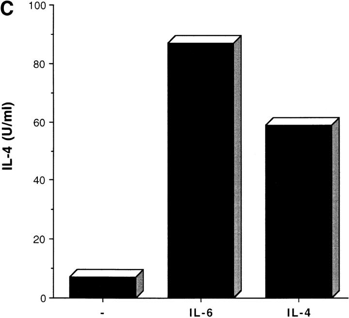
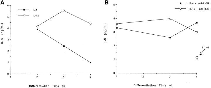
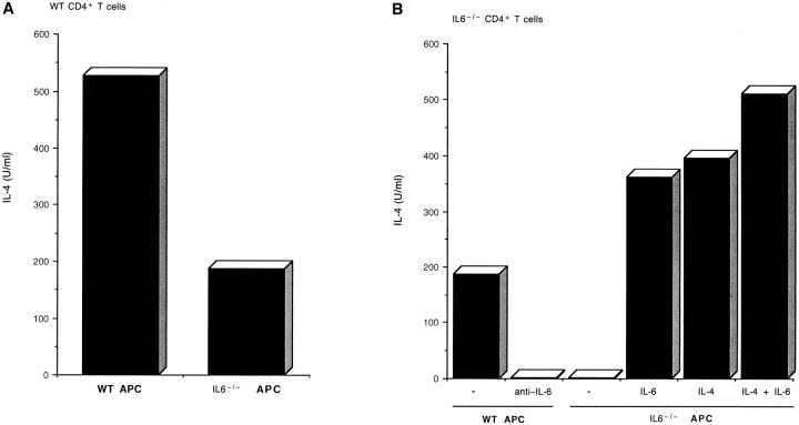
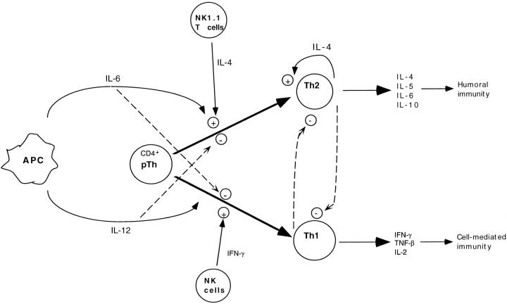
Interleukin (IL)-4 is the most potent factor that causes naive CD4+ T cells to differentiate to the T helper cell (Th) 2 phenotype, while IL-12 and interferon γ trigger the differentiation of Th1 cells. However, the source of the initial polarizing IL-4 remains unclear. Here, we show that IL-6, probably secreted by antigen-presenting cells, is able to polarize naive CD4+ T cells to effector Th2 cells by inducing the initial production of IL-4 in CD4+ T cells. These results show that the nature of the cytokine (IL-12 or IL-6), which is produced by antigen-presenting cells in response to a particular pathogen, is a key factor in determining the nature of the immune response.
Overview
The claims and statistics of this paper haven’t been checked yet.
Run the check — free, no account needed. It reads the open-access text and checks the sources it cites.
Integrity signals
Registry
Cites
Statistics
Figures
Verification
Claims the paper makes
The claims and statistics of this paper haven’t been checked yet.
Run the check — free, no account needed. It reads the open-access text and checks the sources it cites.
Claims other papers make about this one
Each is checked against this paper’s own text: does it say what the citing paper says it does?
The cited source says what the claim attributes to it.
In the citing paper
“In addition to the aforementioned role of IL-6 on Tfh cells, IL-6 is known to direct the differentiation of IL-4-producing T helper type 2 (Th2) cells, while it inhibits the differentiation of IFNγ-producing Th1 cells (155).”
From IL-6 in inflammation, autoimmunity and cancer (2021) · cites this paper as [155]
Self-check
Percentages, sums and reported differences re-computed from the paper’s own numbers.
The self-check needs the paper’s full text.
Citations
Cites
Single-Step Method of RNA Isolation by Acid Guanidinium Thiocyanate–Phenol–Chloroform Extraction
Chomczynski, P., & Sacchi, N. (1987) · Analytical Biochemistry · doi:10.1006/abio.1987.9999 · Cited by 65,515
Development of T H 1 CD4 + T Cells Through IL-12 Produced by Listeria -Induced Macrophages
Hsieh, C.-S., Macatonia, S. E., Tripp, C. S., Wolf, S. F., O'Garra, A., & Murphy, K. M. (1993) · Science · doi:10.1126/science.8097338 · Cited by 3,134
IL-10 inhibits cytokine production by activated macrophages
Fiorentino, D. F., Zlotnik, A., Mosmann, T. R., Howard, M., & O’Garra, A. (1991) · The Journal of Immunology · doi:10.4049/jimmunol.147.11.3815 · Cited by 2,888
Van Snick, J. (1990) · Annual Review of Immunology · doi:10.1146/annurev.iy.08.040190.001345 · Cited by 1,890
Lymphocyte responses and cytokines
Paul, W. E., & Seder, R. A. (1994) · Cell · doi:10.1016/0092-8674(94)90332-8 · Cited by 1,832
Impaired immune and acute-phase responses in interleukin-6-deficient mice
Kopf, M., Baumann, H., Freer, G., Freudenberg, M., Lamers, M., Kishimoto, T., Zinkernagel, R., Bluethmann, H., & Köhler, G. (1994) · Nature · doi:10.1038/368339a0 · Cited by 1,768 · cited 1×
“In correlation with the previous characterization of IL-62/2 mice (32, 33), analysis of cellular populations in the spleen”
Regulated expression of three C/EBP isoforms during adipose conversion of 3T3-L1 cells.
Cao, Z., Umek, R. M., & McKnight, S. L. (1991) · Genes & Development · doi:10.1101/gad.5.9.1538 · Cited by 1,663
Kuchroo, V. K., Das, M. P., Brown, J. A., Ranger, A. M., Zamvil, S. S., Sobel, R. A., Weiner, H. L., Nabavi, N., & Glimcher, L. H. (1995) · Cell · doi:10.1016/0092-8674(95)90349-6 · Cited by 1,629
A nuclear factor for IL‐6 expression (NF‐IL6) is a member of a C/EBP family.
Akira, S., Isshiki, H., Sugita, T., Tanabe, O., Kinoshita, S. M., Nishio, Y., Nakajima, T., Hirano, T., & Kishimoto, T. (1990) · The EMBO Journal · doi:10.1002/j.1460-2075.1990.tb08316.x · Cited by 1,450
Interleukin-6 triggers the association of its receptor with a possible signal transducer, gp130
Taga, T., Hibi, M., Hirata, Y., Yamasaki, K., Yasukawa, K., Matsuda, T., Hirano, T., & Kishimoto, T. (1989) · Cell · doi:10.1016/0092-8674(89)90438-8 · Cited by 1,396
D’Andrea, A., Aste-Amézaga, M., Valiante, N. M., Ma, X., Kubin, M. Z., & Trinchieri, G. (1993) · The Journal of Experimental Medicine · doi:10.1084/jem.178.3.1041 · Cited by 1,367
IL‐4 directs the development of Th2‐like helper effectors
Swain, S. L., Weinberg, A. D., English, M., & Huston, G. (1990) · The Journal of Immunology · doi:10.4049/jimmunol.145.11.3796 · Cited by 1,254
D’Andrea, A., Rengaraju, M., Valiante, N. M., Chehimi, J., Kubin, M. Z., Aste-Amézaga, M., Chan, S. H., Kobayashi, M., Young, D., & Nickbarg, E. (1992) · The Journal of Experimental Medicine · doi:10.1084/jem.176.5.1387 · Cited by 1,143
Production of hybridoma growth factor by human monocytes
Aarden, L. A., de Groot, E. R., Schaap, O. L., & Lansdorp, P. M. (1987) · European Journal of Immunology · doi:10.1002/eji.1830171004 · Cited by 1,091
Cloning and Expression of the Human Interleukin-6 (BSF-2/IFNβ 2) Receptor
Yamasaki, K., Taga, T., Hirata, Y., Yawata, H., Kawanishi, Y., Seed, B., Taniguchi, T., Hirano, T., & Kishimoto, T. (1988) · Science · doi:10.1126/science.3136546 · Cited by 1,059
Molecular cloning and expression of an IL-6 signal transducer, gp130
Hibi, M., Murakami, M., Saito, M., Hirano, T., Taga, T., & Kishimoto, T. (1990) · Cell · doi:10.1016/0092-8674(90)90411-7 · Cited by 1,039
Seder, R. A., Gazzinelli, R. T., Sher, A., & Paul, W. E. (1993) · Proceedings of the National Academy of Sciences · doi:10.1073/pnas.90.21.10188 · Cited by 976
Le Gros, G., Ben-Sasson, S. Z., Seder, R., Finkelman, F. D., & Paul, W. E. (1990) · The Journal of experimental medicine · doi:10.1084/jem.172.3.921 · Cited by 954
Gearing, D. P., Comeau, M. R., Friend, D. J., Gimpel, S. D., Thut, C. J., McGourty, J., Brasher, K. K., King, J. A., Gillis, S. A., Mosley, B., et al. (1992) · Science · doi:10.1126/science.1542794 · Cited by 889
Yoshimoto, T., & Paul, W. E. (1994) · The Journal of Experimental Medicine · doi:10.1084/jem.179.4.1285 · Cited by 841
Tripp, C. S., Wolf, S. F., & Unanue, E. R. (1993) · Proceedings of the National Academy of Sciences · doi:10.1073/pnas.90.8.3725 · Cited by 768
Interleukin‐6 deficient mice are protected from bone loss caused by estrogen depletion.
Poli, V., Balena, R. F., Fattori, E., Markatos, A., Yamamoto, M., Tanaka, H., Ciliberto, G., Rodan, G. A., & Costantini, F. (1994) · The EMBO Journal · doi:10.1002/j.1460-2075.1994.tb06368.x · Cited by 745
Constant, S. L., Pfeiffer, C., Woodard, A., Pasqualini, T., & Bottomly, K. (1995) · The Journal of Experimental Medicine · doi:10.1084/jem.182.5.1591 · Cited by 677
Interleukin-6 and its receptor: a paradigm for cytokines
Kishimoto, T., Akira, S., & Taga, T. (1992) · Science · doi:10.1126/science.1411569 · Cited by 665
Kaye, J. G., Hsu, M.-L., Sauron, M.-E., Jameson, S. C., Gascoigne, N. R. J., & Hedrick, S. Μ. (1989) · Nature · doi:10.1038/341746a0 · Cited by 632
Poli, V., Mancini, F. P., & Cortese, R. (1990) · Cell · doi:10.1016/0092-8674(90)90459-r · Cited by 598
Lenschow, D. J., Ho, S. C., Sattar, H. A., Rhee, L., Gray, G. S., Nabavi, N., Herold, K. C., & Bluestone, J. A. (1995) · The Journal of Experimental Medicine · doi:10.1084/jem.181.3.1145 · Cited by 582
Descombes, P., Chojkier, M., Lichtsteiner, S., Falvey, E., & Schibler, U. (1990) · Genes & Development · doi:10.1101/gad.4.9.1541 · Cited by 575
Nakajima, T., Kinoshita, S. M., Sasagawa, T., Sasaki, K., Naruto, M., Kishimoto, T., & Akira, S. (1993) · Proceedings of the National Academy of Sciences · doi:10.1073/pnas.90.6.2207 · Cited by 573
Bretscher, P. A., Wei, G., Menon, J. N., & Bielefeldt‐Ohmann, H. (1992) · Science · doi:10.1126/science.1636090 · Cited by 499
Nuclear factor of activated T cells contains Fos and Jun
Jain, J., McCaffrey, P. G., Valge-archer, V. E., & Rao, A. (1992) · Nature · doi:10.1038/356801a0 · Cited by 497
Altered peptide ligands can control CD4 T lymphocyte differentiation in vivo.
Pfeiffer, C., Stein, J., Southwood, S., Ketelaar, H., Sette, A., & Bottomly, K. (1995) · The Journal of Experimental Medicine · doi:10.1084/jem.181.4.1569 · Cited by 388
Northrop, J. P., Ullman, K. S., & Crabtree, G. R. (1993) · Journal of Biological Chemistry · doi:10.1016/s0021-9258(18)53861-8 · Cited by 345
Dalrymple, S., Lucian, L., Slattery, R., McNeil, T., Aud, D., Fuchino, S., Lee, F., & Murray, R. (1995) · Infection and Immunity · doi:10.1128/iai.63.6.2262-2268.1995 · Cited by 330
Constructing polycompetitor cDNAs for quantitative PCR
Reiner, S. L., Zheng, S., Corry, D. B., & Locksley, R. M. (1993) · Journal of Immunological Methods · doi:10.1016/0022-1759(93)90104-f · Cited by 312
Romani, L. R., Mencacci, A., Cenci, E., Spaccapelo, R., Toniatti, C., Puccetti, P., Bistoni, F., & Poli, V. (1996) · The Journal of Experimental Medicine · doi:10.1084/jem.183.4.1345 · Cited by 308
Molecular Cloning of a Transcription Factor, AGP/EBP, That Belongs to Members of the C/EBP Family
Chang, C., Chen, T. T., Lei, H., Chen, D., & Lee, S.-C. (1990) · Molecular and Cellular Biology · doi:10.1128/mcb.10.12.6642 · Cited by 264
Involvement of IL-6 signal transducer gp130 in IL-11-mediated signal transduction.
Yin, T., Taga, T., Tsang, M., Yasukawa, K., Kishimoto, T., & Yang, Y. (1993) · The Journal of Immunology · doi:10.4049/jimmunol.151.5.2555 · Cited by 262
Seder, R. A., Paul, W. E., Dvořàk, A. M., Sharkis, S. J., Kagey‐Sobotka, A., Niv, Y., Finkelman, F. D., Barbieri, S. A., Galli, S. J., & Plaut, M. (1991) · Proceedings of the National Academy of Sciences · doi:10.1073/pnas.88.7.2835 · Cited by 258
Kamogawa, Y., Minasi, L. A. E., Carding, S. R., Bottomly, K., & Flavell, R. A. (1993) · Cell · doi:10.1016/0092-8674(93)90542-x · Cited by 257
Self-citationCoordinate and cooperative roles for NF-AT and AP-1 in the regulation of the murine IL-4 gene
Rooney, J. W., Hoey, T. C., & Glimcher, L. H. (1995) · Immunity · doi:10.1016/1074-7613(95)90028-4 · Cited by 255
Szabo, S. J., Gold, J. S., Murphy, T. L., & MURPHY, K. M. M. (1993) · Molecular and Cellular Biology · doi:10.1128/mcb.13.8.4793 · Cited by 240
The NFAT-1 DNA Binding Complex in Activated T Cells Contains Fra-1 and JunB
Boise, L., Petryniak, B., Mao, X., June, C. H., Wang, C.-Y., Lindsten, T., Bravo, R., Kovary, K., Leiden, J. M., & Thompson, C. B. (1993) · Molecular and Cellular Biology · doi:10.1128/mcb.13.3.1911-1919.1993 · Cited by 216
Taga, T., Narazaki, M., Yasukawa, K., Saito, T., Miki, D., Hamaguchi, M., Davis, S. H., SHOYAB, M., Yancopoulos, G. D., & Kishimoto, T. (1992) · Proceedings of the National Academy of Sciences · doi:10.1073/pnas.89.22.10998 · Cited by 198
Chuvpilo, S. A., Schomberg, C., Gerwig, R., Heinfling, A., Reeves, R., Grummt, F., & ling, E. S. (1993) · Nucleic Acids Research · doi:10.1093/nar/21.24.5694 · Cited by 186
Transcription of the interleukin 4 gene is regulated by multiple promoter elements.
Todd, M., Grusby, M. J., Lederer, J. A., Lacy, E., Lichtman, A. H., & Glimcher, L. H. (1993) · The Journal of Experimental Medicine · doi:10.1084/jem.177.6.1663 · Cited by 101
Bradley, L. M., Duncan, D. D., Yoshimoto, K., & Swain, S. L. (1993) · The Journal of Immunology · doi:10.4049/jimmunol.150.8.3119 · Cited by 98
Nuclear factor-IL6 activates the human IL-4 promoter in T cells
Davydov, I. V., Krammer, P. H., & Li-Weber, M. (1995) · The Journal of Immunology · doi:10.4049/jimmunol.155.11.5273 · Cited by 85
High- and low-affinity receptors for murine interleukin 6. distinct distribution on b and t cells
Coulie, P. G., Stevens, M., & Snick, J. V. (1989) · European Journal of Immunology · doi:10.1002/eji.1830191121 · Cited by 83
Szabo, S. J., Gold, J. S., Murphy, T. L., & MURPHY, K. M. M. (1993) · Molecular and Cellular Biology · doi:10.1128/mcb.13.8.4793-4805.1993 · Cited by 62
Molecular dissection of the mouse interleukin-4 promoter.
Bruhn, K. W., Nelms, K. A., Boulay, J., Paul, W. E., & Lenardo, M. J. (1993) · Proceedings of the National Academy of Sciences · doi:10.1073/pnas.90.20.9707 · Cited by 58
Research works printed but not linked: 1
- Chomczynski, 1987, Single-step method of RNA extraction by acid guanidinium thiocyanate-phenolchloroform, Anal Biochem, 162, 156, 10.1016/0003-2697(87)90021-2 · DOI
Cited by
Sorted by citations
Immunobiology of Dendritic Cells
Banchereau, J., Brière, F., Caux, C., Davoust, J. M., Lebecque, S., Liu, Y., Pulendran, B., & Palucka, K. (2000) · Annual Review of Immunology · doi:10.1146/annurev.immunol.18.1.767 · Cited by 6,755
A Meta-Analysis of Cytokines in Major Depression
Dowlati, Y., Herrmann, N., Swardfager, W., Liu, H., Sham, L., Reim, E. K., & Lanctôt, K. L. (2010) · Biological Psychiatry · doi:10.1016/j.biopsych.2009.09.033 · Cited by 4,729
IL-6 as a keystone cytokine in health and disease
Hunter, C. A., & Jones, S. A. (2015) · Nature Immunology · doi:10.1038/ni.3153 · Cited by 2,511
Zheng, W.-P., & Flavell, R. A. (1997) · Cell · doi:10.1016/s0092-8674(00)80240-8 · Cited by 2,384
Self-citationREGULATION OF IMMUNE RESPONSES BY TGF-β
Letterio, J. J., & Roberts, A. B. (1998) · Annual Review of Immunology · doi:10.1146/annurev.immunol.16.1.137 · Cited by 1,894
Metadata
- Authors
- Rincón, M., Anguita, J., Nakamura, T., Fikrig, E., & Flavell, R. A.
- Affiliations
- Howard Hughes Medical Institute (US)
- From the *Section of Immunobiology, Yale University School of Medicine, New Haven, Connecticut 06520-8011; ‡Section of Rheumatology, Department of Internal Medicine, Yale University School of Medicine, New Haven, Connecticut 06520-9031; and §Howard Hughes Medical Institute, Yale University School of Medicine, New Haven, Connecticut 06520-8011
- Venue
- The Journal of Experimental Medicine
- Publisher
- Rockefeller University Press
- Published
- 1997-02-03
- Volume
- 185
- Issue
- 3
- Pages
- 461-470
- ISSN
- 0022-1007 (ISSN-L)
- 1540-9538
- PMC
- PMC2196041
- OpenAlex
- W2150763544
- Cited by
- 810
- References
- 55
- Citations by year
- 2026: 3 · 2025: 11 · 2024: 14 · 2023: 18 · 2022: 20 · 2021: 23 · 2020: 23 · 2019: 19 · 2018: 19 · 2017: 15 · 2016: 18 · 2015: 16 · 2014: 30 · 2013: 9 · 2012: 25
- FWCI
- 29.64
- Type
- journal article
- Language
- en
- Open access
- green
- www.ncbi.nlm.nih.gov
- Licence
- cc-by-nc-sa
- Locations
- The Journal of Experimental Medicine (publishedVersion)
- PubMed Central (publishedVersion, cc-by-nc-sa)
- PubMed (publishedVersion)
- Funders
- National Institutes of Health (5p01ai030548-09)
- National Cancer Institute (CA-65861, R01CA065861)
- National Institute of Allergy and Infectious Diseases (1-P01-AI30548)
- Topics
- Life Sciences › Immunology and Microbiology › Immunology › T-cell and B-cell Immunology
- Life Sciences › Immunology and Microbiology › Immunology › Psoriasis: Treatment and Pathogenesis
- Life Sciences › Immunology and Microbiology › Immunology › Cytokine Signaling Pathways and Interactions
- Keywords
- interleukin-6, IL-4, CD4+ T cells, Th2 differentiation, Th1 differentiation, antigen-presenting cells, immune regulation
- MeSH terms
- Animals, Antigen-Presenting Cells, Cell Differentiation, Mice, Inbred C57BL, CD4-Positive T-Lymphocytes, Interleukin-4, Interleukin-6, Mice
About these checks
Registry, retraction, Cites and statistics results are computed, not judged, with one exception: a printed reference that no index lists is read once by a model before it counts as a citation error. Claims are extracted by a model and kept only when their quote appears verbatim in the text; each cited source is then checked, with a model’s help, for whether it says what the claim relies on. Those verdicts can be wrong — read the claim’s quote and the cited source before relying on one.
Statistics and arithmetic are recomputed, not judged.
Confidence bands are shown only for fields calibrated on our annotated test set. None is calibrated yet, so no band is shown.
What counts as a claim ›How we measure confidence ›How we check this ›
Are you an author, or do you see a problem with this page? Request a review
