

The DOI matches its Crossref record; 41 of the 44 references it cites were found in a registry or an index.
Synergism of cytotoxic T lymphocyte-associated antigen 4 blockade and depletion of CD25+ regulatory T cells in antitumor therapy reveals alternative pathways for suppression of autoreactive cytotoxic T lymphocyte responses
Open accessAbstract
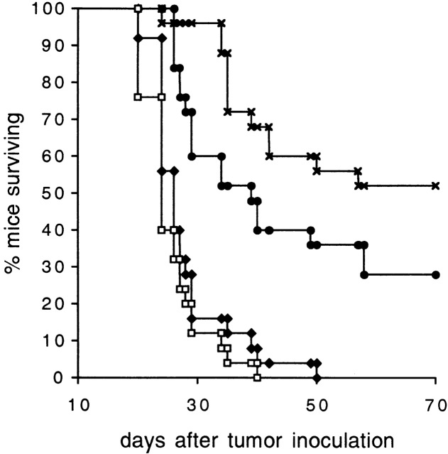
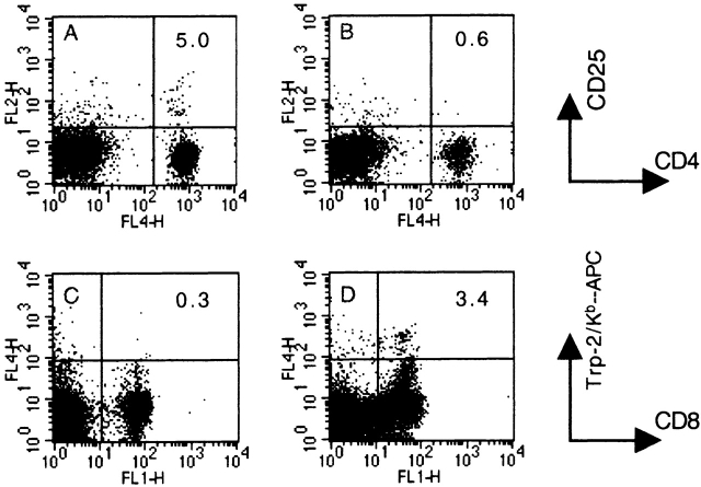
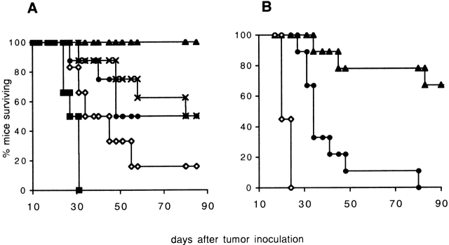
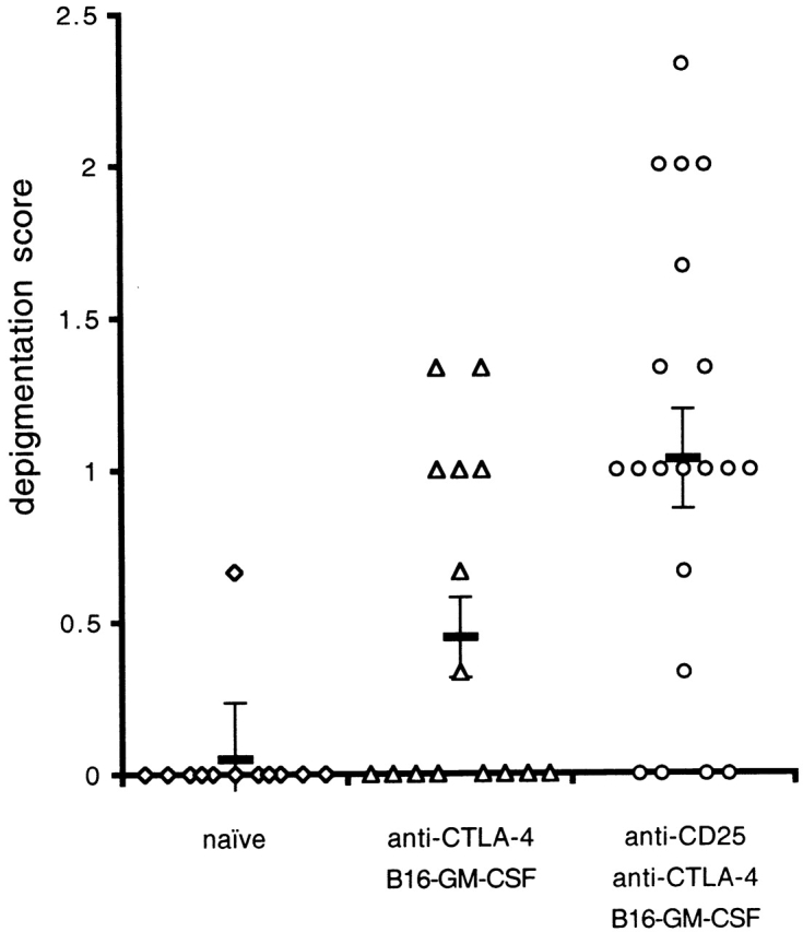
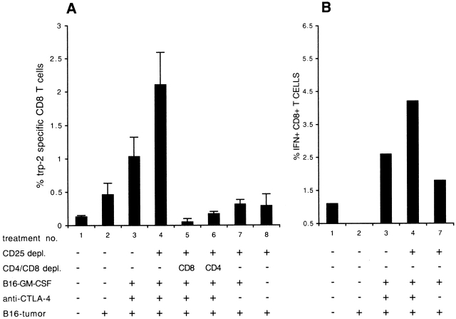
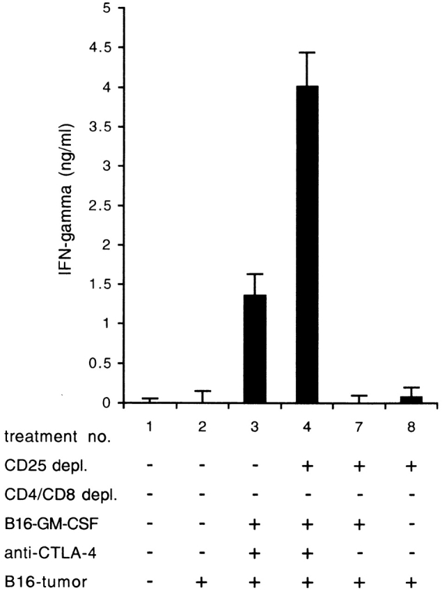
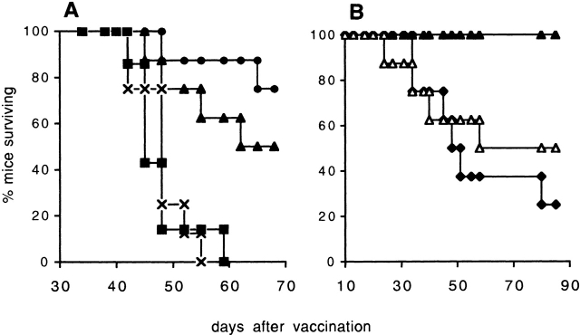
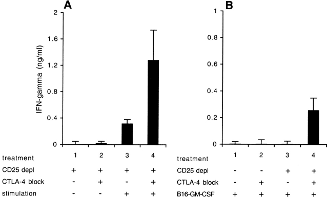
Therapeutic efficacy of a tumor cell–based vaccine against experimental B16 melanoma requires the disruption of either of two immunoregulatory mechanisms that control autoreactive T cell responses: the cytotoxic T lymphocyte–associated antigen (CTLA)-4 pathway or the CD25+ regulatory T (Treg) cells. Combination of CTLA-4 blockade and depletion of CD25+ Treg cells results in maximal tumor rejection. Efficacy of the antitumor therapy correlates with the extent of autoimmune skin depigmentation as well as with the frequency of tyrosinase-related protein 2180–188–specific CTLs detected in the periphery. Furthermore, tumor rejection is dependent on the CD8+ T cell subset. Our data demonstrate that the CTL response against melanoma antigens is an important component of the therapeutic antitumor response and that the reactivity of these CTLs can be augmented through interference with immunoregulatory mechanisms. The synergism in the effects of CTLA-4 blockade and depletion of CD25+ Treg cells indicates that CD25+ Treg cells and CTLA-4 signaling represent two alternative pathways for suppression of autoreactive T cell immunity. Simultaneous intervention with both regulatory mechanisms is therefore a promising concept for the induction of therapeutic antitumor immunity.
Overview
The claims and statistics of this paper haven’t been checked yet.
Run the check — free, no account needed. It reads the open-access text and checks the sources it cites.
Integrity signals
Registry
Cites
Statistics
Figures
Verification
Claims the paper makes
The claims and statistics of this paper haven’t been checked yet.
Run the check — free, no account needed. It reads the open-access text and checks the sources it cites.
Claims other papers make about this one
Each is checked against this paper’s own text: does it say what the citing paper says it does?
In the citing paper
“CD8+ T cells and CD4+ effector T cells have demonstrated anti-cancer potential, while regulatory T cells (CD4+ CD25+ Tregs) have been shown to be able to contribute to the attenuated immune response exhibited in cancer [65,66].”
From Current and Future Trends of Colorectal Cancer Treatment: Exploring Advances in Immunotherapy (2024) · cites this paper as [66]
Self-check
Percentages, sums and reported differences re-computed from the paper’s own numbers.
The self-check needs the paper’s full text.
Citations
Cites
Park, J., Adoro, S., Guinter, T. I., Erman, B., Alag, A., Catálfamo, M., Kimura, M. Y., Cui, Y., Lucas, P. J., Gress, R. E., et al. (2010) · Nature Immunology · doi:10.1038/ni.1840 · Cited by 5,835
Sakaguchi, S., Sakaguchi, N., Asano, M., Itoh, M., & Toda, M. (1995) · The Journal of Immunology · doi:10.4049/jimmunol.155.3.1151 · Cited by 5,608
Phenotypic Analysis of Antigen-Specific T Lymphocytes
Altman, J. D., Moss, P. A. H., Goulder, P. J. R., Barouch, D. H., McHeyzer-Williams, M. G., Bell, J. I., McMichael, A. J., & Davis, M. M. (1996) · Science · doi:10.1126/science.274.5284.94 · Cited by 3,544 · cited 1×
“H-2K b tetramers were produced as described previously (20, 21) and stored frozen in Tris-buffered saline/16% glycerol/0.”
MethodThornton, A. M., & Shevach, E. M. (1998) · The Journal of Experimental Medicine · doi:10.1084/jem.188.2.287 · Cited by 2,637
CD28 and CTLA-4 have opposing effects on the response of T cells to stimulation.
Krummel, M. F., & Allison, J. P. (1995) · The Journal of experimental medicine · doi:10.1084/jem.182.2.459 · Cited by 2,245 · cited 2×
“Anderson Cancer Center, Houston, TX), GM-CSF‐producing B16 cell lines BL6/GM-E, BL6/GM18 (18), B16/B7.1 (18), 9H10 ( 19 ), and PC61 (American Type Culture Collection) were cultured in Iscove’s IMDM (BioWhittaker) supplemented with 1 U/ml penicillin, 1 � g/ml streptomycin, 2 � M L -glutamine, 20 � M � -mercaptoethanol…”
MethodSelf-citationTakahashi, T., Tagami, T., Yamazaki, S., Uede, T., Shimizu, J., Sakaguchi, N., Mak, T. W., & Sakaguchi, S. (2000) · The Journal of Experimental Medicine · doi:10.1084/jem.192.2.303 · Cited by 2,233
CTLA-4 can function as a negative regulator of T cell activation
Walunas, T. L., Lenschow, D. J., Bakker, C. Y., Linsley, P. S., Freeman, G. J., Green, J. M., Thompson, C. B., & Bluestone, J. A. (1994) · Immunity · doi:10.1016/1074-7613(94)90071-x · Cited by 2,222
Read, S., Malmström, V., & Powrie, F. M. (2000) · The Journal of Experimental Medicine · doi:10.1084/jem.192.2.295 · Cited by 2,206
Salomon, B. L., Lenschow, D. J., Rhee, L., Ashourian, N., Singh, B., Sharpe, A. H., & Bluestone, J. A. (2000) · Immunity · doi:10.1016/s1074-7613(00)80195-8 · Cited by 2,058
Takahashi, T., Kuniyasu, Y., Toda, M., Sakaguchi, N., Itoh, M., Iwata, M., Shimizu, J., & Sakaguchi, S. (1998) · International Immunology · doi:10.1093/intimm/10.12.1969 · Cited by 1,566
Cited by
Sorted by citations
Items 901–1,000 of 1,000
Active specific immunotherapy for solid tumors
Baars, A. (2007) · Digital Academic REpository of VU University Amsterdam (Vrije Universiteit Amsterdam)
The tumour microenvironment influences antigen specific T cell transmigration
Popple, A. L. (2012) · Nottingham ePrints (University of Nottingham)
Frequenzevaluation von Ras- und Influenza-spezifischen T-Zellen bei Pankreaskarzinompatienten
Wiefelspütz, A. (2009) · Universitätsbibliothek der FU Berlin Hochschulschriftenstelle u. Dokumentenserver · doi:10.17169/refubium-12436
Strategies for exploiting the immune system to achieve prevention and improve therapy of cancer
Hamilton, D. H. (2007) · University Library - University of Saskatchewan (University of Saskatchewan)
Multiple costimulatory modalities enhance CTL avidity.
Garnett‐Benson, C., Chakraborty, M., Kudo‐Saito, C., Schlom, J., & Hodge, J. W. (2006) · Cancer Research
Developing effective tumor vaccines: basis, challenges and perspectives
Xu, Q., & Chen, W. (2007) · Frontiers of Medicine · doi:10.1007/s11684-007-0003-9
Merkel cell polyomavirus infection and host defence in patients with Merkel cell carcinoma
Sihto, H. J. (2012) · Työväentutkimus Vuosikirja
Amplification of Anti-Tumor Immunity Without Autoimmune Complications
Wei, W. (2006) · Defense Technical Information Center (DTIC)
Dendritic Cell Subsets as Targets and Vectors for Vaccination
Ueno, H., Fay, J. W., Banchereau, J., & Palucka, K. (2006) · Wiley eBooks · doi:10.1002/9783527619696.ch52
Lejeune, M., de la Cruz‐Merino, L., & lvaro, T. (2012) · InTech eBooks · doi:10.5772/34028
Targeted and Untargeted Fusion Proteins: Current Approaches to Cancer Immunotherapy
Khawli, L. A., Hu, P., & Epstein, A. L. (2013) · Wiley eBooks · doi:10.1002/9781118354599.ch19
Adjuvant Effect of Chaperone-Rich Cell Lysate: The Effects of CRCL on the Activation of Immune Cells
Cantrell, J. (2009) · UA Campus Repository (The University of Arizona)
Polarized Dendritic Cells for Tumor Immunotherapy
Hokey, D. A. (2005) · D-Scholarship@Pitt (University of Pittsburgh)
Allogeneic Antitumor Vaccine Strategies
Laport, G. G., & June, C. H. (2003) · Humana Press eBooks · doi:10.1007/978-1-59259-333-0_18
Untersuchungen zur Funktion von dendritischen Zellen in einem Spontan-Tumormodell
Naujoks, M. (2011) · Site cant be reached · doi:10.5282/edoc.13953
Activation of Immune-Mediated Tumor Cell Death by Chemotherapy
McCoy, M. J., Nowak, A. K., & Lake, R. (2012) · Springer eBooks · doi:10.1007/978-1-4614-5847-0_14
Promises and Challenges with Immunomodulatory Biologics
Ponce, R. A. (2014) · Wiley eBooks · doi:10.1002/9783527676965.ch8
Review on Immunotherapy Strategies against Infectious and Noninfectious Diseases
Sisay, A. B. A. T. (2015) · Journal of Health, Medicine and Nursing
Straathof, K. C. M. (2006) · Leiden Repository (Leiden University)
Effects of Regulatory T Cell–Dendritic Cell Interactions on Adaptive Immune Responses
Bopp, T., Probst, H. C., Radsak, M. P., Schmitt, E., Stassen, M., Schild, H., & Tenzer, S. (2014) · Springer eBooks · doi:10.1007/978-3-319-05104-8_3
Chambers, I., Blanpain, C., Watt, F. M., Tamai, K., Jensen, K. B., Jaks, V., Cotsarellis, G., Aractingi, S., & del Rio, M. (2013) · Experimental Dermatology · doi:10.1111/exd.12211
The association between CD4+ CD25+ regulatory T cells and non-small cell lung carcinoma
Abul-dahab, M., Elhoseiny, S. M., & Sabry, I. M. (2013) · Comparative Clinical Pathology · doi:10.1007/s00580-013-1825-2
Harnessing the regulatory feedback to T cells to enhance antitumor immune responses
Qiao, Y., Zheng, P., & Liu, Y. (2006) · Drug Discovery Today Therapeutic Strategies · doi:10.1016/j.ddstr.2006.02.005
Monocytes and Dendritic Cells in Sjögren’s Syndrome
Wildenberg, M. E. (2008) · EUR Research Repository (Erasmus University Rotterdam)
Self-citationCOMBINATIONAL CYTOKINE THERAPY OF CANCER: PLEIOTROPIC IMPACT WITHIN THE TUMOR MICROENVIRONMENT
Berhanu, A. (2005) · D-Scholarship@Pitt (University of Pittsburgh)
Influencia de la catepsina L en la población de células T regulatorias CD4+: rol del estroma
Camicia, G. (2011) · Repositorio Digital Institucional de la Universidad de Buenos Aires (Universidad de Buenos Aires)
In situ tumor destruction: towards in vivo modulation of immune responses by dendritic cells.
den Brok, M. H. M. G. M. (2006) · Radboud Repository (Radboud University)
Homeostatic T Cell Expansion to Induce Anti-Tumor Autoimmunity in Breast Cancer
Baccalà, R. (2006) · Defense Technical Information Center (DTIC) · doi:10.21236/ada456894
Promoting Type-1 CD4 + T Cell Immune Responses Against Tumor-Associated Antigen MAGE-A6
Vujanovic, L. N. (2006) · D-Scholarship@Pitt (University of Pittsburgh)
Targeting Cancer Associated Fibroblasts for Cancer Immunotherapy
(2014) · Cancer Cell & Microenvironment · doi:10.14800/ccm.80
Allison, J. P. (2003) · Cancer immunity
Self-citationEnvironmental modulation of mucosal immunity. Opportunities in respiratory viral infections
Schijf, M. A. (2013) · Utrecht University Repository (Utrecht University)
Novel Immunotherapy to Eliminate Minimal Residual Disease in AML
Kline, J. P. (2013)
Melero, I., & Bendandi, M. (2017) · Revista de medicina de la Universidad de Navarra · doi:10.15581/021.7425
T Cells and Antigen Recognition
Emens, L. A. (2007) · Springer eBooks · doi:10.1007/978-1-4020-6087-8_3
Dendritic Cell-Based Cancer Immunotherapy: Achievements and Novel Concepts
Bol, K. F., Schreibelt, G., Aarntzen, E. H. J. G., de Vries, I. J. M., & Figdor, C. G. (2012) · Springer eBooks · doi:10.1007/978-1-4614-4732-0_4
Regulatory T Cells in Hepatitis and Hepatocellular Carcinoma
Wang, F., & Gao, G. F. (2008) · Springer eBooks · doi:10.1007/978-0-387-77909-6_21
Managing Regulatory T Cells to Improve Cancer Immunotherapy
Curiel, T. J. (2012) · Springer eBooks · doi:10.1007/978-1-4614-4732-0_9
SA-4-1BBL as a modulator of T effector to T regulatory cell conversion.
Madireddi, S. (2010) · ThinkIR: The University of Louisville's Institutional Repository (University of Louisville) · doi:10.18297/etd/880
Galani, I. E. (2008) · heiDOK (Heidelberg University) · doi:10.11588/heidok.00008497
TRP-1 AS A MODEL TUMOR ANTIGEN FOR IMMUNOTHERAPY AND IMMUNE TOLERANCE IN THE THYMUS
Brandmaier, A. (2011) · IUScholarWorks (Indiana University) · doi:10.7912/c2/1708
иммунорегуляторные С убпопуляции т-КлетоК при опухолевом роС те и аллергичеСКих заболеваниях
Чердынцева, Н. В., & Rams, S. (2010) · CyberLeninK (CyberLeninka)
Vaccines and Their Role in CD8 T Cell-Mediated Antitumor Immunity
Kinkead, H. L., Jaffee, E. M., Lutz, E. R., & Armstrong, T. D. (2016) · Elsevier eBooks · doi:10.1016/b978-0-12-374279-7.17019-5
TGF-β Dependent T-Cell Regulation in Colitis and Colon Cancer
Becker, C., & Neurath, M. F. (2008) · Humana Press eBooks · doi:10.1007/978-1-59745-293-9_11
Fondation Rene Touraine pour la Recherche en Dermatologie
(2009) · Experimental Dermatology · doi:10.1111/j.1600-0625.2009.00873.x
Alfaro, C., Murillo, O., Tirapu, I., Azpilicueta, A., Huarte, E., Arina, A., Arribillaga, L., Bendandi, M., Prìeto, J., Lasarte, J. J., et al. (2006)
Immunological Strategies to Fight Skin Cancer
Schadendorf, D., Paschen, A., & Eichmüller, S. B. (2004) · CRC Press eBooks · doi:10.1201/b14248-46
Post-translational regulation of FOXP3
Coffer, P. J., Prakken, A. B. J., & van Loosdregt, J. (2006)
Therapeutic brain cancer targeting by gene therapy and immunomodulation
Savelkoul, H. F. J., & Schijns, V. E. C. J. (2012)
The Immune Response to Non-Small Cell Lung Cancer
Ohri, C. M. (2010) · Figshare
HPV16-specific cellular immunity in health and disease
van Poelgeest, & Elizabeth, M. I. (2007)
Immune monitoring of kidney transplant recipients with post-transplant malignancy.
Hope, C. M. (2014) · Adelaide Research & Scholarship (AR&S) (University of Adelaide)
Donnou, S., & Couez, D. (2007) · OpenGrey (Institut de l'Information Scientifique et Technique)
Application of Antigen Cross-Presentation Research into Patient Care
(2017) · Frontiers research topics · doi:10.3389/978-2-88945-191-3
Vitiligo bij melanoom: een gunstig teken
Luiten, R. M. (2006) · Data Archiving and Networked Services (DANS)
Cancer Vaccines: Some Basic Considerations
Rammensee, H., Singh‐Jasuja, H., Emmerich, N., & Pascolo, S. (2009) · Elsevier eBooks · doi:10.1016/b978-0-12-369420-1.00050-0
Morrison, L. J., Alter, J. S., Ginsburg, M., Marx, J. H., & Blee, K. M. (2003)
Identification d'Epitopes T CD4+ d'Antigènes tumoraux
Wang, X.-F. (2007) · HAL
Robbins, P. F., & Lu, Y. (2019) · Springer eBooks · doi:10.1007/978-3-319-46029-1_44-1
Immunotherapy strategies against infectious and noninfectious diseases: Review
Beyene, A., & Sisay, T. (2017) · Journal of Emerging Technologies and Innovative Research
McNamara, A. (2019) · ERA: Education and Research Archive (University of Alberta) · doi:10.7939/r3-5838-dr52
Immunopathology as a Basis for Immunotherapy of Head and Neck Squamous Cell Carcinoma
Qian, X., Hoffmann, T. K., Kaufmann, A. M., & Albers, A. E. (2020) · Springer eBooks · doi:10.1007/978-3-030-57949-4_13
Rao, A. (2012) · D-Scholarship@Pitt (University of Pittsburgh)
Manipulating Immunological Checkpoints to Maximize Antitumor Immunity
Emens, L. A., Reilly, R. T., & Jaffee, E. M. (2006) · Humana Press eBooks · doi:10.1385/1-59745-011-1:331
Cellular immunotherapy (CI), where have we been and where are we going?
Yannelli, J. R. (2009) · Springer eBooks · doi:10.1007/978-90-481-2289-9_15
Chikileva, I. O., Anisimova, N. Y., Olga, L., Kiselevsky, M. V., & Abramov, V. M. (2008) · Springer eBooks · doi:10.1007/978-1-4020-6931-4_8
Checkpoint Blockade and Combinatorial Immunotherapies
Peggs, K. S., Quezada, S. A., & Allison, J. P. (2007) · Springer eBooks · doi:10.1007/978-1-4020-6087-8_16
Self-citationImproving immunotherapy for high grade glioma
Hunn, M. K. (2015) · Open Access Victoria University of Wellington | Te Herenga Waka (Figshare) · doi:10.26686/wgtn.17013347
Schäffer, A. A. (2020) · Faculty Opinions – Post-Publication Peer Review of the Biomedical Literature · doi:10.3410/f.737383720.793571667
Sharma, P., Subudhi, S. K., Peggs, K. S., Goswami, S., Gao, J., Quezada, S. A., & Allison, J. P. (2017) · Wiley eBooks · doi:10.1002/9781119000822.hfcm068
Self-citationVaccine Therapy and Immunotherapy for Pancreatic Cancer
Zheng, L., & Jaffee, E. M. (2017) · Springer eBooks · doi:10.1007/978-1-4939-6631-8_53-2
Vaccine Therapy and Immunotherapy for Pancreatic Cancer
Zheng, L., & Jaffee, E. M. (2009) · Springer eBooks · doi:10.1007/978-0-387-77498-5_53
Robbins, P. F., & Lu, Y. (2020) · Springer eBooks · doi:10.1007/978-3-030-05070-2_44
Frontiers in Cancer Immunotherapy
Murphy, J. F. (2020) · Springer eBooks · doi:10.1007/978-3-030-50287-4_1
Alexander, M. A. (2011) · Science Publishers eBooks · doi:10.1201/b10503-18
Lambe, T., & Cornall, R. J. (2008) · Experimental Dermatology · doi:10.1111/j.1600-0625.2007.00666_12.x
Development of novel immune interventions for genito-urinary cancers
Agarwal, N., & Vogelzang, N. J. (2011) · Informa Healthcare eBooks · doi:10.3109/9781841848303.003
(2004) · Encyclopedic Dictionary of Genetics, Genomics and Proteomics · doi:10.1002/0471684228.egp01871
Reddy, S. P. (2019) · Faculty Opinions – Post-Publication Peer Review of the Biomedical Literature · doi:10.3410/f.734139155.793555121
Leonard, W. J., & Spolski, R. J. (2017) · Faculty Opinions – Post-Publication Peer Review of the Biomedical Literature · doi:10.3410/f.727509038.793537551
Kolar, P., Hebel, K., Knieke, K., & Brunner-Weinzierl, M. C. (2009) · Arthritis & Rheumatism · doi:10.1002/art.24769
Therapeutic Targeting Regulatory T Cells in Tumor
Wang, W., & Zou, W. (2013) · Springer eBooks · doi:10.1007/978-94-007-6217-6_25
Mitsui, J., Nishikawa, H., Muraoka, D., Wang, L., Noguchi, T., Sato, E., Kondo, S., Allison, J. P., Sakaguchi, S., Old, L. J., et al. (2023) · doi:10.1158/1078-0432.c.6519084.v1
Self-citationLee, C.-H., Chiang, Y.-H., Chang, S., Chong, C.-L., Cheng, B.-M., & Roffler, S. R. (2023) · doi:10.1158/1078-0432.c.6517468
Mitsui, J., Nishikawa, H., Muraoka, D., Wang, L., Noguchi, T., Sato, E., Kondo, S., Allison, J. P., Sakaguchi, S., Old, L. J., et al. (2023) · doi:10.1158/1078-0432.c.6519084
Self-citationLee, C.-H., Chiang, Y.-H., Chang, S., Chong, C.-L., Cheng, B.-M., & Roffler, S. R. (2023) · doi:10.1158/1078-0432.c.6517468.v1
Rahman, E. N. S. E. A., Irekeola, A. A., Shueb, R. H., Lazim, N. M., Mohamud, R., Chen, X., Ghazali, L., Awang, N. M. S. H., Haron, A., & Yean, C. Y. (2023) · Cytokine · doi:10.1016/j.cyto.2023.156341
Additional file 1 of Hyperfunction of CD4 CD25 regulatory T cells in de novo acute myeloid leukemia
Wan, Y., Zhang, C., Xu, Y., Wang, M., Rao, Q., Xing, H., Tian, Z. R., Tang, K., Mi, Y., Wang, Y., et al. (2020) · Figshare · doi:10.6084/m9.figshare.12375575
Additional file 3 of Hyperfunction of CD4 CD25 regulatory T cells in de novo acute myeloid leukemia
Wan, Y., Zhang, C., Xu, Y., Wang, M., Rao, Q., Xing, H., Tian, Z., Tang, K., Mi, Y., Wang, Y., et al. (2020) · Figshare · doi:10.6084/m9.figshare.12375587
A Systemic Review of T Regulatory Cells and Their Role In Hodgkin and Non-Hodgkin Lymphoma
Taur, N., Kushwah, R., Jain, M., Singh, A., & Mittal, R. (2024) · Annals of Pathology and Laboratory Medicine · doi:10.21276/apalm.3258
Immune Checkpoint Inhibitors in Therapeutics and their Adverse Effect Profile: A Review
Adiga, S., Kishore, B. A., & Vasishta, S. (2025) · The Open Biomedical Engineering Journal · doi:10.2174/0118741207369739250423073032
Role of gene therapy and immunotherapy in precision medicine
Khan, I. A., Khatun, H., & Viswa, P. D. K. (2025) · Elsevier eBooks · doi:10.1016/b978-0-443-33302-6.00011-1
Additional file 2 of Hyperfunction of CD4 CD25 regulatory T cells in de novo acute myeloid leukemia
Wan, Y., Zhang, C., Xu, Y., Wang, M., Rao, Q., Xing, H., Tian, Z., Tang, K., Mi, Y., Wang, Y., et al. (2020) · Figshare · doi:10.6084/m9.figshare.12375578
Metadata
- Authors
- Sutmuller, R. P. M., van Duivenvoorde, L. M., van Elsas, A., Schumacher, T. N. M., Wildenberg, M. E., Allison, J. P., Toes, R. E. M., Offringa, R., & Melief, C. J. M.
- Affiliations
- Leiden University Medical Center (NL)
- aDepartment of Immunohematology and Blood Transfusion, Tumor Immunology Lab, E3-Q, Leiden University Medical Center, 2300 RC Leiden, Netherlands
- The Netherlands Cancer Institute (NL)
- University of California, Berkeley (US)
- Venue
- The Journal of Experimental Medicine
- Publisher
- Rockefeller University Press
- Published
- 2001-09-17
- Volume
- 194
- Issue
- 6
- Pages
- 823-832
- ISSN
- 0022-1007 (ISSN-L)
- 1540-9538
- PMC
- PMC2195955
- OpenAlex
- W2148911309
- Cited by
- 1,008
- References
- 44
- Citations by year
- 2026: 3 · 2025: 7 · 2024: 8 · 2023: 12 · 2022: 15 · 2021: 15 · 2020: 18 · 2019: 11 · 2018: 12 · 2017: 25 · 2016: 22 · 2015: 36 · 2014: 26 · 2013: 48 · 2012: 41
- FWCI
- 13.30
- Type
- journal article
- Language
- en
- Open access
- green
- www.ncbi.nlm.nih.gov
- Licence
- cc-by-nc-sa
- Locations
- The Journal of Experimental Medicine (publishedVersion)
- PubMed Central (publishedVersion, cc-by-nc-sa)
- PubMed (publishedVersion)
- Data Archiving and Networked Services (DANS) (submittedVersion)
- Pure Amsterdam UMC (submittedVersion)
- Topics
- Life Sciences › Immunology and Microbiology › Immunology › T-cell and B-cell Immunology
- Health Sciences › Medicine › Oncology › Cancer Immunotherapy and Biomarkers
- Life Sciences › Immunology and Microbiology › Immunology › Immunotherapy and Immune Responses
- Keywords
- CTLA-4 blockade, CD25+ regulatory T cells, Treg depletion, antitumor immunity, B16 melanoma, CD8+ T cells, tyrosinase-related protein 2, immune regulation, tumor rejection
- MeSH terms
- Abatacept, Animals, Antibodies, Monoclonal, Antigens, Differentiation, Antigens, Neoplasm, Female, Immunotherapy, Lymphocyte Depletion, Melanoma, Experimental, Mice, Inbred C57BL, Mice, Nude, T-Lymphocytes, Cytotoxic, Tumor Cells, Cultured, Receptors, Interleukin-2, Biomarkers, CD4-Positive T-Lymphocytes, Antigens, CD, Immunoconjugates, Intramolecular Oxidoreductases, Mice, CTLA-4 Antigen
About these checks
Registry, retraction, Cites and statistics results are computed, not judged, with one exception: a printed reference that no index lists is read once by a model before it counts as a citation error. Claims are extracted by a model and kept only when their quote appears verbatim in the text; each cited source is then checked, with a model’s help, for whether it says what the claim relies on. Those verdicts can be wrong — read the claim’s quote and the cited source before relying on one.
Statistics and arithmetic are recomputed, not judged.
Confidence bands are shown only for fields calibrated on our annotated test set. None is calibrated yet, so no band is shown.
What counts as a claim ›How we measure confidence ›How we check this ›
Are you an author, or do you see a problem with this page? Request a review
