Cited by 970

The DOI matches its Crossref record.

Rapid mobilization of murine and human hematopoietic stem and progenitor cells with AMD3100, a CXCR4 antagonist

Broxmeyer, Hal E., Orschell, Christie M., Clapp, D. Wade, Hangoc, Giao, Cooper, Scott, Plett, P. Artur, Liles, W. Conrad, Li, Xiaxin, Graham-Evans, Barbara, Campbell, Timothy B., Calandra, Gary, Bridger, Gary, Dale, David C., Srour, Edward F.·2005·The Journal of Experimental Medicine

Abstract

Improving approaches for hematopoietic stem cell (HSC) and hematopoietic progenitor cell (HPC) mobilization is clinically important because increased numbers of these cells are needed for enhanced transplantation. Chemokine stromal cell derived factor-1 (also known as CXCL12) is believed to be involved in retention of HSCs and HPCs in bone marrow. AMD3100, a selective antagonist of CXCL12 that binds to its receptor, CXCR4, was evaluated in murine and human systems for mobilizing capacity, alone and in combination with granulocyte colony-stimulating factor (G-CSF). AMD3100 induced rapid mobilization of mouse and human HPCs and synergistically augmented G-CSF–induced mobilization of HPCs. AMD3100 also mobilized murine long-term repopulating (LTR) cells that engrafted primary and secondary lethally-irradiated mice, and human CD34+ cells that can repopulate nonobese diabetic-severe combined immunodeficiency (SCID) mice. AMD3100 synergized with G-CSF to mobilize murine LTR cells and human SCID repopulating cells (SRCs). Human CD34+ cells isolated after treatment with G-CSF plus AMD3100 expressed a phenotype that was characteristic of highly engrafting mouse HSCs. Synergy of AMD3100 and G-CSF in mobilization was due to enhanced numbers and perhaps other characteristics of the mobilized cells. These results support the hypothesis that the CXCL12-CXCR4 axis is involved in marrow retention of HSCs and HPCs, and demonstrate the clinical potential of AMD3100 for HSC mobilization.

Overview

The claims and statistics of this paper haven’t been checked yet.

Run the check — free, no account needed. It reads the open-access text and checks the sources it cites.

A few free checks a day · papers already verified open instantly · results are public

Integrity signals

Registry

Crossref · 10.1084/jem.20041385

Cites

Statistics

Figures

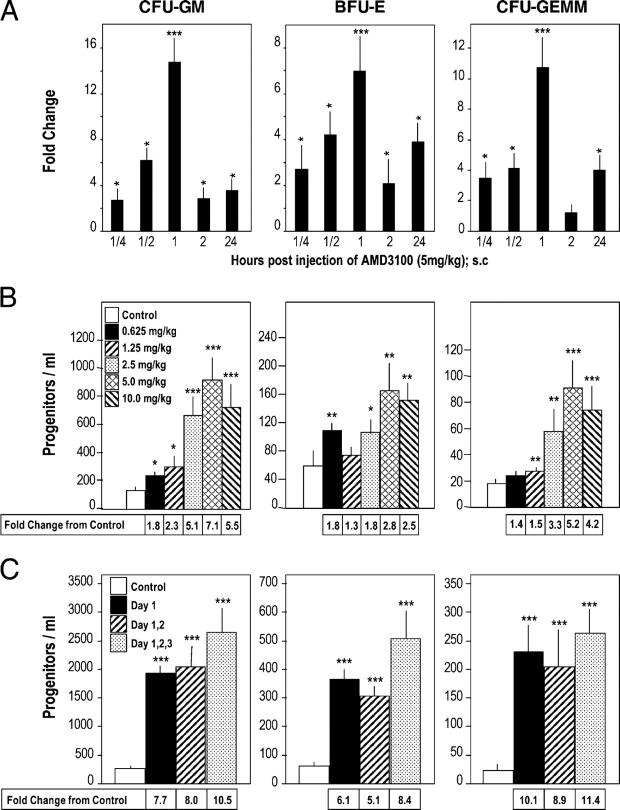
Figure 1. Time and dose response effects of AMD3100 as a mobilizer of HPC (CFU-GM, BFU-E and CFU-GEMM) to the blood of mice. (A) Time course of mobilization in response to a single s.c. injection of 5 mg/kg AMD3100 into C3H/HeJ mice; results are for one full experiment (5 mice/group and time 0 control numbers of CFU-GM, BFU-E, and CFU-GEMM per ml of blood were 78 ± 18, 17 ± 2, and 10 ± 2, respectively). (B) Dose response analysis at 1 h post s.c. injections of 0.625, 1.25, 2.5, 5.0, or 10 mg/kg AMD3100 into C3H/HeJ mice; results are average of 7 mice/group from a total of two separate experiments. (C) Influence of multiple injections of 5 mg/kg AMD3100 or saline given on day 1; days 1 and 2; or days 1, 2, and 3 given 24 h apart, and analyzed 1 h after the last injection into C3H/HeJ mice; results are average of 14 mice for control group for different timed saline injections and 8 mice for each test group from a total of two complete experiments. *P < 0.05; **P < 0.01; ***P < 0.001.
Figure 1. Time and dose response effects of AMD3100 as a mobilizer of HPC (CFU-GM, BFU-E and CFU-GEMM) to the blood of mice. (A) Time course of mobilization in response to a single s.c. injection of 5 mg/kg AMD3100 into C3H/HeJ mice; results are for one full experiment (5 mice/group and time 0 control numbers of CFU-GM, BFU-E, and CFU-GEMM per ml of blood were 78 ± 18, 17 ± 2, and 10 ± 2, respectively). (B) Dose response analysis at 1 h post s.c. injections of 0.625, 1.25, 2.5, 5.0, or 10 mg/kg AMD3100 into C3H/HeJ mice; results are average of 7 mice/group from a total of two separate experiments. (C) Influence of multiple injections of 5 mg/kg AMD3100 or saline given on day 1; days 1 and 2; or days 1, 2, and 3 given 24 h apart, and analyzed 1 h after the last injection into C3H/HeJ mice; results are average of 14 mice for control group for different timed saline injections and 8 mice for each test group from a total of two complete experiments. *P < 0.05; **P < 0.01; ***P < 0.001.CC BY-NC-SA · PubMed Central
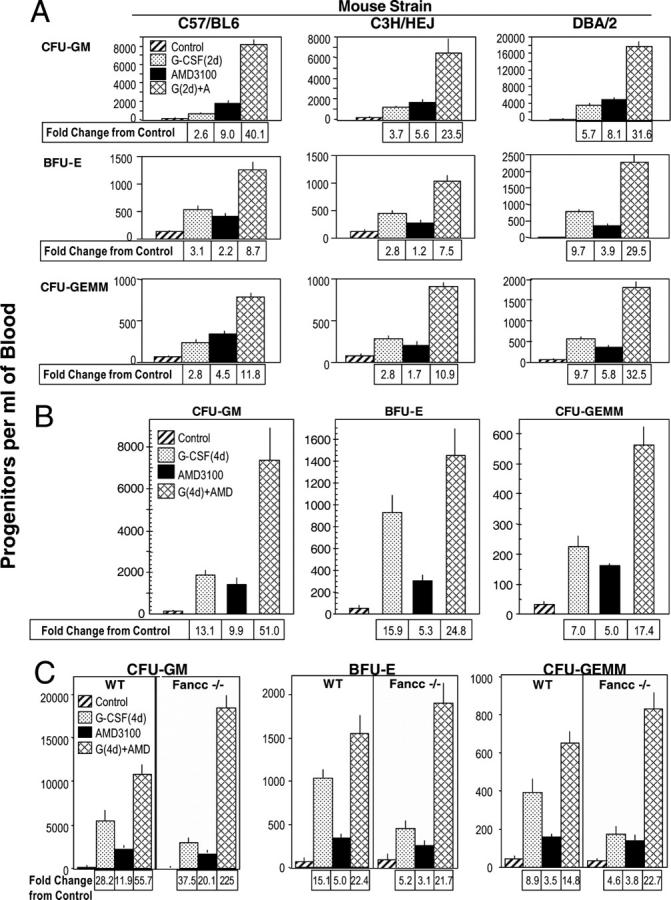
Figure 2. Comparative AMD3100 enhancement of G-CSF–induced mobilization of CFU-GM, BFU-E, and CFU-GEMM in: (A) 2 d G-CSF: C57Bl/6, C3H/HeJ, DBA/2, (B) 4 d G-CSF: C57Bl/6, and (C) 4 d of G-CSF: Fancc−/− and +/+ mice. The results shown in A are an average from a total of three experiments of 15 C57Bl/6, 15 DBA/2, and 11 C3H/HeJ mice. Results in B are for one experiment of 5 mice/group. Results shown in C are the averages of 15 wild-type and 8 Fancc−/− mice per group averaged from a total of two experiments. For parts A–C, all experimental points within a group are at least P < 0.001 compared with the control for the specific progenitor cell of each mouse strain.
Figure 2. Comparative AMD3100 enhancement of G-CSF–induced mobilization of CFU-GM, BFU-E, and CFU-GEMM in: (A) 2 d G-CSF: C57Bl/6, C3H/HeJ, DBA/2, (B) 4 d G-CSF: C57Bl/6, and (C) 4 d of G-CSF: Fancc−/− and +/+ mice. The results shown in A are an average from a total of three experiments of 15 C57Bl/6, 15 DBA/2, and 11 C3H/HeJ mice. Results in B are for one experiment of 5 mice/group. Results shown in C are the averages of 15 wild-type and 8 Fancc−/− mice per group averaged from a total of two experiments. For parts A–C, all experimental points within a group are at least P < 0.001 compared with the control for the specific progenitor cell of each mouse strain.CC BY-NC-SA · PubMed Central
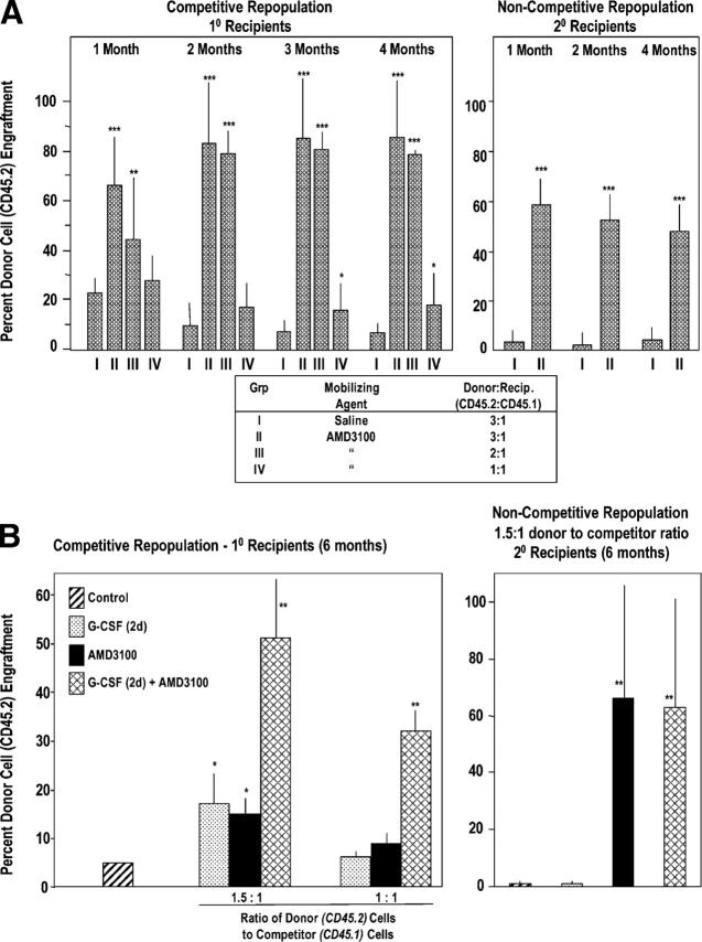
Figure 3. AMD3100 mobilizes competitive repopulating mouse HSCs with self-renewal capacity (A) and G-CSF synergizes this effect (B). (A, left) 18 C57Bl/6 mice (CD45.2) serving as donors were injected s.c. with 0.1 ml saline, and 36 C57Bl/6 mice were injected with 0.1 ml AMD3100 at 5 mg/kg (=∼100 μg/mouse). Blood was collected 1 h later and LDMNCs isolated. BM from nonirradiated B6.BoyJ (CD45.1) mice served as competitor cells. The ratio of donor (CD45.2) blood cells to competitor (CD45.1) BM cells was set as the number of LDMNC blood cells in 3 donor mice to a constant number of 0.5 × 10⁶ competitor BM cells equaling 3:1. A 2:1 ratio was the number of LDMNCs in blood of 2 donor mice to 0.5 × 10⁶ competitor cells, and a 1:1 ratio was LDMNC in 1 donor mouse to 0.5 × 10⁶ competitor cells. Results are based on analysis of transplantation of 6 lethally irradiated recipients per test group of i.v. infused cells. Right panel: BM cells were removed from femurs of primary mice 4 mo after injection of 3:1 saline or 3:1 AMD3100:competitor cell mixture and 2.5 × 10⁶ marrow cells from 3 mice of each group injected separately into 3 lethally secondary irradiated B6.BoyJ mice in a noncompetitive assay. Results are shown as percent CD45.2 C57Bl/6 donor cell chimerism in CD45.1 B6.BoyJ recipients. *P < 0.05; **P < 0.01; ***P < 0.001 compared with group 1 at each time point. (B) G-CSF and AMD3100 were administered as in Figure 2, part A, with G-CSF given twice a day for 2 d. Left panel: competitive repopulation in primary recipients 6 mo post transplant. The results are an average of mouse cells from five donors into six recipients each. Right panel: secondary repopulation in a noncompetitive assay 6 mo after transplant. Results are an average of 3 donor mice with cells transplanted into three recipients each. *P < 0.01 or **P < 0.001 compared with control.
Figure 3. AMD3100 mobilizes competitive repopulating mouse HSCs with self-renewal capacity (A) and G-CSF synergizes this effect (B). (A, left) 18 C57Bl/6 mice (CD45.2) serving as donors were injected s.c. with 0.1 ml saline, and 36 C57Bl/6 mice were injected with 0.1 ml AMD3100 at 5 mg/kg (=∼100 μg/mouse). Blood was collected 1 h later and LDMNCs isolated. BM from nonirradiated B6.BoyJ (CD45.1) mice served as competitor cells. The ratio of donor (CD45.2) blood cells to competitor (CD45.1) BM cells was set as the number of LDMNC blood cells in 3 donor mice to a constant number of 0.5 × 10⁶ competitor BM cells equaling 3:1. A 2:1 ratio was the number of LDMNCs in blood of 2 donor mice to 0.5 × 10⁶ competitor cells, and a 1:1 ratio was LDMNC in 1 donor mouse to 0.5 × 10⁶ competitor cells. Results are based on analysis of transplantation of 6 lethally irradiated recipients per test group of i.v. infused cells. Right panel: BM cells were removed from femurs of primary mice 4 mo after injection of 3:1 saline or 3:1 AMD3100:competitor cell mixture and 2.5 × 10⁶ marrow cells from 3 mice of each group injected separately into 3 lethally secondary irradiated B6.BoyJ mice in a noncompetitive assay. Results are shown as percent CD45.2 C57Bl/6 donor cell chimerism in CD45.1 B6.BoyJ recipients. *P < 0.05; **P < 0.01; ***P < 0.001 compared with group 1 at each time point. (B) G-CSF and AMD3100 were administered as in Figure 2, part A, with G-CSF given twice a day for 2 d. Left panel: competitive repopulation in primary recipients 6 mo post transplant. The results are an average of mouse cells from five donors into six recipients each. Right panel: secondary repopulation in a noncompetitive assay 6 mo after transplant. Results are an average of 3 donor mice with cells transplanted into three recipients each. *P < 0.01 or **P < 0.001 compared with control.CC BY-NC-SA · PubMed Central
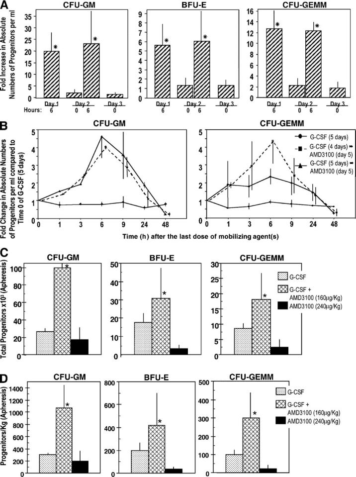
Figure 4. Influence of AMD3100, alone and with G-CSF, on circulating HPCs in healthy human volunteers. (A) Influence of multiple injections of AMD3100 alone. Three volunteers were injected i.v. with AMD3100 (80 μg/kg) on the first day (day 1) and again at 24 h. Progenitors/ml blood were assessed preinjection (day 1, 0 h) and at the noted time intervals. Fold changes are calculated based on the following day 1, 0 h progenitors/ml for three donors: CFU-GM (387, 78, 201), BFU-E (712, 78, 217), and CFU-GEMM (77, 29, 93) grown in methylcellulose cultures in the presence of Epo, stem cell factor, and interleukin-3. The fold changes for CFU-GM also include progenitor cells/ml as calculated in agar culture medium with GM-CSF and stem cell factor (control numbers for three donors: 108, 18, 54). *P < 0.001 compared with 0 h counts of that particular day. (B) Influence of AMD3100 on mobilization of circulating HPCs in healthy human volunteers receiving G-CSF or G-CSF plus AMD3100 (160 μg/kg). The results shown are the mean plus range of fold increases of the absolute numbers of CFU-GM and CFU-GEMM per ml of blood for two healthy volunteers each without apheresis. Fold changes are based on the following numbers of G-CSF day 5, 0 h control progenitor cells/ml: group I: G-CSF (CFU-GM: 34403 and 27194; CFU-GEMM: 14363 and 10083); group II: G-CSF (4 d) plus AMD3100 (day 5; CFU-GM: 11968 and 2675; CFU-GEMM: 3366 and 510), and group III: G-CSF (5 d) plus AMD3100 (day 5; CFU-GM: 2698 and 2380; CFU-GEMM: 1028 and 1238). (C) Results of apheresis after mobilization of circulating HPCs. Results are expressed as total progenitors collected (in thousands) for donors receiving G-CSF (n = 3), G-CSF + AMD3100 (160 μg/kg; n = 3), or AMD3100 (240 μg/kg; n = 4). *P < 0.01 for G-CSF– or AMD3100 versus G-CSF + AMD3100-mobilized cells. (D) Results of apheresis shown in C but expressed as progenitors mobilized per kg donor. *P < 0.05 for same comparisons as in C.
Figure 4. Influence of AMD3100, alone and with G-CSF, on circulating HPCs in healthy human volunteers. (A) Influence of multiple injections of AMD3100 alone. Three volunteers were injected i.v. with AMD3100 (80 μg/kg) on the first day (day 1) and again at 24 h. Progenitors/ml blood were assessed preinjection (day 1, 0 h) and at the noted time intervals. Fold changes are calculated based on the following day 1, 0 h progenitors/ml for three donors: CFU-GM (387, 78, 201), BFU-E (712, 78, 217), and CFU-GEMM (77, 29, 93) grown in methylcellulose cultures in the presence of Epo, stem cell factor, and interleukin-3. The fold changes for CFU-GM also include progenitor cells/ml as calculated in agar culture medium with GM-CSF and stem cell factor (control numbers for three donors: 108, 18, 54). *P < 0.001 compared with 0 h counts of that particular day. (B) Influence of AMD3100 on mobilization of circulating HPCs in healthy human volunteers receiving G-CSF or G-CSF plus AMD3100 (160 μg/kg). The results shown are the mean plus range of fold increases of the absolute numbers of CFU-GM and CFU-GEMM per ml of blood for two healthy volunteers each without apheresis. Fold changes are based on the following numbers of G-CSF day 5, 0 h control progenitor cells/ml: group I: G-CSF (CFU-GM: 34403 and 27194; CFU-GEMM: 14363 and 10083); group II: G-CSF (4 d) plus AMD3100 (day 5; CFU-GM: 11968 and 2675; CFU-GEMM: 3366 and 510), and group III: G-CSF (5 d) plus AMD3100 (day 5; CFU-GM: 2698 and 2380; CFU-GEMM: 1028 and 1238). (C) Results of apheresis after mobilization of circulating HPCs. Results are expressed as total progenitors collected (in thousands) for donors receiving G-CSF (n = 3), G-CSF + AMD3100 (160 μg/kg; n = 3), or AMD3100 (240 μg/kg; n = 4). *P < 0.01 for G-CSF– or AMD3100 versus G-CSF + AMD3100-mobilized cells. (D) Results of apheresis shown in C but expressed as progenitors mobilized per kg donor. *P < 0.05 for same comparisons as in C.CC BY-NC-SA · PubMed Central
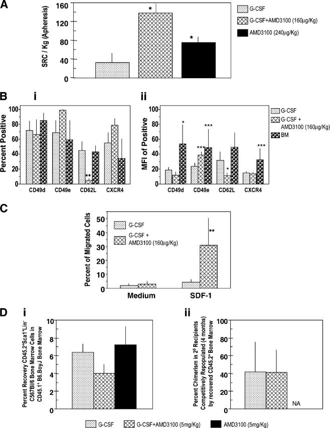
Figure 5. Influence of AMD3100, G-CSF, and the combination of G-CSF plus AMD3100 on mobilization of NOD-SCID SRCs from normal human volunteers (apheresis samples), surface expression of adhesion molecules and chemotaxis of CD34+ cells, and homing of mobilized murine Sca1+Lin− cells. (A) SRCs per kg in apheresis samples from G-CSF– (n = 3), G-CSF + AMD3100- (160 μg/kg; n = 3), and AMD3100- (240 μg/kg; n = 4) mobilized circulating blood. Each set of test samples was assayed simultaneously in limiting dilutions in conditioned NOD-SCID mice. For every sample, four different cell concentrations were used and four mice were transplanted with each cell concentration. Mice were assayed for chimerism 8 wk later; those that demonstrated >0.2% chimerism (total CD45+ cells in BM) were considered to be positive. Percentage of negative mice were used to calculate SRC frequencies. (B, i) Expression of CD49d (VLA-4), CD49e (VLA-5), CD26L (L-selectin), and CXCR4 on mobilized CD34+ cells (mean ± 1SEM of percent positive cells of five to seven different G-CSF samples, three AMD3100 plus G-CSF samples, and four BM samples). (B, ii) Mean fluorescent intensity (MFI) of positive samples. Same number of samples evaluated as in B, i except G-CSF group has a different number (n =7). (C) CD34+ cells isolated from G-CSF (n = 6) and G-CSF plus AMD3100 (n = 3) mobilized peripheral blood were assessed for chemotaxis to SDF-1/CXCL12 (100 ng/ml) and results expressed as percentage of migrated cells. Data for each sample were collected in duplicate. (D, i) Homing of CD45.2+ Sca1+Lin− BM cells from C57Bl/6 mice into lethally irradiated CD45.1+ B6.BoyJ BM from samples mobilized with G-CSF (2 times/d for 4 d as in Fig. 2B; n = 3), G-CSF + AMD3100 (5 mg/kg; n = 4), and AMD3100 (5 mg/kg; n = 3). Homing was assessed as in Materials and methods. (D, ii) Competitive repopulation of CD45.2+ BM cells recovered after homing shown in D, i. Results of chimerism are after 4 mo in 2° irradiated B6.BoyJ recipients. For A–D, significant differences compared with G-CSF: *P < 0.001; **P < 0.01; ***P < 0.05; all other values are P > 0.05. NA, data not depicted.
Figure 5. Influence of AMD3100, G-CSF, and the combination of G-CSF plus AMD3100 on mobilization of NOD-SCID SRCs from normal human volunteers (apheresis samples), surface expression of adhesion molecules and chemotaxis of CD34+ cells, and homing of mobilized murine Sca1+Lin− cells. (A) SRCs per kg in apheresis samples from G-CSF– (n = 3), G-CSF + AMD3100- (160 μg/kg; n = 3), and AMD3100- (240 μg/kg; n = 4) mobilized circulating blood. Each set of test samples was assayed simultaneously in limiting dilutions in conditioned NOD-SCID mice. For every sample, four different cell concentrations were used and four mice were transplanted with each cell concentration. Mice were assayed for chimerism 8 wk later; those that demonstrated >0.2% chimerism (total CD45+ cells in BM) were considered to be positive. Percentage of negative mice were used to calculate SRC frequencies. (B, i) Expression of CD49d (VLA-4), CD49e (VLA-5), CD26L (L-selectin), and CXCR4 on mobilized CD34+ cells (mean ± 1SEM of percent positive cells of five to seven different G-CSF samples, three AMD3100 plus G-CSF samples, and four BM samples). (B, ii) Mean fluorescent intensity (MFI) of positive samples. Same number of samples evaluated as in B, i except G-CSF group has a different number (n =7). (C) CD34+ cells isolated from G-CSF (n = 6) and G-CSF plus AMD3100 (n = 3) mobilized peripheral blood were assessed for chemotaxis to SDF-1/CXCL12 (100 ng/ml) and results expressed as percentage of migrated cells. Data for each sample were collected in duplicate. (D, i) Homing of CD45.2+ Sca1+Lin− BM cells from C57Bl/6 mice into lethally irradiated CD45.1+ B6.BoyJ BM from samples mobilized with G-CSF (2 times/d for 4 d as in Fig. 2B; n = 3), G-CSF + AMD3100 (5 mg/kg; n = 4), and AMD3100 (5 mg/kg; n = 3). Homing was assessed as in Materials and methods. (D, ii) Competitive repopulation of CD45.2+ BM cells recovered after homing shown in D, i. Results of chimerism are after 4 mo in 2° irradiated B6.BoyJ recipients. For A–D, significant differences compared with G-CSF: *P < 0.001; **P < 0.01; ***P < 0.05; all other values are P > 0.05. NA, data not depicted.CC BY-NC-SA · PubMed Central

Verification

Claims the paper makes

The claims and statistics of this paper haven’t been checked yet.

Run the check — free, no account needed. It reads the open-access text and checks the sources it cites.

A few free checks a day · papers already verified open instantly · results are public

Claims other papers make about this one

Each is checked against this paper’s own text: does it say what the citing paper says it does?

Self-check

Percentages, sums and reported differences re-computed from the paper’s own numbers.

The self-check needs the paper’s full text.

Citations

Cites

No references could be resolved yet.

Research works printed but not linked: 47

  1. 2003, Annu. Rev. Immunol., 21, 759, 10.1146/annurev.immunol.21.120601.141007 · DOI
  2. 2001, Science., 294, 1933, 10.1126/science.1064081 · DOI · Paper page
  3. 2003, Blood., 102, 1249, 10.1182/blood-2003-01-0318 · DOI
  4. 2004, Blood., 103, 1580, 10.1182/blood-2003-05-1595 · DOI
  5. 1995, Stem Cells., 13, 512, 10.1002/stem.5530130508 · DOI
  6. 1997, Blood., 9, 2736
  7. 2000, Br. J. Haematol., 110, 638, 10.1046/j.1365-2141.2000.02252.x · DOI
  8. 2001, Blood., 98, 2917, 10.1182/blood.V98.10.2917 · DOI · Paper page
  9. 2002, Curr. Opin. Mol. Ther., 4, 482
  10. 1997, J. Exp. Med., 185, 111, 10.1084/jem.185.1.111 · DOI · Paper page
  11. 1998, Blood., 91, 100, 10.1182/blood.V91.1.100 · DOI
  12. 2000, J. Clin. Invest., 105, 101, 10.1172/JCI7954 · DOI
  13. 2002, J. Exp. Med., 195, 1145, 10.1084/jem.20011284 · DOI
  14. 1999, Science, 283, 845, 10.1126/science.283.5403.845 · DOI · Paper page
  15. 2002, Leukemia., 16, 1992, 10.1038/sj.leu.2402684 · DOI
  16. 2004, Science., 305, 1000, 10.1126/science.1097071 · DOI · Paper page
  17. 2003, J. Immunol., 170, 421, 10.4049/jimmunol.170.1.421 · DOI
  18. 2003, J. Leuk. Biol., 73, 630, 10.1189/jlb.1002495 · DOI · Paper page
  19. 2001, J. Immunol., 166, 5027, 10.4049/jimmunol.166.8.5027 · DOI
  20. 2001, Blood., 97, 3354, 10.1182/blood.V97.11.3354 · DOI
  21. 2002, Blood., 99, 44, 10.1182/blood.V99.1.44 · DOI
  22. 2002, Nat. Immunol., 3, 687, 10.1038/ni813 · DOI
  23. 2003, Nat. Rev. Drug Discov, 2, 581, 10.1038/nrd1134 · DOI
  24. 2002, FEBS Lett., 527, 255, 10.1016/S0014-5793(02)03143-5 · DOI
  25. 2004, Stem Cells Dev., 13, 563, 10.1089/scd.2004.13.563 · DOI
  26. 2003, Blood., 102, 2728, 10.1182/blood-2003-02-0663 · DOI
  27. 2004, Supportive Cancer Therapy, 1, 165, 10.3816/SCT.2004.n.008 · DOI
  28. 2004, J. Clin. Oncol., 22, 1095, 10.1200/JCO.2004.07.131 · DOI · Paper page
  29. 2004, Transfusion., 45, 295
  30. 1999, J. Exp. Med., 189, 1987, 10.1084/jem.189.12.1987 · DOI
  31. 1980, Blood., 55, 77, 10.1182/blood.V55.1.77.77 · DOI
  32. 1998, Blood., 91, 4092, 10.1182/blood.V91.11.4092 · DOI · Paper page
  33. 1997, Stem Cells., 15, 199, 10.1002/stem.5530150826 · DOI
  34. 2000, Blood., 96, 1380, 10.1182/blood.V96.4.1380 · DOI
  35. 1995, Blood., 86, 4437, 10.1182/blood.V86.12.4437.bloodjournal86124437 · DOI
  36. 1997, Transfusion., 37, 507, 10.1046/j.1537-2995.1997.37597293882.x · DOI
  37. 1998, J. Hematol., 7, 111
  38. 2002, Transfusion., 42, 4, 10.1046/j.1537-2995.2002.00010.x · DOI
  39. 1997, Proc. Natl. Acad. Sci. USA., 94, 5320, 10.1073/pnas.94.10.5320 · DOI
  40. 1996, Nat. Genet., 12, 448, 10.1038/ng0496-448 · DOI
  41. 2000, Blood., 96, 2100, 10.1182/blood.V96.6.2100 · DOI
  42. 1981, J. Immunol., 126, 1614, 10.4049/jimmunol.126.4.1614 · DOI
  43. 2002, Exp. Hematol., 30, 1061, 10.1016/S0301-472X(02)00880-9 · DOI
  44. 2002, Blood., 100, 3545, 10.1182/blood.V100.10.3545 · DOI
  45. 2003, Blood., 102, 2285, 10.1182/blood-2002-12-3742 · DOI
  46. 1984, Am. J. Hematol, 16, 277, 10.1002/ajh.2830160309 · DOI
  47. 2002, Blood., 99, 1585, 10.1182/blood.V99.5.1585 · DOI

Cited by

Metadata

Authors
Broxmeyer, H. E., Orschell, C. M., Clapp, D. W., Hangoc, G., Cooper, S., Plett, P. A., Liles, W. C., Li, X., Graham-Evans, B., Campbell, T. B., Calandra, G., Bridger, G., Dale, D. C., & Srour, E. F.
Affiliations
  • 1Department of Microbiology and Immunology, Indiana University School of Medicine
  • 2Department of Medicine, Indiana University School of Medicine
  • 7Department of Medicine, University of Washington, Seattle, WA 98195
  • 3Department of Pediatrics, Indiana University School of Medicine
  • 8AnorMed Inc., Langley, British Columbia, Canada, V2Y 1N5
Venue
The Journal of Experimental Medicine
Published
2005-04-18
Volume
201
Issue
8
Pages
1307-1318
ISSN
  • 1540-9538
  • 0022-1007
Cited by
970
References
0
Language
en

About these checks

Registry, retraction, Cites and statistics results are computed, not judged, with one exception: a printed reference that no index lists is read once by a model before it counts as a citation error. Claims are extracted by a model and kept only when their quote appears verbatim in the text; each cited source is then checked, with a model’s help, for whether it says what the claim relies on. Those verdicts can be wrong — read the claim’s quote and the cited source before relying on one.

Statistics and arithmetic are recomputed, not judged.

Confidence bands are shown only for fields calibrated on our annotated test set. None is calibrated yet, so no band is shown.

What counts as a claim ›How we measure confidence ›How we check this ›

Are you an author, or do you see a problem with this page? Request a review