Cited by 191

The DOI matches its Crossref record; 9 of the 9 references it cites were found in a registry or an index.

The Human papillomavirus type 16 E7 oncoprotein induces a transcriptional repressor complex on the Toll-like receptor 9 promoter

Hasan, Uzma A., Zannetti, Claudia, Parroche, Peggy, Goutagny, Nadège, Malfroy, Marine, Roblot, Guillaume, Carreira, Christine, Hussain, Ishraq, Müller, Martin, Taylor-Papadimitriou, Joyce, Picard, Didier, Sylla, Bakary S., Trinchieri, Giorgio, Medzhitov, Ruslan, Tommasino, Massimo·2013·Journal of Experimental Medicine

Abstract

Human papillomavirus type 16 (HPV16) and other oncogenic viruses have been reported to deregulate immunity by suppressing the function of the double-stranded DNA innate sensor TLR9. However, the mechanisms leading to these events remain to be elucidated. We show that infection of human epithelial cells with HPV16 promotes the formation of an inhibitory transcriptional complex containing NF-κBp50–p65 and ERα induced by the E7 oncoprotein. The E7-mediated transcriptional complex also recruited the histone demethylase JARID1B and histone deacetylase HDAC1. The entire complex bound to a specific region on the TLR9 promoter, which resulted in decreased methylation and acetylation of histones upstream of the TLR9 transcriptional start site. The involvement of NF-κB and ERα in the TLR9 down-regulation by HPV16 E7 was fully confirmed in cervical tissues from human patients. Importantly, we present evidence that the HPV16-induced TLR9 down-regulation affects the interferon response which negatively regulates viral infection. Our studies highlight a novel HPV16-mediated mechanism that combines epigenetic and transcriptional events to suppress a key innate immune sensor.

Overview

The claims and statistics of this paper haven’t been checked yet.

Run the check — free, no account needed. It reads the open-access text and checks the sources it cites.

A few free checks a day · papers already verified open instantly · results are public

Integrity signals

Registry

Crossref · 10.1084/jem.20122394

Cites

9 of 9 references verified0 citation errors · 0 not checkable

Statistics

Figures

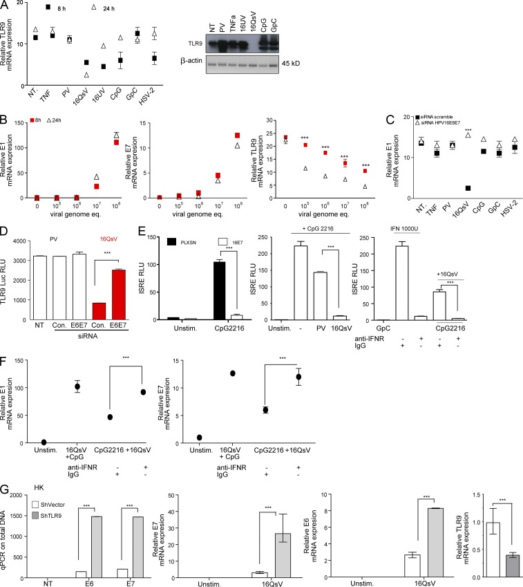
Figure 1. TLR9 expression is suppressed 24 h after infection by native 16QsV. (A, left) C33A cells were not treated (NT) or treated for 8 or 24 h with TNF, PV, 16QsV (at 107 viral concentrations genome equivalents), 16UV (rendered replication incompetent using UV), CpG 2006, GpC 2006, or infected with HSV-2. TLR9 mRNA levels were determined by qPCR. Shown are data from four independent experiments performed in triplicate. Error bars indicate SEM. (A, right) TLR9 protein was examined by immunoblotting in C33A cells. Cells were harvested after 24 h treatment with PV, TNF, 16UV, 16QsV CpG 2006, and GpC control. (B) C33A cells were treated with increasing viral concentrations genome equivalents (v.g.e.; as measured by qPCR on the viral DNA of infected cells) of 16QsV for 8 or 24 h). E1, E7 (left and middle), and TLR9 mRNA levels (right) were determined for their relative expression by qPCR. Shown are data from five independent experiments performed in duplicate with ***, P < 0.0001, based on an unpaired Student’s t test. (C) C33A cells were infected as indicated for 24 h. siRNA against HPV16E6E7 was transfected 24 h after stimulation and TLR9 mRNA levels were determined by qPCR. Shown are data from four independent experiments performed in triplicate and error bars indicate SEM with ***, P < 0.0001, based on an unpaired Student’s t test. (D) C33A cells were transfected with the TLR9 luciferase promoter and, 24 h later, treated with 16 or control PV. siRNA against HPV16E6E7 was transfected 24 h after infection and TLR9 luciferase levels were measured 18 h later. Shown are data from four independent experiments performed in triplicate, and error bars indicate SEM with ***, P < 0.0001, based on an unpaired Student’s t test. (E, left) HK transduced with the pLXSN vector control or HPV16E7 were stimulated for 24 h with TLR9 agonist CpG ODN 2216 (type A IFN inducer) and the IFN bioassay was performed. (E, middle) C33A cells were unstimulated or infected with 16QsV or PV for 36 h, and cells were washed and then treated with CpG 2216 for 24 h. Supernatants were collected and the type I IFN bioassay was performed. (E, right) C33A cells were stimulated with GpC or CpG 2216 for 24 h and then infected with 16QsV for 36 h. Supernatants were harvested and treated with anti-IFNR or IgG control and the type I IFN bioassay was performed as described. (F) C33A cells were unstimulated and infected with 16QsV for 36 h + CpG 2216 stimulation for 24 h or vice versa ± anti-IFNR or IgG control. Cells were harvested for RNA extraction and qRT-PCR was performed for E1 (left) or E7 (right) relative gene expression. Shown are data from four independent experiments performed in triplicate, and error bars indicate SEM with ***, P < 0.0001, based on an unpaired Student’s t test. (G) HK cells were transfected for a total of 48 h with shTLR9 and shControl (scramble sequence). 20 h after transfection, HK cells were infected for 24 h with 16QsV at a viral load of 107 v.g.e. Viral load was measured by qPCR using E6- and E7-specific primers. HPV16 E6 and E7 transcripts were also measured by qPCR and TLR9 transcripts were assessed by qPCR after 24 h transfection with shRNA. Data are representative of six independent experiments performed in triplicate. Shown are the mean ± SEM with ***, P < 0.0001, based on an unpaired Student’s t test.
Figure 1. TLR9 expression is suppressed 24 h after infection by native 16QsV. (A, left) C33A cells were not treated (NT) or treated for 8 or 24 h with TNF, PV, 16QsV (at 107 viral concentrations genome equivalents), 16UV (rendered replication incompetent using UV), CpG 2006, GpC 2006, or infected with HSV-2. TLR9 mRNA levels were determined by qPCR. Shown are data from four independent experiments performed in triplicate. Error bars indicate SEM. (A, right) TLR9 protein was examined by immunoblotting in C33A cells. Cells were harvested after 24 h treatment with PV, TNF, 16UV, 16QsV CpG 2006, and GpC control. (B) C33A cells were treated with increasing viral concentrations genome equivalents (v.g.e.; as measured by qPCR on the viral DNA of infected cells) of 16QsV for 8 or 24 h). E1, E7 (left and middle), and TLR9 mRNA levels (right) were determined for their relative expression by qPCR. Shown are data from five independent experiments performed in duplicate with ***, P < 0.0001, based on an unpaired Student’s t test. (C) C33A cells were infected as indicated for 24 h. siRNA against HPV16E6E7 was transfected 24 h after stimulation and TLR9 mRNA levels were determined by qPCR. Shown are data from four independent experiments performed in triplicate and error bars indicate SEM with ***, P < 0.0001, based on an unpaired Student’s t test. (D) C33A cells were transfected with the TLR9 luciferase promoter and, 24 h later, treated with 16 or control PV. siRNA against HPV16E6E7 was transfected 24 h after infection and TLR9 luciferase levels were measured 18 h later. Shown are data from four independent experiments performed in triplicate, and error bars indicate SEM with ***, P < 0.0001, based on an unpaired Student’s t test. (E, left) HK transduced with the pLXSN vector control or HPV16E7 were stimulated for 24 h with TLR9 agonist CpG ODN 2216 (type A IFN inducer) and the IFN bioassay was performed. (E, middle) C33A cells were unstimulated or infected with 16QsV or PV for 36 h, and cells were washed and then treated with CpG 2216 for 24 h. Supernatants were collected and the type I IFN bioassay was performed. (E, right) C33A cells were stimulated with GpC or CpG 2216 for 24 h and then infected with 16QsV for 36 h. Supernatants were harvested and treated with anti-IFNR or IgG control and the type I IFN bioassay was performed as described. (F) C33A cells were unstimulated and infected with 16QsV for 36 h + CpG 2216 stimulation for 24 h or vice versa ± anti-IFNR or IgG control. Cells were harvested for RNA extraction and qRT-PCR was performed for E1 (left) or E7 (right) relative gene expression. Shown are data from four independent experiments performed in triplicate, and error bars indicate SEM with ***, P < 0.0001, based on an unpaired Student’s t test. (G) HK cells were transfected for a total of 48 h with shTLR9 and shControl (scramble sequence). 20 h after transfection, HK cells were infected for 24 h with 16QsV at a viral load of 107 v.g.e. Viral load was measured by qPCR using E6- and E7-specific primers. HPV16 E6 and E7 transcripts were also measured by qPCR and TLR9 transcripts were assessed by qPCR after 24 h transfection with shRNA. Data are representative of six independent experiments performed in triplicate. Shown are the mean ± SEM with ***, P < 0.0001, based on an unpaired Student’s t test.CC BY-NC-SA · PubMed Central
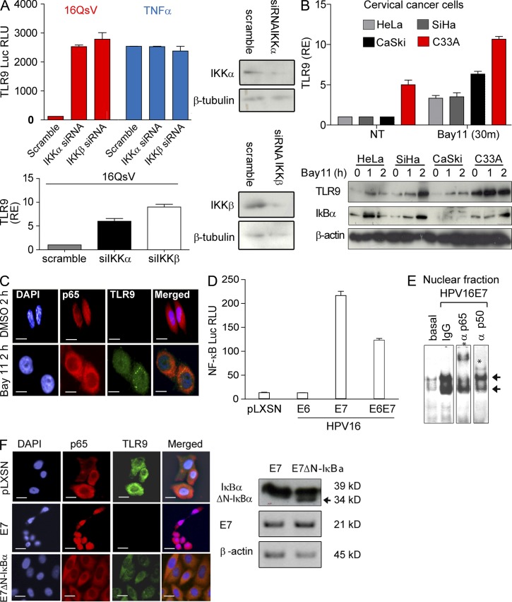
Figure 2. HPV16E7 activates the NF-κB pathway that leads to the suppression of TLR9. (A, top) C33A cells were treated with siRNA for IKKα or β for 16 h, and then transfected with TLR9 luciferase promoter. After 24 h, cells were exposed to the indicated treatments and harvested 24 h later to measure activity (left). IKKα or β levels as determined by immunoblotting (right) were analyzed. (A, bottom) C33A cells were treated with siRNA for IKKα or β for 24 h and TLR9 mRNA levels were measured by qPCR. Shown are data from six independent experiments performed in triplicate and error bars indicate SEM. (B) SiHa (HPV16+), HeLa (HPV18+), CaSki (HPV16+), and C33A control cells were treated with the IKKβ inhibitor Bay11. At the indicated times, mRNA and protein expression of TLR9 as well as protein levels of IκBα was determined by immunoblotting. Shown are data from seven independent experiments performed in triplicate and error bars indicate SEM. RE, mRNA relative expression. (C) SiHa cells were treated with Bay11 for the indicated time and immunofluorescence was performed to determine NF-κBp65 cellular localization (red) and TLR9 expression (green). Nuclear staining was controlled using DAPI. Shown are data from one out of four examined fields and one out of three independent experiments. Bars, 10 µm. (D) Human female primary keratinocytes (HK) were transduced with the indicated retroviruses and transfected with the NF-κB reporter gene construct. At 48 h after transfection, luciferase activity was determined. Data are from one assay representative of seven independent experiments; shown are mean ± SEM from triplicate values in three. (E) EMSA was performed on HPV16E7-transduced HK using the NF-κB EMSA kit (Panomics) according to the manufacturer’s instructions. For the supershift analyses, nuclear extracts were incubated with NF-κBp50 or p65 or IgG control. The arrow indicates NF-κB complexes, and the asterisk indicates a supershift. Shown are data from one out of four independent experiments that gave identical results. (F, left) HKs were retrovirally transduced with HPV16E7, empty vector (pLXSN), or HPV16E7ΔN-IκBα and were harvested at the doubling population 7 and stained by immunofluorescence for TLR9 and NF-κBp65. Either Alexa Fluor 488 nm (green) or 594 nm (red) was used as secondary antibodies. Nuclear staining was controlled using DAPI. Shown are data from one out of six examined fields and one out of three independent experiments. Bars, 10 µm. (F, right) Western blot for ΔN-IκBα (marked with an arrow), E7, and β-actin. Shown are data from one out of five independent experiments that gave identical results.
Figure 2. HPV16E7 activates the NF-κB pathway that leads to the suppression of TLR9. (A, top) C33A cells were treated with siRNA for IKKα or β for 16 h, and then transfected with TLR9 luciferase promoter. After 24 h, cells were exposed to the indicated treatments and harvested 24 h later to measure activity (left). IKKα or β levels as determined by immunoblotting (right) were analyzed. (A, bottom) C33A cells were treated with siRNA for IKKα or β for 24 h and TLR9 mRNA levels were measured by qPCR. Shown are data from six independent experiments performed in triplicate and error bars indicate SEM. (B) SiHa (HPV16+), HeLa (HPV18+), CaSki (HPV16+), and C33A control cells were treated with the IKKβ inhibitor Bay11. At the indicated times, mRNA and protein expression of TLR9 as well as protein levels of IκBα was determined by immunoblotting. Shown are data from seven independent experiments performed in triplicate and error bars indicate SEM. RE, mRNA relative expression. (C) SiHa cells were treated with Bay11 for the indicated time and immunofluorescence was performed to determine NF-κBp65 cellular localization (red) and TLR9 expression (green). Nuclear staining was controlled using DAPI. Shown are data from one out of four examined fields and one out of three independent experiments. Bars, 10 µm. (D) Human female primary keratinocytes (HK) were transduced with the indicated retroviruses and transfected with the NF-κB reporter gene construct. At 48 h after transfection, luciferase activity was determined. Data are from one assay representative of seven independent experiments; shown are mean ± SEM from triplicate values in three. (E) EMSA was performed on HPV16E7-transduced HK using the NF-κB EMSA kit (Panomics) according to the manufacturer’s instructions. For the supershift analyses, nuclear extracts were incubated with NF-κBp50 or p65 or IgG control. The arrow indicates NF-κB complexes, and the asterisk indicates a supershift. Shown are data from one out of four independent experiments that gave identical results. (F, left) HKs were retrovirally transduced with HPV16E7, empty vector (pLXSN), or HPV16E7ΔN-IκBα and were harvested at the doubling population 7 and stained by immunofluorescence for TLR9 and NF-κBp65. Either Alexa Fluor 488 nm (green) or 594 nm (red) was used as secondary antibodies. Nuclear staining was controlled using DAPI. Shown are data from one out of six examined fields and one out of three independent experiments. Bars, 10 µm. (F, right) Western blot for ΔN-IκBα (marked with an arrow), E7, and β-actin. Shown are data from one out of five independent experiments that gave identical results.CC BY-NC-SA · PubMed Central
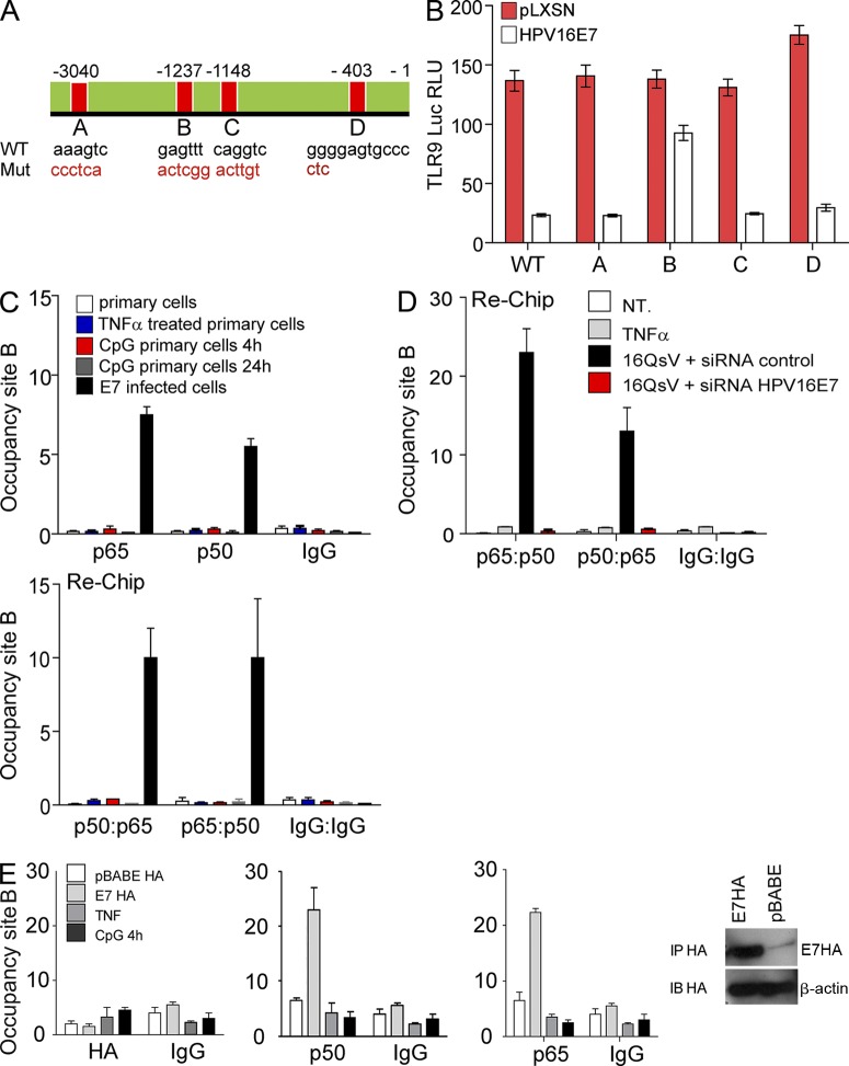
Figure 3. TLR9 promoter regulation by HPV16E7 depends on the locality of the NF-κB complex at a specific cis site. (A) Predicted NF-κB binding sites (A, B, C, and D) in the TLR9 promoter. Sequence mutations were made at site A, B, C, and D to inactivate the binding sites. Top sequences are the native form; bottom sequences indicate the mutations made. (B) HKs were transduced with HPV16E7 or empty vector. Transduced cells were then transfected with TLR9 promoter luciferase expression vectors containing either a wt promoter sequence or sequences with the mutation in the A, B, C, or D site indicated in A. 40 h after transfection, luciferase activity was measured. Shown are data from six independent experiments performed in triplicate and error bars indicate SEM. (C) The binding of NF-κB complexes to the TLR9 promoter in HK was determined by ChIP and ReChIP assays. Sheared chromatin from HK transduced with HPV16E7 or stimulated with TNF or CpG 2006 (4 and 24 h) was immunoprecipitated with antibodies to NF-κBp50 or p65. Site B on the TLR9 promoter was amplified by qPCR to determine the specific binding of transcription factors bound to DNA. Immunoprecipitated DNA and input DNA was amplified with gene-specific and β-globin (Hbb) primers by qPCR, using input DNA to generate a standard curve. ChIP data are represented as % input (gene-specific)/% input(β-globin) = occupancy site B. Shown are data from six independent experiments performed in triplicate and error bars indicate SEM. (D) Re-ChIP analysis was performed for p50–p65 NF-κB complexes using C33A cells first infected with 16QsV for 36 h. Cells were then transfected with siRNA for HPV16E7 and harvested 24 h later. Shown are data from five independent experiments performed in triplicate and error bars indicate SEM. (E) E7 does not bind to the NF-kB cis element site B. ChIP was performed using human epithelial cells transfected with pbabe HA, pbabe HPV16E7-HA, or stimulated with CpG (4 h) or TNF (8 h). ChIP was performed using HA, NF-κBp50 or p65, or an IgG control antibody to examine occupancy of site B. Shown are data from six independent experiments performed in triplicate, and error bars indicate SEM. Shown is an immunoblot to control the ChIP for HA-E7.
Figure 3. TLR9 promoter regulation by HPV16E7 depends on the locality of the NF-κB complex at a specific cis site. (A) Predicted NF-κB binding sites (A, B, C, and D) in the TLR9 promoter. Sequence mutations were made at site A, B, C, and D to inactivate the binding sites. Top sequences are the native form; bottom sequences indicate the mutations made. (B) HKs were transduced with HPV16E7 or empty vector. Transduced cells were then transfected with TLR9 promoter luciferase expression vectors containing either a wt promoter sequence or sequences with the mutation in the A, B, C, or D site indicated in A. 40 h after transfection, luciferase activity was measured. Shown are data from six independent experiments performed in triplicate and error bars indicate SEM. (C) The binding of NF-κB complexes to the TLR9 promoter in HK was determined by ChIP and ReChIP assays. Sheared chromatin from HK transduced with HPV16E7 or stimulated with TNF or CpG 2006 (4 and 24 h) was immunoprecipitated with antibodies to NF-κBp50 or p65. Site B on the TLR9 promoter was amplified by qPCR to determine the specific binding of transcription factors bound to DNA. Immunoprecipitated DNA and input DNA was amplified with gene-specific and β-globin (Hbb) primers by qPCR, using input DNA to generate a standard curve. ChIP data are represented as % input (gene-specific)/% input(β-globin) = occupancy site B. Shown are data from six independent experiments performed in triplicate and error bars indicate SEM. (D) Re-ChIP analysis was performed for p50–p65 NF-κB complexes using C33A cells first infected with 16QsV for 36 h. Cells were then transfected with siRNA for HPV16E7 and harvested 24 h later. Shown are data from five independent experiments performed in triplicate and error bars indicate SEM. (E) E7 does not bind to the NF-kB cis element site B. ChIP was performed using human epithelial cells transfected with pbabe HA, pbabe HPV16E7-HA, or stimulated with CpG (4 h) or TNF (8 h). ChIP was performed using HA, NF-κBp50 or p65, or an IgG control antibody to examine occupancy of site B. Shown are data from six independent experiments performed in triplicate, and error bars indicate SEM. Shown is an immunoblot to control the ChIP for HA-E7.CC BY-NC-SA · PubMed Central
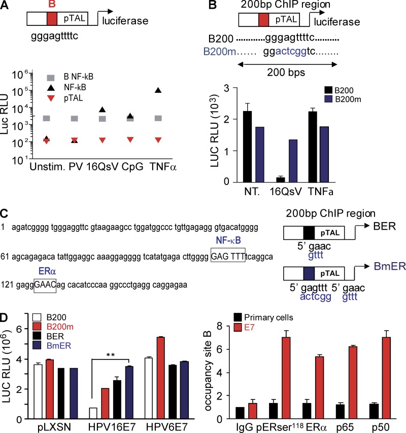
Figure 4. The negative regulatory effect of HPV16 on the TLR9 promoter requires NF-κB and ERα binding to site B flanking sequences in human epithelial cells and HK. (A, top) Diagram illustrates the minimal B site promoter. (A, bottom) Human epithelial cervical cells were transfected with the NF-κB luciferase consensus site, the B minimal promoter or the control vector pTAL. 24 h after transfection 16QsV, control PV (native), CpG 2006, or TNF were added. Cells were harvested and luciferase activity was measured after 24 h. (B, top) Diagram depicting constructs including a 200 bp sequence of the TLR9 promoter containing the NF-κB cis element on site B WT (B200) or mutated (Bm). (B, bottom) C33A cells were transfected with B200 or B200m. 24 h after transfection, 16QsV or TNF were added. Cells were harvested and luciferase activity was measured after 24 h (right). (C, left) The 200 bp sequence around site B on the TLR9 promoter with mapped NF-κB and ERα cis sites. (C, right) Site B was mutated at the cis ERα site (BER) and double mutation for NF-κB and ERα cis elements (BmER). (D, left) HK cells transduced with pLXSN, HPV16E7, or HPV6E7 were transfected with B200, B200m, BER, or BmER. 24 h after transfection luciferase activity was measured. (D, right) ChIP analyses of ERα phosphorylated and total forms and NF-κBp65 or p50 binding to site B on the TLR9 promoter in HK transduced ± HPV16E7. Data are representative of the mean of five or more independent experiments performed in triplicate, and error bars indicate SEM. **, P < 0.001 based on an unpaired Student’s t test.
Figure 4. The negative regulatory effect of HPV16 on the TLR9 promoter requires NF-κB and ERα binding to site B flanking sequences in human epithelial cells and HK. (A, top) Diagram illustrates the minimal B site promoter. (A, bottom) Human epithelial cervical cells were transfected with the NF-κB luciferase consensus site, the B minimal promoter or the control vector pTAL. 24 h after transfection 16QsV, control PV (native), CpG 2006, or TNF were added. Cells were harvested and luciferase activity was measured after 24 h. (B, top) Diagram depicting constructs including a 200 bp sequence of the TLR9 promoter containing the NF-κB cis element on site B WT (B200) or mutated (Bm). (B, bottom) C33A cells were transfected with B200 or B200m. 24 h after transfection, 16QsV or TNF were added. Cells were harvested and luciferase activity was measured after 24 h (right). (C, left) The 200 bp sequence around site B on the TLR9 promoter with mapped NF-κB and ERα cis sites. (C, right) Site B was mutated at the cis ERα site (BER) and double mutation for NF-κB and ERα cis elements (BmER). (D, left) HK cells transduced with pLXSN, HPV16E7, or HPV6E7 were transfected with B200, B200m, BER, or BmER. 24 h after transfection luciferase activity was measured. (D, right) ChIP analyses of ERα phosphorylated and total forms and NF-κBp65 or p50 binding to site B on the TLR9 promoter in HK transduced ± HPV16E7. Data are representative of the mean of five or more independent experiments performed in triplicate, and error bars indicate SEM. **, P < 0.001 based on an unpaired Student’s t test.CC BY-NC-SA · PubMed Central
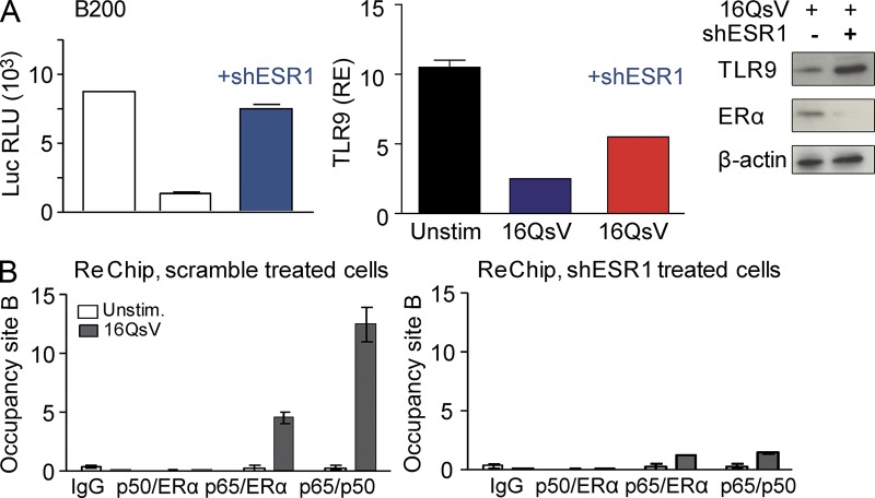
Figure 5. ERα complexes with NF-κBp65 to suppress TLR9 transcription by 16QsV. (A, left) C33A cells were transfected with B200 and, 24 h later, shESR1 was introduced. 12 h after shESR1 introduction, cells were infected with 16QsV. Cells were harvested and luciferase activity was measured after 24 h. (A, right) qPCR and immunoblot analysis for TLR9 and ERα in the human epithelial cervical cells treated with shESR1. (B) C33A cells were transfected with a shRNA scramble for ERα (left) or with an shRNA for ERα (shESR1; right) for 24 h. Cells were then infected for 12 h with 16QsV and analyzed by ReChIP for NF-κBp50–ERα or NF-κBp65–ERα or NF-κBp65–p50 occupancy on site B of the TLR9 promoter. Data are representative of the mean of five or more independent experiments performed in triplicate; graphs show the mean ± SEM.
Figure 5. ERα complexes with NF-κBp65 to suppress TLR9 transcription by 16QsV. (A, left) C33A cells were transfected with B200 and, 24 h later, shESR1 was introduced. 12 h after shESR1 introduction, cells were infected with 16QsV. Cells were harvested and luciferase activity was measured after 24 h. (A, right) qPCR and immunoblot analysis for TLR9 and ERα in the human epithelial cervical cells treated with shESR1. (B) C33A cells were transfected with a shRNA scramble for ERα (left) or with an shRNA for ERα (shESR1; right) for 24 h. Cells were then infected for 12 h with 16QsV and analyzed by ReChIP for NF-κBp50–ERα or NF-κBp65–ERα or NF-κBp65–p50 occupancy on site B of the TLR9 promoter. Data are representative of the mean of five or more independent experiments performed in triplicate; graphs show the mean ± SEM.CC BY-NC-SA · PubMed Central
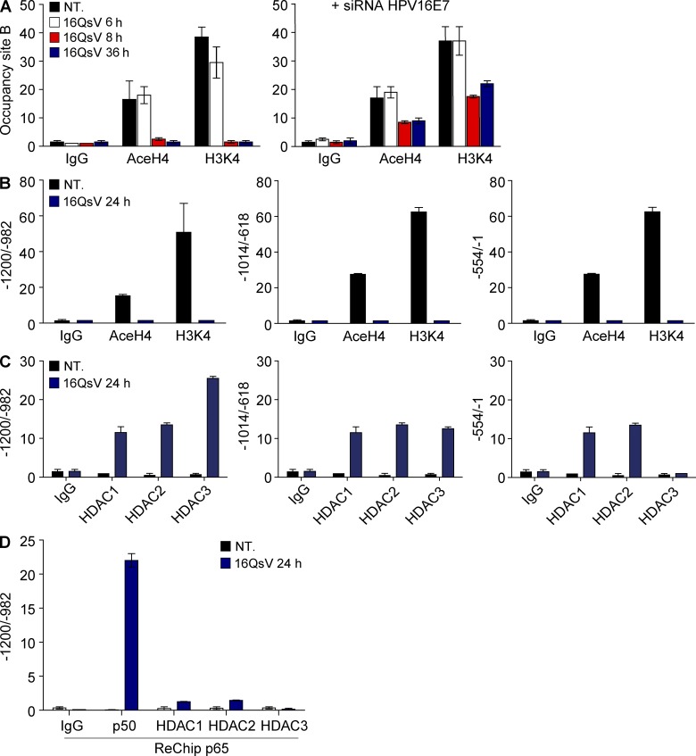
Figure 6. 16QsV induces closing of the chromatin structure on the TLR9 promoter from site B until the transcription start site. (A, left) ChIP using anti AceH4 or H3K4me3 histone antibodies was performed for site B using C33A cells infected with 16QsV for 6, 8, and 36 h. (A, right) C33A cells were treated as in A (left) except that 1 h after incubation with 16QsV, cells were treated with siRNA against HPV16E7. (B) Chromatin from C33A cells that had been incubated ± 16QsV for 24 h was analyzed by ChIP for AceH4 or H3K4me3 histones interacting with chromatin DNA along the TLR9 promoter. AceH4 or H3K4me3 binding to histones associated DNA upstream of site B on the TLR9 promoter were amplified by qPCR along the regions −1200 until −1. (C) HDAC1-3 binding to histone associated DNA of upstream of site B on the TLR9 promoter were amplified by qPCR along the regions −1200 until −1. (D) ReChIP for NF-κBp65–p50 or NF-κBp65–HDAC1-3 was performed on C33A cells treated with 16QsV for 36 h. Data are representative of three or more independent experiments; graphs shown are mean ± SEM from triplicate values.
Figure 6. 16QsV induces closing of the chromatin structure on the TLR9 promoter from site B until the transcription start site. (A, left) ChIP using anti AceH4 or H3K4me3 histone antibodies was performed for site B using C33A cells infected with 16QsV for 6, 8, and 36 h. (A, right) C33A cells were treated as in A (left) except that 1 h after incubation with 16QsV, cells were treated with siRNA against HPV16E7. (B) Chromatin from C33A cells that had been incubated ± 16QsV for 24 h was analyzed by ChIP for AceH4 or H3K4me3 histones interacting with chromatin DNA along the TLR9 promoter. AceH4 or H3K4me3 binding to histones associated DNA upstream of site B on the TLR9 promoter were amplified by qPCR along the regions −1200 until −1. (C) HDAC1-3 binding to histone associated DNA of upstream of site B on the TLR9 promoter were amplified by qPCR along the regions −1200 until −1. (D) ReChIP for NF-κBp65–p50 or NF-κBp65–HDAC1-3 was performed on C33A cells treated with 16QsV for 36 h. Data are representative of three or more independent experiments; graphs shown are mean ± SEM from triplicate values.CC BY-NC-SA · PubMed Central
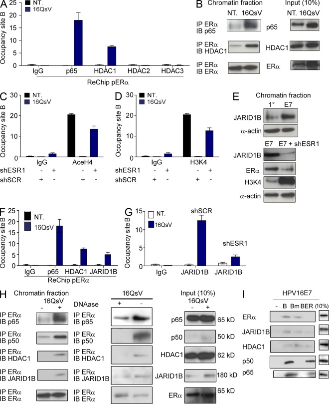
Figure 7. Recruitment of epigenetic demethylating and deacetylating enzymes by ERα to site B on the TLR9 promoter in 16QsV human epithelial cells. (A) ReChIP for pERα–p65 or HDAC1-3 was performed on C33A cells treated with 16QsV for 24 h. (B, left) Immunoprecipation for ERα interactions with NF-κBp65 or HDAC1 was performed on chromatin fraction of C33A infected for 36 h with 16QsV. (B, right) Input controls. (C) ChIP using anti AceH4 histone antibodies was performed for site B on C33A cells infected with 16QsV for 24 h ± shESR1. (D) ChIP using anti H3K4me3 histone antibodies was performed for site B on C33A cells infected with 16QsV for 24 h ± shESR1. (E) Chromatin fraction Western blot analysis of JARID1B, ERα, or H3K4me3 expression in pLXSN or HPV16E7 ± shESR1-transduced HK. (F) ReChIP for pERα–p65 or pERα/HDAC1 or pERα/JARID1B was performed on C33A cells treated with 16QsV for 36 h. (G) ChIP using anti-JARID1B antibody was performed for site B on C33A cells infected with 16QsV for 24 h ± shScramble control sequence (shSCR) or shESR1. (H, left) Immunoprecipation for ERα interactions with NF-κBp65, p50, JARID1B, or HDAC1 was performed on chromatin fractions of C33A infected for 36 h with 16QsV ± DNase I treatment. (H, right) 10% of input loaded protein. (I, left) Oligo pulldown assay for site B, Bm, and BER using protein lysates from pLXSN or HPV16E7-transduced cells. Bound proteins were assessed by immunoblotting for NF-κBp65, p50, JARID1B, HDAC1, or ERα. (I, right) Input controls (10%). Data are representative of three or more independent experiments; graphs shown are the mean ± SEM from triplicate values.
Figure 7. Recruitment of epigenetic demethylating and deacetylating enzymes by ERα to site B on the TLR9 promoter in 16QsV human epithelial cells. (A) ReChIP for pERα–p65 or HDAC1-3 was performed on C33A cells treated with 16QsV for 24 h. (B, left) Immunoprecipation for ERα interactions with NF-κBp65 or HDAC1 was performed on chromatin fraction of C33A infected for 36 h with 16QsV. (B, right) Input controls. (C) ChIP using anti AceH4 histone antibodies was performed for site B on C33A cells infected with 16QsV for 24 h ± shESR1. (D) ChIP using anti H3K4me3 histone antibodies was performed for site B on C33A cells infected with 16QsV for 24 h ± shESR1. (E) Chromatin fraction Western blot analysis of JARID1B, ERα, or H3K4me3 expression in pLXSN or HPV16E7 ± shESR1-transduced HK. (F) ReChIP for pERα–p65 or pERα/HDAC1 or pERα/JARID1B was performed on C33A cells treated with 16QsV for 36 h. (G) ChIP using anti-JARID1B antibody was performed for site B on C33A cells infected with 16QsV for 24 h ± shScramble control sequence (shSCR) or shESR1. (H, left) Immunoprecipation for ERα interactions with NF-κBp65, p50, JARID1B, or HDAC1 was performed on chromatin fractions of C33A infected for 36 h with 16QsV ± DNase I treatment. (H, right) 10% of input loaded protein. (I, left) Oligo pulldown assay for site B, Bm, and BER using protein lysates from pLXSN or HPV16E7-transduced cells. Bound proteins were assessed by immunoblotting for NF-κBp65, p50, JARID1B, HDAC1, or ERα. (I, right) Input controls (10%). Data are representative of three or more independent experiments; graphs shown are the mean ± SEM from triplicate values.CC BY-NC-SA · PubMed Central
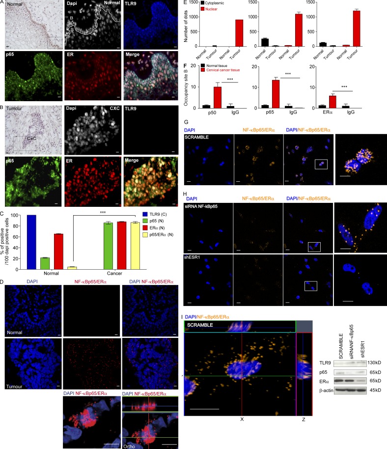
Figure 8. HPV16-positive cervical cancer lesions contain NF-κBp65–ERα nuclear complexes which bind to site B on the TLR9 promoter. Histology and immunofluorescence (IF) of normal cervical issue (A) and HPV16+ cervical cancer biopsies (B). Nuclear (white), TLR9 (blue), ERα (red), or NF-κBp65 (green). The box in the histology staining indicates which part of the slide was examined by IF. Normal (HPV−) and a neoplastic biopsy (HPV16+) from one representative patient out of eight tested with similar results is shown. Bars, 10 µm. (C) Histograms representing the cellular distribution for TLR9, ERα, and NF-κBp65 in normal (HPV−) and cancer cervical tissue (HPV16 positive by Apex screening). For each stained biopsy, six fields were examined and cytoplasmic or nuclear staining was counted manually and the percentage scored out of 100 cells. Data are representative of three independent experiments performed on 16 biopsies (eight normal and eight neoplastic); graphs shown are the mean ± SEM. ***, P < 0.0001. (D) Duolink analysis of NF-κBp65–ERα in normal versus cervical cancer tissue. DAPI marks the nucleus and the red dots represent NF-κBp65–ERα proximity ligation <40 nm distance. Shown are data from one out of five independent experiments that gave identical results. Represented here is one out of six fields examined for each section. Bars, 10 µm. (E) NF-κBp65 and ERα proximity interaction locality were estimated by counting the number of red dots manually and using the Duolink Image tool (Olink, Biosciences) present in three field sections on three normal versus three cervical cancer tissues. Data are representative of three independent experiments performed on six biopsies (normal and neoplastic); graphs shown are the mean ± SEM. (F) ChIP analysis was performed on normal cervical tissue and cervical cancer biopsies for NF-κBp65, p50, or ERα binding to site B on the TLR9 promoter (represented one experiment out of three ChIP, and qPCR for occupancy on site B was performed in duplicate three times on three normal vs. four cervical cancer tissues). Shown are data from six independent experiments performed in triplicate, and error bars indicate SEM. ***, P < 0.0001 based on an unpaired Student’s t test. (G) Duolink analysis of NF-κBp65–ERα in the cervical cancer cell line CaSki. DAPI marks the nucleus and the orange dots represent NF-κBp65–ERα proximity ligation <40 nm distance. Shown are data from one out of three independent experiments that gave identical results in which one of five fields were examined. Bars, 20 µm. (H) As in G, however cells were treated with a siRNA sequence for NF-κBp65 or small hairpin for ERα (as described in Fig. 5). Shown are data from one out of five independent experiments that gave identical results. Bars, 20 µm. (I, left) 63× zoom × 2 and ortho Z slices (3.3 µM) show nuclear penetration of the NF-κBp65 and ERα proximity ligation of <40 nm in scramble short hairpin for ERα-treated cells. Shown are data from one out of five fields examined and one out of five independent experiments that gave identical results. Bar, 20 µm. (I, right) Immunoblot demonstrating the efficiency of siRNA knockdown of NF-κBp65 and shESR1. Shown are data from one out three immunoblots performed.
Figure 8. HPV16-positive cervical cancer lesions contain NF-κBp65–ERα nuclear complexes which bind to site B on the TLR9 promoter. Histology and immunofluorescence (IF) of normal cervical issue (A) and HPV16+ cervical cancer biopsies (B). Nuclear (white), TLR9 (blue), ERα (red), or NF-κBp65 (green). The box in the histology staining indicates which part of the slide was examined by IF. Normal (HPV−) and a neoplastic biopsy (HPV16+) from one representative patient out of eight tested with similar results is shown. Bars, 10 µm. (C) Histograms representing the cellular distribution for TLR9, ERα, and NF-κBp65 in normal (HPV−) and cancer cervical tissue (HPV16 positive by Apex screening). For each stained biopsy, six fields were examined and cytoplasmic or nuclear staining was counted manually and the percentage scored out of 100 cells. Data are representative of three independent experiments performed on 16 biopsies (eight normal and eight neoplastic); graphs shown are the mean ± SEM. ***, P < 0.0001. (D) Duolink analysis of NF-κBp65–ERα in normal versus cervical cancer tissue. DAPI marks the nucleus and the red dots represent NF-κBp65–ERα proximity ligation <40 nm distance. Shown are data from one out of five independent experiments that gave identical results. Represented here is one out of six fields examined for each section. Bars, 10 µm. (E) NF-κBp65 and ERα proximity interaction locality were estimated by counting the number of red dots manually and using the Duolink Image tool (Olink, Biosciences) present in three field sections on three normal versus three cervical cancer tissues. Data are representative of three independent experiments performed on six biopsies (normal and neoplastic); graphs shown are the mean ± SEM. (F) ChIP analysis was performed on normal cervical tissue and cervical cancer biopsies for NF-κBp65, p50, or ERα binding to site B on the TLR9 promoter (represented one experiment out of three ChIP, and qPCR for occupancy on site B was performed in duplicate three times on three normal vs. four cervical cancer tissues). Shown are data from six independent experiments performed in triplicate, and error bars indicate SEM. ***, P < 0.0001 based on an unpaired Student’s t test. (G) Duolink analysis of NF-κBp65–ERα in the cervical cancer cell line CaSki. DAPI marks the nucleus and the orange dots represent NF-κBp65–ERα proximity ligation <40 nm distance. Shown are data from one out of three independent experiments that gave identical results in which one of five fields were examined. Bars, 20 µm. (H) As in G, however cells were treated with a siRNA sequence for NF-κBp65 or small hairpin for ERα (as described in Fig. 5). Shown are data from one out of five independent experiments that gave identical results. Bars, 20 µm. (I, left) 63× zoom × 2 and ortho Z slices (3.3 µM) show nuclear penetration of the NF-κBp65 and ERα proximity ligation of <40 nm in scramble short hairpin for ERα-treated cells. Shown are data from one out of five fields examined and one out of five independent experiments that gave identical results. Bar, 20 µm. (I, right) Immunoblot demonstrating the efficiency of siRNA knockdown of NF-κBp65 and shESR1. Shown are data from one out three immunoblots performed.CC BY-NC-SA · PubMed Central

Verification

Claims the paper makes

The claims and statistics of this paper haven’t been checked yet.

Run the check — free, no account needed. It reads the open-access text and checks the sources it cites.

A few free checks a day · papers already verified open instantly · results are public

Claims other papers make about this one

Each is checked against this paper’s own text: does it say what the citing paper says it does?

Self-check

Percentages, sums and reported differences re-computed from the paper’s own numbers.

The self-check needs the paper’s full text.

Citations

Cites

Show all 9 references

Research works printed but not linked: 76

  1. Accardi, 2011, IkappaB kinase beta promotes cell survival by antagonizing p53 functions through DeltaNp73alpha phosphorylation and stabilization, Mol. Cell. Biol., 31, 2210, 10.1128/MCB.00964-10 · DOI
  2. Adams, 2008, Immunization of malignant melanoma patients with full-length NY-ESO-1 protein using TLR7 agonist imiquimod as vaccine adjuvant, J. Immunol., 181, 776, 10.4049/jimmunol.181.1.776 · DOI
  3. Avalos, 2011, Competition by inhibitory oligonucleotides prevents binding of CpG to C-terminal TLR9, Eur. J. Immunol., 41, 2820, 10.1002/eji.201141563 · DOI
  4. Bangert, 2011, Immune functions of the skin, Clin. Dermatol., 29, 360, 10.1016/j.clindermatol.2011.01.006 · DOI
  5. Brake, 2005, Estrogen contributes to the onset, persistence, and malignant progression of cervical cancer in a human papillomavirus-transgenic mouse model, Proc. Natl. Acad. Sci. USA., 102, 2490, 10.1073/pnas.0409883102 · DOI
  6. Buck, 2005, Generation of HPV pseudovirions using transfection and their use in neutralization assays, Methods Mol. Med., 119, 445
  7. Catchpole, 2011, PLU-1/JARID1B/KDM5B is required for embryonic survival and contributes to cell proliferation in the mammary gland and in ER+ breast cancer cells, Int. J. Oncol., 38, 1267
  8. Chung, 2009, Prevention and treatment of cervical cancer in mice using estrogen receptor antagonists, Proc. Natl. Acad. Sci. USA., 106, 19467, 10.1073/pnas.0911436106 · DOI
  9. Chung, 2008, Requirement for estrogen receptor alpha in a mouse model for human papillomavirus-associated cervical cancer, Cancer Res., 68, 9928, 10.1158/0008-5472.CAN-08-2051 · DOI
  10. Chung, 2010, Estrogen and ERalpha: culprits in cervical cancer?, Trends Endocrinol. Metab., 21, 504, 10.1016/j.tem.2010.03.005 · DOI
  11. Coban, 2005, Toll-like receptor 9 mediates innate immune activation by the malaria pigment hemozoin, J. Exp. Med., 201, 19, 10.1084/jem.20041836 · DOI
  12. Daud, 2008, Validation of reference genes in cervical cell samples from human papillomavirus-infected and -uninfected women for quantitative reverse transcription-PCR assays, Clin. Vaccine Immunol., 15, 1369, 10.1128/CVI.00074-08 · DOI
  13. Daud, 2011, Association between toll-like receptor expression and human papillomavirus type 16 persistence, Int. J. Cancer., 128, 879, 10.1002/ijc.25400 · DOI
  14. Debenedictis, 2001, Immune functions of the skin, Clin. Dermatol., 19, 573, 10.1016/S0738-081X(00)00173-5 · DOI
  15. DeCarlo, 2012, Toll-like receptor transcriptome in the HPV-positive cervical cancer microenvironment, Clin. Dev. Immunol., 2012, 785825, 10.1155/2012/785825 · DOI
  16. Dey, 2008, The histone demethylase KDM5b/JARID1b plays a role in cell fate decisions by blocking terminal differentiation, Mol. Cell. Biol., 28, 5312, 10.1128/MCB.00128-08 · DOI
  17. Ewald, 2011, Nucleic acid recognition by Toll-like receptors is coupled to stepwise processing by cathepsins and asparagine endopeptidase, J. Exp. Med., 208, 643, 10.1084/jem.20100682 · DOI
  18. Fathallah, 2010, EBV latent membrane protein 1 is a negative regulator of TLR9, J. Immunol., 185, 6439, 10.4049/jimmunol.0903459 · DOI
  19. Feldman, 2007, Identification of proteins within the nuclear factor-kappa B transcriptional complex including estrogen receptor-alpha, Am. J. Obstet. Gynecol., 196, 10.1016/j.ajog.2006.12.033 · DOI
  20. Fiola, 2010, TLR9 contributes to the recognition of EBV by primary monocytes and plasmacytoid dendritic cells, J. Immunol., 185, 3620, 10.4049/jimmunol.0903736 · DOI
  21. Flores, 1999, Establishment of the human papillomavirus type 16 (HPV-16) life cycle in an immortalized human foreskin keratinocyte cell line, Virology., 262, 344, 10.1006/viro.1999.9868 · DOI
  22. Gibson, 2012, Estrogen dependent signaling in reproductive tissues - a role for estrogen receptors and estrogen related receptors, Mol. Cell. Endocrinol., 348, 361, 10.1016/j.mce.2011.09.026 · DOI · Paper page
  23. Gonda, 1996, The c-myb negative regulatory domain, Curr. Top. Microbiol. Immunol., 211, 99
  24. Gondois-Rey, 2009, Hepatitis C virus is a weak inducer of interferon alpha in plasmacytoid dendritic cells in comparison with influenza and human herpesvirus type-1, PLoS ONE., 4, e4319, 10.1371/journal.pone.0004319 · DOI
  25. Häcker, 2006, Regulation and function of IKK and IKK-related kinases, Sci. STKE., 2006, re13, 10.1126/stke.3572006re13 · DOI
  26. Hasan, 2004, Differential induction of gene promoter constructs by constitutively active human TLRs, Biochem. Biophys. Res. Commun., 321, 124, 10.1016/j.bbrc.2004.06.134 · DOI
  27. Hasan, 2005, Human TLR10 is a functional receptor, expressed by B cells and plasmacytoid dendritic cells, which activates gene transcription through MyD88, J. Immunol., 174, 2942, 10.4049/jimmunol.174.5.2942 · DOI
  28. Hasan, 2007, TLR9 expression and function is abolished by the cervical cancer-associated human papillomavirus type 16, J. Immunol., 178, 3186, 10.4049/jimmunol.178.5.3186 · DOI
  29. Hasan, 2007, Cell proliferation and survival induced by Toll-like receptors is antagonized by type I IFNs, Proc. Natl. Acad. Sci. USA., 104, 8047, 10.1073/pnas.0700664104 · DOI
  30. Hirsch, 2010, Impaired Toll-like receptor 7 and 9 signaling: from chronic viral infections to cancer, Trends Immunol., 31, 391, 10.1016/j.it.2010.07.004 · DOI
  31. Hudak, 2002, Immune surveillance and effector functions of CCR10(+) skin homing T cells, J. Immunol., 169, 1189, 10.4049/jimmunol.169.3.1189 · DOI
  32. Ito, 2003, Alterations of immune functions in barrier disrupted skin by UVB irradiation, J. Dermatol. Sci., 33, 151, 10.1016/S0923-1811(03)00177-4 · DOI
  33. Joel, 1998, pp90rsk1 regulates estrogen receptor-mediated transcription through phosphorylation of Ser-167, Mol. Cell. Biol., 18, 1978, 10.1128/MCB.18.4.1978 · DOI
  34. Kaplan, 2012, Early immune events in the induction of allergic contact dermatitis, Nat. Rev. Immunol., 12, 114, 10.1038/nri3150 · DOI
  35. Karim, 2011, Human papillomavirus deregulates the response of a cellular network comprising of chemotactic and proinflammatory genes, PLoS ONE., 6, e17848, 10.1371/journal.pone.0017848 · DOI
  36. Kim, 2010, Histone demethylase JARID1B/KDM5B is a corepressor of TIEG1/KLF10, Biochem. Biophys. Res. Commun., 401, 412, 10.1016/j.bbrc.2010.09.068 · DOI
  37. Kim, 2003, Methylation patterns of papillomavirus DNA, its influence on E2 function, and implications in viral infection, J. Virol., 77, 12450, 10.1128/JVI.77.23.12450-12459.2003 · DOI
  38. Lebre, 2007, Human keratinocytes express functional Toll-like receptor 3, 4, 5, and 9, J. Invest. Dermatol., 127, 331, 10.1038/sj.jid.5700530 · DOI
  39. Lin, 2009, Combination of proteasome and HDAC inhibitors for uterine cervical cancer treatment, Clin. Cancer Res., 15, 570, 10.1158/1078-0432.CCR-08-1813 · DOI
  40. Lund, 2003, Toll-like receptor 9–mediated recognition of Herpes simplex virus-2 by plasmacytoid dendritic cells, J. Exp. Med., 198, 513, 10.1084/jem.20030162 · DOI
  41. Malanchi, 2004, Human papillomavirus type 16 E6 promotes retinoblastoma protein phosphorylation and cell cycle progression, J. Virol., 78, 13769, 10.1128/JVI.78.24.13769-13778.2004 · DOI
  42. Mansour, 2007, E7 properties of mucosal human papillomavirus types 26, 53 and 66 correlate with their intermediate risk for cervical cancer development, Virology., 367, 1, 10.1016/j.virol.2007.05.005 · DOI
  43. Marur, 2010, HPV-associated head and neck cancer: a virus-related cancer epidemic, Lancet Oncol., 11, 781, 10.1016/S1470-2045(10)70017-6 · DOI
  44. McCloskey, 2010, Human papillomavirus type 16 E6/E7 upregulation of nucleophosmin is important for proliferation and inhibition of differentiation, J. Virol., 84, 5131, 10.1128/JVI.01965-09 · DOI
  45. Méndez, 2000, Chromatin association of human origin recognition complex, cdc6, and minichromosome maintenance proteins during the cell cycle: assembly of prereplication complexes in late mitosis, Mol. Cell. Biol., 20, 8602, 10.1128/MCB.20.22.8602-8612.2000 · DOI
  46. Metz, 2008, Mast cell functions in the innate skin immune system, Immunobiology., 213, 251, 10.1016/j.imbio.2007.10.017 · DOI
  47. Moore, 2010, Why do viruses cause cancer? Highlights of the first century of human tumour virology, Nat. Rev. Cancer., 10, 878, 10.1038/nrc2961 · DOI
  48. Morizane, 2012, Cathelicidin antimicrobial peptide LL-37 in psoriasis enables keratinocyte reactivity against TLR9 ligands, J. Invest. Dermatol., 132, 135, 10.1038/jid.2011.259 · DOI
  49. Nelson, 2011, Some highlights of the first century of immunotherapy, Ann. Allergy Asthma Immunol., 107, 417, 10.1016/j.anai.2011.05.013 · DOI
  50. Nijwening, 2011, The histone demethylase Jarid1b (Kdm5b) is a novel component of the Rb pathway and associates with E2f-target genes in MEFs during senescence, PLoS ONE., 6, e25235, 10.1371/journal.pone.0025235 · DOI
  51. Nusinzon, 2006, Positive and negative regulation of the innate antiviral response and beta interferon gene expression by deacetylation, Mol. Cell. Biol., 26, 3106, 10.1128/MCB.26.8.3106-3113.2006 · DOI
  52. Pedersen, 2010, Histone demethylases in development and disease, Trends Cell Biol., 20, 662, 10.1016/j.tcb.2010.08.011 · DOI
  53. Pivarcsi, 2007, Tumor immune escape by the loss of homeostatic chemokine expression, Proc. Natl. Acad. Sci. USA., 104, 19055, 10.1073/pnas.0705673104 · DOI
  54. Pyeon, 2005, Production of infectious human papillomavirus independently of viral replication and epithelial cell differentiation, Proc. Natl. Acad. Sci. USA., 102, 9311, 10.1073/pnas.0504020102 · DOI
  55. Rampias, 2009, E6 and e7 gene silencing and transformed phenotype of human papillomavirus 16-positive oropharyngeal cancer cells, J. Natl. Cancer Inst., 101, 412, 10.1093/jnci/djp017 · DOI
  56. Reiser, 2011, High-risk human papillomaviruses repress constitutive kappa interferon transcription via E6 to prevent pathogen recognition receptor and antiviral-gene expression, J. Virol., 85, 11372, 10.1128/JVI.05279-11 · DOI · Paper page
  57. Rincon-Orozco, 2009, Epigenetic silencing of interferon-kappa in human papillomavirus type 16-positive cells, Cancer Res., 69, 8718, 10.1158/0008-5472.CAN-09-0550 · DOI
  58. Ronco, 1998, Human papillomavirus 16 E6 oncoprotein binds to interferon regulatory factor-3 and inhibits its transcriptional activity, Genes Dev., 12, 2061, 10.1101/gad.12.13.2061 · DOI · Paper page
  59. Roszak, 2012, Involvement of Toll-like Receptor 9 polymorphism in cervical cancer development, Mol. Biol. Rep., 39, 8425, 10.1007/s11033-012-1695-8 · DOI
  60. Ryerson, 2008, Burden of potentially human papillomavirus-associated cancers of the oropharynx and oral cavity in the US, 1998-2003, Cancer., 113, 2901, 10.1002/cncr.23745 · DOI
  61. Sasai, 2010, Bifurcation of Toll-like receptor 9 signaling by adaptor protein 3, Science., 329, 1530, 10.1126/science.1187029 · DOI
  62. Scibetta, 2007, Functional analysis of the transcription repressor PLU-1/JARID1B, Mol. Cell. Biol., 27, 7220, 10.1128/MCB.00274-07 · DOI
  63. Sepulveda, 2009, Critical role for asparagine endopeptidase in endocytic Toll-like receptor signaling in dendritic cells, Immunity., 31, 737, 10.1016/j.immuni.2009.09.013 · DOI
  64. Shai, 2008, p53 Loss synergizes with estrogen and papillomaviral oncogenes to induce cervical and breast cancers, Cancer Res., 68, 2622, 10.1158/0008-5472.CAN-07-5266 · DOI
  65. Takai, 2011, Novel chemotherapy using histone deacetylase inhibitors in cervical cancer, Asian Pac. J. Cancer Prev., 12, 575
  66. Takeshita, 2004, Transcriptional regulation of the human TLR9 gene, J. Immunol., 173, 2552, 10.4049/jimmunol.173.4.2552 · DOI
  67. Tang, 2006, Short-term induction and long-term suppression of HPV16 oncogene silencing by RNA interference in cervical cancer cells, Oncogene., 25, 2094, 10.1038/sj.onc.1209244 · DOI
  68. Tay, 2012, Cervical cancer in the human papillomavirus vaccination era, Curr. Opin. Obstet. Gynecol., 24, 3, 10.1097/GCO.0b013e32834daed9 · DOI
  69. Tommasino, 2003, The role of TP53 in Cervical carcinogenesis, Hum. Mutat., 21, 307, 10.1002/humu.10178 · DOI
  70. van Gent, 2011, EBV lytic-phase protein BGLF5 contributes to TLR9 downregulation during productive infection, J. Immunol., 186, 1694, 10.4049/jimmunol.0903120 · DOI
  71. Vincent, 2011, Hepatitis B virus impairs TLR9 expression and function in plasmacytoid dendritic cells, PLoS ONE., 6, e26315, 10.1371/journal.pone.0026315 · DOI
  72. Woltman, 2011, Hepatitis B virus lacks immune activating capacity, but actively inhibits plasmacytoid dendritic cell function, PLoS ONE., 6, e15324, 10.1371/journal.pone.0015324 · DOI
  73. Xu, 2009, HBsAg inhibits TLR9-mediated activation and IFN-alpha production in plasmacytoid dendritic cells, Mol. Immunol., 46, 2640, 10.1016/j.molimm.2009.04.031 · DOI
  74. Yu, 2010, Expression of toll-like receptor 4 is down-regulated during progression of cervical neoplasia, Cancer Immunol. Immunother., 59, 1021, 10.1007/s00262-010-0825-1 · DOI
  75. Zauner, 2010, TLR9 triggering in Burkitt’s lymphoma cell lines suppresses the EBV BZLF1 transcription via histone modification, Oncogene., 29, 4588, 10.1038/onc.2010.203 · DOI
  76. Zhang, 2004, Human papillomavirus type 16 E7 protein increases acetylation of histone H3 in human foreskin keratinocytes, Virology., 329, 189, 10.1016/j.virol.2004.08.009 · DOI

Metadata

Authors
Hasan, U. A., Zannetti, C., Parroche, P., Goutagny, N., Malfroy, M., Roblot, G., Carreira, C., Hussain, I., Müller, M., Taylor-Papadimitriou, J., Picard, D., Sylla, B. S., Trinchieri, G., Medzhitov, R., & Tommasino, M.
Affiliations
  • Infections and Cancer Biology Group, International Agency for Research on Cancer, Lyon 69008, France 1
  • CIRI, Oncoviruses and Innate Immunity, INSERM U1111, Ecole Normale Supérieure, Université de Lyon, CNRS-UMR5308, Hospices Civils de Lyon, Lyon 69000, France 2
  • CRCL, UMR INSERM 1052 - CNRS 5286, Centre Léon Bérard, Lyon 69008, France 3
  • Division of Veterinary Biochemistry FVSc & AH, SK Alusteng, Kashmir 190006, India 4
  • Programme of Infection & Cancer, DKFZ, Heidelberg 69120, Germany 5
  • Breast Cancer Biology Group, King’s College London School of Medicine, Thomas Guy House, Guy’s Hospital, London SE1 9RT, England, UK 6
  • University of Geneva, 1211 Genève 4, Switzerland 7
  • Cancer and Inflammation Program, Center for Cancer Research, National Cancer Institute, Frederick, MD 21702 8
  • Yale University School of Medicine, New Haven, CT 06510 9
Venue
Journal of Experimental Medicine
Published
2013-06-10
Volume
210
Issue
7
Pages
1369-1387
ISSN
  • 1540-9538
  • 0022-1007
Cited by
191
References
9
Language
en

About these checks

Registry, retraction, Cites and statistics results are computed, not judged, with one exception: a printed reference that no index lists is read once by a model before it counts as a citation error. Claims are extracted by a model and kept only when their quote appears verbatim in the text; each cited source is then checked, with a model’s help, for whether it says what the claim relies on. Those verdicts can be wrong — read the claim’s quote and the cited source before relying on one.

Statistics and arithmetic are recomputed, not judged.

Confidence bands are shown only for fields calibrated on our annotated test set. None is calibrated yet, so no band is shown.

What counts as a claim ›How we measure confidence ›How we check this ›

Are you an author, or do you see a problem with this page? Request a review