Cited by 305

The DOI matches its Crossref record; 12 of the 12 references it cites were found in a registry or an index.

Tissue‐resident macrophages in omentum promote metastatic spread of ovarian cancer

Etzerodt, Anders, Moulin, Morgane, Doktor, Thomas Koed, Delfini, Marcello, Mossadegh-Keller, Noushine, Bajenoff, Marc, Sieweke, Michael H., Moestrup, Søren Kragh, Auphan-Anezin, Nathalie, Lawrence, Toby·2020·Journal of Experimental Medicine

Abstract

Experimental and clinical evidence suggests that tumor-associated macrophages (TAMs) play important roles in cancer progression. Here, we have characterized the ontogeny and function of TAM subsets in a mouse model of metastatic ovarian cancer that is representative for visceral peritoneal metastasis. We show that the omentum is a critical premetastatic niche for development of invasive disease in this model and define a unique subset of CD163+ Tim4+ resident omental macrophages responsible for metastatic spread of ovarian cancer cells. Transcriptomic analysis showed that resident CD163+ Tim4+ omental macrophages were phenotypically distinct and maintained their resident identity during tumor growth. Selective depletion of CD163+ Tim4+ macrophages in omentum using genetic and pharmacological tools prevented tumor progression and metastatic spread of disease. These studies describe a specific role for tissue-resident macrophages in the invasive progression of metastatic ovarian cancer. The molecular pathways of cross-talk between tissue-resident macrophages and disseminated cancer cells may represent new targets to prevent metastasis and disease recurrence.

Overview

The claims and statistics of this paper haven’t been checked yet.

Run the check — free, no account needed. It reads the open-access text and checks the sources it cites.

A few free checks a day · papers already verified open instantly · results are public

Integrity signals

Registry

Crossref · 10.1084/jem.20191869

Cites

12 of 12 references verified0 citation errors · 0 not checkable

Statistics

Figures

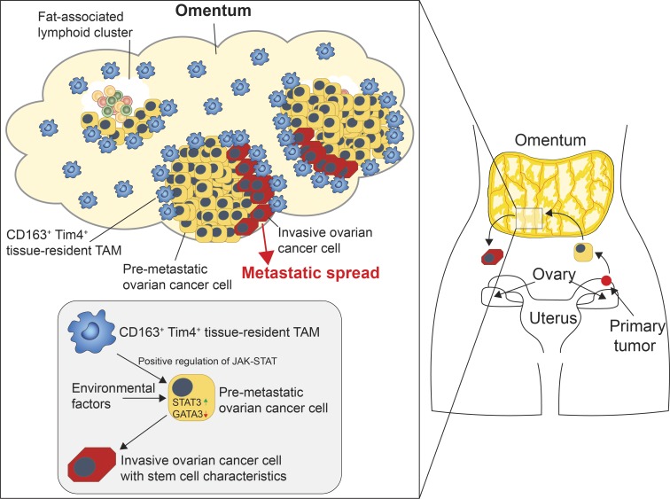
Graphical abstract
Graphical abstract CC BY-NC-SA · PubMed Central
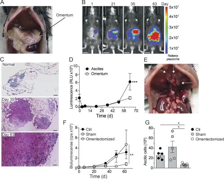
Figure 1. Omentum is a critical premetastatic niche for ovarian cancer cells. (A) Location of the omentum in the abdominal cavity of mice. (B) In vivo bioluminescence imaging of mice at 1, 21, 35, and 63 d after i.p. injection of 106 ID8-Luc cells; scale bar: 1 cm. (C) H&E stain of frozen omental sections from naive mice or 35 and 63 d after injection of ID8-Luc cells; scale bar: 100 µm. (D) Ex vivo luminescence analysis of omentum and ascites. RLU, relative luminescent unit. Data are represented as mean ± SEM of n = 5, and statistically significant difference was calculated using two-way ANOVA followed by Tukey post hoc test; ***, P < 0.001. (E) Tumor nodules on the diaphragm of mice 10 wk after injection of ID8-Luc cells. (F) In vivo bioluminescence analysis of omentectomized, sham-operated, or control (Ctrl) mice from 0–63 d after injection of ID8-Luc cells. cps, counts per second. Data are represented as mean ± SEM of n = 5, and statistically significant difference was calculated using two-way ANOVA followed by Tukey post hoc test; **, P < 0.01. (G) End-point analysis of malignant ascites in omentectomized or control mice 63 d after injection of ID8-Luc cells. Data are represented as mean ± SEM of n = 5, and statistically significant difference was calculated using Kruskal-Wallis one-way ANOVA followed by Dunn’s multiple comparisons test; *, P < 0.05. All data are representative of two independent experiments.
Figure 1. Omentum is a critical premetastatic niche for ovarian cancer cells. (A) Location of the omentum in the abdominal cavity of mice. (B) In vivo bioluminescence imaging of mice at 1, 21, 35, and 63 d after i.p. injection of 106 ID8-Luc cells; scale bar: 1 cm. (C) H&E stain of frozen omental sections from naive mice or 35 and 63 d after injection of ID8-Luc cells; scale bar: 100 µm. (D) Ex vivo luminescence analysis of omentum and ascites. RLU, relative luminescent unit. Data are represented as mean ± SEM of n = 5, and statistically significant difference was calculated using two-way ANOVA followed by Tukey post hoc test; ***, P < 0.001. (E) Tumor nodules on the diaphragm of mice 10 wk after injection of ID8-Luc cells. (F) In vivo bioluminescence analysis of omentectomized, sham-operated, or control (Ctrl) mice from 0–63 d after injection of ID8-Luc cells. cps, counts per second. Data are represented as mean ± SEM of n = 5, and statistically significant difference was calculated using two-way ANOVA followed by Tukey post hoc test; **, P < 0.01. (G) End-point analysis of malignant ascites in omentectomized or control mice 63 d after injection of ID8-Luc cells. Data are represented as mean ± SEM of n = 5, and statistically significant difference was calculated using Kruskal-Wallis one-way ANOVA followed by Dunn’s multiple comparisons test; *, P < 0.05. All data are representative of two independent experiments.CC BY-NC-SA · PubMed Central
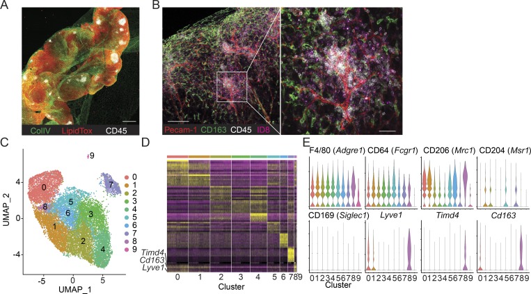
Figure 2. Single-cell transcriptional profiling of omental macrophages after tumor colonization. (A) Whole-mount confocal imaging of omentum showing the location of FALCs by visualizing collagen IV (ColIV, green), adipocytes (LipidTox, red), and leukocyte clusters (CD45, white); scale bar: 0.5 mm. (B) Whole-mount imaging of ID8-Luc infiltration in the omentum 24 h after injection of ID8 cells. Pecam-1 staining for blood vessels (red), CD163 for macrophages (green), CD45 for FALCs (white), and Qdot705 for ID8 cells (magenta); scale bar: 100 µm and 50 µm in magnification. (C) Two-dimensional representation and clustering of single-cell TAM transcriptomes using UMAP (n = 16,514). (D) Heatmap showing the top 10 DEGs in each UMAP cluster. (E) Violin plots showing expression of commonly used macrophage markers.
Figure 2. Single-cell transcriptional profiling of omental macrophages after tumor colonization. (A) Whole-mount confocal imaging of omentum showing the location of FALCs by visualizing collagen IV (ColIV, green), adipocytes (LipidTox, red), and leukocyte clusters (CD45, white); scale bar: 0.5 mm. (B) Whole-mount imaging of ID8-Luc infiltration in the omentum 24 h after injection of ID8 cells. Pecam-1 staining for blood vessels (red), CD163 for macrophages (green), CD45 for FALCs (white), and Qdot705 for ID8 cells (magenta); scale bar: 100 µm and 50 µm in magnification. (C) Two-dimensional representation and clustering of single-cell TAM transcriptomes using UMAP (n = 16,514). (D) Heatmap showing the top 10 DEGs in each UMAP cluster. (E) Violin plots showing expression of commonly used macrophage markers.CC BY-NC-SA · PubMed Central
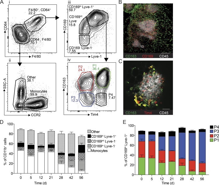
Figure 3. Characterization of macrophage subsets in omentum by flow cytometry and confocal microscopy. (A) Flow cytometry analysis of the myeloid cell compartment in omentum. Myeloid cells are gated as (i) Live, CD45.2+, singlets, Linneg (CD5, CD19, NK1.1, Ly6G, Siglec F), CD11b+, and subsequently macrophages were gated as F4/80+, CD64+. Monocytes were gated as (ii) F4/80−, CD64−, CCR2+. F4/80−, CD64−, and CCR2− CD11b+ myeloid cells were designated as other. F4/80+ CD64+ macrophages were further gated based on (iii) CD169 and Lyve-1 expression, and finally CD169hi Lyve-1+ cells were further divided into four subpopulations based on (iv) CD163 and Tim4 expression. SSC-A, side scatter area. (B) Whole-mount imaging of FALCs (CD45, white; CD169+, red; CD163+, green) in the omentum; scale bar: 100 µm. (C) Whole-mount imaging of FALCs (CD45, white; CD163+, green; Tim4+, red) in omentum; scale bar: 50 µm. (D) Flow cytometry analysis and relative distribution of CD11b+ myeloid cell populations shown in A, i–iii during tumor growth. (E) Flow cytometry analysis and relative distribution of CD169hi Lyve-1+ macrophage subsets shown in A, iv during tumor growth. Whole-mount confocal imaging analysis is representative of n = 4 in two independent experiments. Flow cytometric data are represented as mean ± SEM of n = 4 and are representative of two independent experiments.
Figure 3. Characterization of macrophage subsets in omentum by flow cytometry and confocal microscopy. (A) Flow cytometry analysis of the myeloid cell compartment in omentum. Myeloid cells are gated as (i) Live, CD45.2+, singlets, Linneg (CD5, CD19, NK1.1, Ly6G, Siglec F), CD11b+, and subsequently macrophages were gated as F4/80+, CD64+. Monocytes were gated as (ii) F4/80−, CD64−, CCR2+. F4/80−, CD64−, and CCR2− CD11b+ myeloid cells were designated as other. F4/80+ CD64+ macrophages were further gated based on (iii) CD169 and Lyve-1 expression, and finally CD169hi Lyve-1+ cells were further divided into four subpopulations based on (iv) CD163 and Tim4 expression. SSC-A, side scatter area. (B) Whole-mount imaging of FALCs (CD45, white; CD169+, red; CD163+, green) in the omentum; scale bar: 100 µm. (C) Whole-mount imaging of FALCs (CD45, white; CD163+, green; Tim4+, red) in omentum; scale bar: 50 µm. (D) Flow cytometry analysis and relative distribution of CD11b+ myeloid cell populations shown in A, i–iii during tumor growth. (E) Flow cytometry analysis and relative distribution of CD169hi Lyve-1+ macrophage subsets shown in A, iv during tumor growth. Whole-mount confocal imaging analysis is representative of n = 4 in two independent experiments. Flow cytometric data are represented as mean ± SEM of n = 4 and are representative of two independent experiments.CC BY-NC-SA · PubMed Central
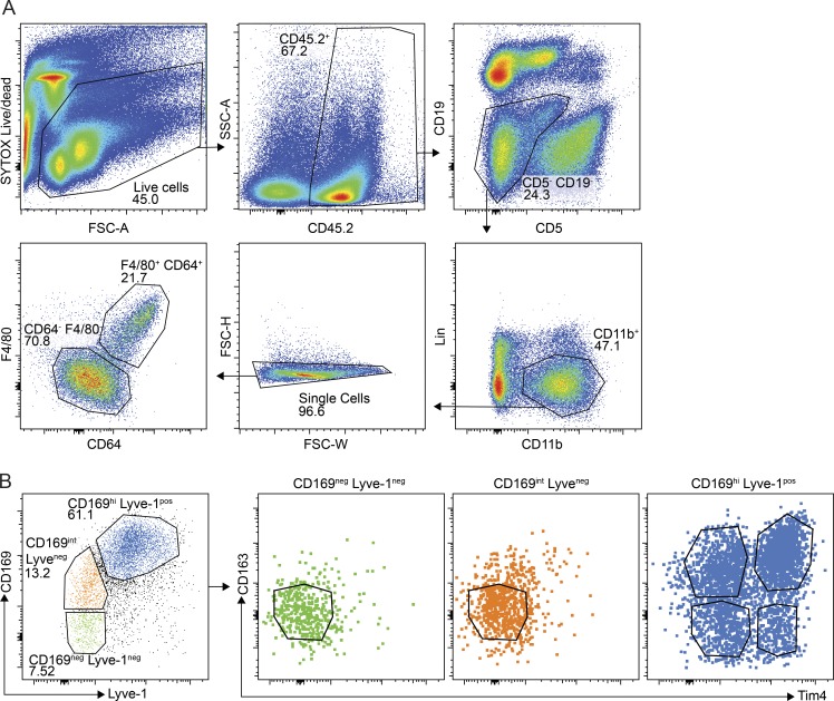
Figure S1. Gating strategy and expression of Tim4 and CD163 on omental macrophages. (A) Gating strategy for the myeloid cell compartment excluding neutrophils. Myeloid cells were gated as Live (forward scatter area [FSC-A] versus Sytox Blue), CD45.2+ (CD45.2 versus SSC-A), CD5−, CD19− (CD5 versus CD19), Linneg(NK1.1, Ly6G), CD11b+ (CD11b versus Lineage), single cells (forward scatter height [FSC-H] versus forward scatter width [FSC-W]), and CD64+ and F4/80+ (F4/80 versus CD64). (B) Expression of CD163 and Tim4 among F4/80+ CD64+ macrophages that are CD169negLyve-1neg (green), CD169int Lyve-1neg (red), or CD169hi Lyve-1pos (blue). Data are representative of two independent experiments.
Figure S1. Gating strategy and expression of Tim4 and CD163 on omental macrophages. (A) Gating strategy for the myeloid cell compartment excluding neutrophils. Myeloid cells were gated as Live (forward scatter area [FSC-A] versus Sytox Blue), CD45.2+ (CD45.2 versus SSC-A), CD5−, CD19− (CD5 versus CD19), Linneg(NK1.1, Ly6G), CD11b+ (CD11b versus Lineage), single cells (forward scatter height [FSC-H] versus forward scatter width [FSC-W]), and CD64+ and F4/80+ (F4/80 versus CD64). (B) Expression of CD163 and Tim4 among F4/80+ CD64+ macrophages that are CD169negLyve-1neg (green), CD169int Lyve-1neg (red), or CD169hi Lyve-1pos (blue). Data are representative of two independent experiments.CC BY-NC-SA · PubMed Central
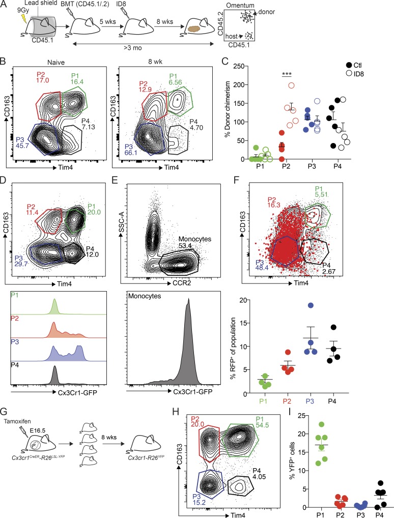
Figure 4. Embryonic origin of tissue-resident CD163+ Tim4+ macrophages in omentum. (A) Analysis of macrophage ontogeny in ID8 tumor-bearing mice using shielded chimeras; host CD45.1 congenic mice were placed in a protective lead shield with only the hind legs exposed, before irradiation at 9 Gy. The following day, mice were reconstituted with donor bone marrow cells from CD45.1/.2 F1 mice. 5 wk after reconstitution, mice were injected with 106 ID8-Luc cells i.p. At 8 wk after tumor injection, omentum was collected for analysis by flow cytometry. (B) Analysis of CD163 and Tim4 expression among CD169hi Lyve-1+ macrophages in omentum from naive and tumor-bearing chimeric mice. (C) Chimerism was calculated as the proportion of CD45.1/.2+ CD169hi Lyve-1+ macrophages relative to CD45.1/.2+ expression among Ly6Chi blood monocytes. Data are represented as mean ± SEM of n = 5, and statistically significant difference was calculated using two-way ANOVA followed by Tukey post hoc test; ***, P < 0.001. (D and E) Flow cytometry analysis of Cx3Cr1-GFP expression in CD169hi Lyve-1+ macrophages (P1–4; D) and CCR2+ monocytes (E). (F) Fate-mapping of Cx3Cr1-expressing monocytes in Cx3cr1-R26tdRFP mice inoculated with ID8-Luc cells. Mice were injected with ID8-Luc i.p., and after 8 wk, 1 mg of tamoxifen was administered by oral gavage, and RFP expression was analyzed in CD169hi Lyve-1+ macrophages 10 d later. Data are represented as mean ± SEM of n = 4. (G) Analysis of CD169hi Lyve-1+ macrophage ontogeny by pulse-labeling of Cx3Cr1−R26YFP embryos with tamoxifen at E16.5. (H) Omentum was harvested and analyzed by flow cytometry 8 wk after birth. (I) The percentage of YFP+ cells was calculated relative to YFP+ microglia. Data are represented as mean ± SEM of n = 6. All data are representative of two independent experiments.
Figure 4. Embryonic origin of tissue-resident CD163+ Tim4+ macrophages in omentum. (A) Analysis of macrophage ontogeny in ID8 tumor-bearing mice using shielded chimeras; host CD45.1 congenic mice were placed in a protective lead shield with only the hind legs exposed, before irradiation at 9 Gy. The following day, mice were reconstituted with donor bone marrow cells from CD45.1/.2 F1 mice. 5 wk after reconstitution, mice were injected with 106 ID8-Luc cells i.p. At 8 wk after tumor injection, omentum was collected for analysis by flow cytometry. (B) Analysis of CD163 and Tim4 expression among CD169hi Lyve-1+ macrophages in omentum from naive and tumor-bearing chimeric mice. (C) Chimerism was calculated as the proportion of CD45.1/.2+ CD169hi Lyve-1+ macrophages relative to CD45.1/.2+ expression among Ly6Chi blood monocytes. Data are represented as mean ± SEM of n = 5, and statistically significant difference was calculated using two-way ANOVA followed by Tukey post hoc test; ***, P < 0.001. (D and E) Flow cytometry analysis of Cx3Cr1-GFP expression in CD169hi Lyve-1+ macrophages (P1–4; D) and CCR2+ monocytes (E). (F) Fate-mapping of Cx3Cr1-expressing monocytes in Cx3cr1-R26tdRFP mice inoculated with ID8-Luc cells. Mice were injected with ID8-Luc i.p., and after 8 wk, 1 mg of tamoxifen was administered by oral gavage, and RFP expression was analyzed in CD169hi Lyve-1+ macrophages 10 d later. Data are represented as mean ± SEM of n = 4. (G) Analysis of CD169hi Lyve-1+ macrophage ontogeny by pulse-labeling of Cx3Cr1−R26YFP embryos with tamoxifen at E16.5. (H) Omentum was harvested and analyzed by flow cytometry 8 wk after birth. (I) The percentage of YFP+ cells was calculated relative to YFP+ microglia. Data are represented as mean ± SEM of n = 6. All data are representative of two independent experiments.CC BY-NC-SA · PubMed Central
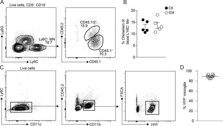
Figure S2. Fate mapping of omental macrophages. (A and B) Flow cytometric analysis of blood monocytes in mice 5 wk (A) or 13 wk (B) after adoptive transfer of CD45.1+/CD45.2+ donor bone marrow in irradiated CD45.1+ host mice that were protected by a lead shield. Monocytes were gated as Live (FSC-A versus Sytox Blue), CD5neg, CD19neg (CD5 versus CD19), NK1.1neg, CD11b+ (CD11b versus NK1.1), and Ly6C+, Ly6Gneg (Ly6C versus Ly6G). Data are represented as mean ± SEM of n = 5. (C) Flow cytometric analysis of YFP+ microglia in Cx3Cr1CreER:R26-yfp mice pulse-labeled with tamoxifen at E16.5. Microglia were gated as Live (FSC-A versus Sytox Blue), Ly6Cneg, CD11cneg (Ly6C versus CD11c), and CD45.2dim, CD11bhi (CD11b versus CD45.2). Data are represented as mean ± SEM of n = 6. All data are representative of two independent experiments.
Figure S2. Fate mapping of omental macrophages. (A and B) Flow cytometric analysis of blood monocytes in mice 5 wk (A) or 13 wk (B) after adoptive transfer of CD45.1+/CD45.2+ donor bone marrow in irradiated CD45.1+ host mice that were protected by a lead shield. Monocytes were gated as Live (FSC-A versus Sytox Blue), CD5neg, CD19neg (CD5 versus CD19), NK1.1neg, CD11b+ (CD11b versus NK1.1), and Ly6C+, Ly6Gneg (Ly6C versus Ly6G). Data are represented as mean ± SEM of n = 5. (C) Flow cytometric analysis of YFP+ microglia in Cx3Cr1CreER:R26-yfp mice pulse-labeled with tamoxifen at E16.5. Microglia were gated as Live (FSC-A versus Sytox Blue), Ly6Cneg, CD11cneg (Ly6C versus CD11c), and CD45.2dim, CD11bhi (CD11b versus CD45.2). Data are represented as mean ± SEM of n = 6. All data are representative of two independent experiments.CC BY-NC-SA · PubMed Central
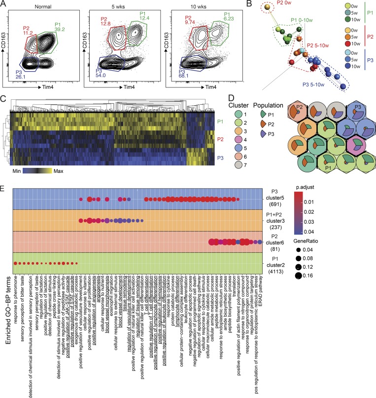
Figure 5. CD163+ Tim4+ tissue-resident macrophages express a unique transcriptional profile. (A) Gating strategy for cell sorting of P1, P2, and P3 populations of omental macrophages from naive (n = 2) and tumor-bearing mice at 5 and 10 wk after injection of ID8 cells (n = 4). 500 cells were sorted for each population and subjected to bulk RNAseq analysis. (B) PCA and network analysis of variable gene expression in P1, P2, and P3 macrophages. PCA analysis was performed on normalized data (mean = 0, and variance = 1), generating a correlation-based PCA plot. Network analysis connects k nearest neighbors (k = 3) based on similarity calculated by Pearson correlation. (C) Heatmap showing hierarchical clustering of the 5,000 most DEGs for each of the three populations at 10 wk of tumor growth. DEGs were identified by pairwise comparison with a cut-off of Padjusted > 0.01. (D) SOMs of identified DEGs using the Kohonen package for R. The SOM analysis found 16 single SOMs that were subsequently grouped by hierarchical clustering to identify population-specific clusters (clusters 1–7). The magnitude of the pie slices indicates the relative proportions of genes enriched in P1 (green), P2 (brown), and P3 (blue) within the SOM. (E) ClusterProfiler enrichment analysis against the GO database on SOM clusters 1–7. Only clusters with significantly enriched pathways are visualized. Libraries were prepared from two independent experiments with n = 2 and sequenced on the same flow cell for a total of four biological replicates. ERAD, endoplasmic reticulum–associated protein degradation; pos, positive.
Figure 5. CD163+ Tim4+ tissue-resident macrophages express a unique transcriptional profile. (A) Gating strategy for cell sorting of P1, P2, and P3 populations of omental macrophages from naive (n = 2) and tumor-bearing mice at 5 and 10 wk after injection of ID8 cells (n = 4). 500 cells were sorted for each population and subjected to bulk RNAseq analysis. (B) PCA and network analysis of variable gene expression in P1, P2, and P3 macrophages. PCA analysis was performed on normalized data (mean = 0, and variance = 1), generating a correlation-based PCA plot. Network analysis connects k nearest neighbors (k = 3) based on similarity calculated by Pearson correlation. (C) Heatmap showing hierarchical clustering of the 5,000 most DEGs for each of the three populations at 10 wk of tumor growth. DEGs were identified by pairwise comparison with a cut-off of Padjusted > 0.01. (D) SOMs of identified DEGs using the Kohonen package for R. The SOM analysis found 16 single SOMs that were subsequently grouped by hierarchical clustering to identify population-specific clusters (clusters 1–7). The magnitude of the pie slices indicates the relative proportions of genes enriched in P1 (green), P2 (brown), and P3 (blue) within the SOM. (E) ClusterProfiler enrichment analysis against the GO database on SOM clusters 1–7. Only clusters with significantly enriched pathways are visualized. Libraries were prepared from two independent experiments with n = 2 and sequenced on the same flow cell for a total of four biological replicates. ERAD, endoplasmic reticulum–associated protein degradation; pos, positive.CC BY-NC-SA · PubMed Central
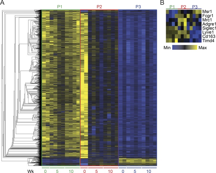
Figure S3. Hierarchical clustering analysis of omental macrophages. (A) Heatmap showing hierarchical clustering analysis of the 5,000 most DEGs in P1 (green), P2 (red), and P3 (blue) CD169hi Lyve-1pos omental macrophages sorted at steady state (0 wk) or 5 wk and 10 wk after infiltration of ID8 ovarian cancer cells. (B) Heatmap showing expression of macrophage markers from transcriptomic analysis of P1 (green), P2 (red), and P3 (blue) CD169hi Lyve-1pos omental macrophages sorted 10 wk after ID8 injection (Fig. 2 E). Libraries were prepared from two independent experiments with n = 2 and sequenced on the same flow cell for a total of four biological replicates.
Figure S3. Hierarchical clustering analysis of omental macrophages. (A) Heatmap showing hierarchical clustering analysis of the 5,000 most DEGs in P1 (green), P2 (red), and P3 (blue) CD169hi Lyve-1pos omental macrophages sorted at steady state (0 wk) or 5 wk and 10 wk after infiltration of ID8 ovarian cancer cells. (B) Heatmap showing expression of macrophage markers from transcriptomic analysis of P1 (green), P2 (red), and P3 (blue) CD169hi Lyve-1pos omental macrophages sorted 10 wk after ID8 injection (Fig. 2 E). Libraries were prepared from two independent experiments with n = 2 and sequenced on the same flow cell for a total of four biological replicates.CC BY-NC-SA · PubMed Central
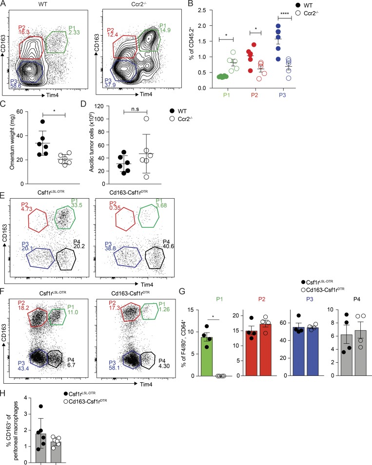
Figure S4. Depletion of monocyte-derived and tissue-resident omental macrophages. (A and B) Flow cytometric analysis of CD169hi Lyve-1pos omental macrophages in Ccr2−/− mice 10 wk after i.p. injection of 106 ID8 ovarian cancer cells. Data are represented as mean ± SEM of n = 5, and statistically significant difference was calculated using two-way ANOVA followed by Tukey post hoc test; *, P < 0.05; ****, P < 0.0001. (C and D) Omentum weight (C) and number of tumor cells in ascites (D) of WT and Ccr2−/− mice 10 wk after injection of ID8 cells. Data are represented as mean ± SEM of n = 6, and statistically significant difference was calculated using Mann-Whitney U test; *, P < 0.05. (E–G) Flow cytometric analysis of CD169hi Lyve-1pos omental macrophages in Cd163-Csf1rDTR mice 1 d (E) or 6 d (F and G) after i.p. injection of 4 ng/kg DT. CD169hi Lyve-1pos omental macrophages were gated as described in Fig. S1 A. (H) Flow cytometric analysis of peritoneal macrophages in Cd163-Csf1rDTR and control mice 6 d after i.p. injection of 4 ng/kg DT. Peritoneal macrophages were gated as Live, CD45.2pos, Linneg(CD5,CD19, Ly6G, NK1.1) CD11b+, F4/80+, and MHCIIneg. Data are represented as mean ± SEM of n = 7, and statistically significant difference was calculated using Mann-Whitney U test; *, P < 0.05. All data are representative of two independent experiments.
Figure S4. Depletion of monocyte-derived and tissue-resident omental macrophages. (A and B) Flow cytometric analysis of CD169hi Lyve-1pos omental macrophages in Ccr2−/− mice 10 wk after i.p. injection of 106 ID8 ovarian cancer cells. Data are represented as mean ± SEM of n = 5, and statistically significant difference was calculated using two-way ANOVA followed by Tukey post hoc test; *, P < 0.05; ****, P < 0.0001. (C and D) Omentum weight (C) and number of tumor cells in ascites (D) of WT and Ccr2−/− mice 10 wk after injection of ID8 cells. Data are represented as mean ± SEM of n = 6, and statistically significant difference was calculated using Mann-Whitney U test; *, P < 0.05. (E–G) Flow cytometric analysis of CD169hi Lyve-1pos omental macrophages in Cd163-Csf1rDTR mice 1 d (E) or 6 d (F and G) after i.p. injection of 4 ng/kg DT. CD169hi Lyve-1pos omental macrophages were gated as described in Fig. S1 A. (H) Flow cytometric analysis of peritoneal macrophages in Cd163-Csf1rDTR and control mice 6 d after i.p. injection of 4 ng/kg DT. Peritoneal macrophages were gated as Live, CD45.2pos, Linneg(CD5,CD19, Ly6G, NK1.1) CD11b+, F4/80+, and MHCIIneg. Data are represented as mean ± SEM of n = 7, and statistically significant difference was calculated using Mann-Whitney U test; *, P < 0.05. All data are representative of two independent experiments.CC BY-NC-SA · PubMed Central
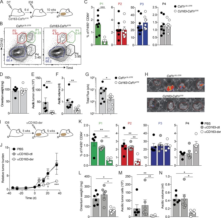
Figure 6. Specific depletion of CD163+ Tim4+ tissue-resident macrophages prevents metastatic spread of ovarian cancer. (A) Cohorts of Cd163-Csf1rDTR or control mice (Csf1rLSL-DTR) were injected with 4 ng/kg DT 6 d before transplantation with ID8 cells. Mice were analyzed for depletion of macrophages and effects on tumor growth at 10 wk. (B and C) Flow cytometry analysis of CD163hi Lyve-1+ macrophages in omentum of Cd163-Csf1rDTR and Csf1rLSL-DTR mice 10 wk after injection of ID8 cells. (D–F) Omentum weight (D), total tumor cells in ascites (E), and ascites volume (F) in Cd163-Csf1rDTR and Csf1rLSL-DTR mice treated with DT. (G and H) Ex vivo bioluminescence analysis of metastases on the diaphragm of Cd163-Csf1rDTR and Csf1rLSL-DTR; scale bar: 0.5 cm. Data are represented as mean ± SEM of n = 7, and statistically significant difference was calculated using Mann-Whitney U test; *, P < 0.05; **, P < 0.01; ***, P < 0.001. (I) Therapeutic depletion of CD163+ macrophages; mice were injected with ID8 cells and after 5 wk randomized into groups and treated with dxr-loaded αCD163-LNPs (αCD163-dxr), empty αCD163-LNPs (αCD163-ctrl), or PBS alone twice a week for 5 wk. (J) Tumor burden monitored by in vivo bioluminescent imaging. Data are represented as mean ± SEM of n = 6, and statistically significant difference was calculated using two-way ANOVA followed by Tukey post hoc test; *, P < 0.05; ***, P < 0.001. (K) Flow cytometry analysis of P1–P4 macrophages in omentum after therapeutic depletion of CD163+ cells. (L–N) Omentum weight (L), total tumor cells in ascites (M), and ascites volume (N). Data are represented as mean ± SEM of n = 6, and statistically significant difference was calculated using Kruskal-Wallis one-way ANOVA followed by Dunn’s multiple comparisons test; *, P < 0.05; **, P < 0.01; ***, P < 0.001. All data are representative of three independent experiments.
Figure 6. Specific depletion of CD163+ Tim4+ tissue-resident macrophages prevents metastatic spread of ovarian cancer. (A) Cohorts of Cd163-Csf1rDTR or control mice (Csf1rLSL-DTR) were injected with 4 ng/kg DT 6 d before transplantation with ID8 cells. Mice were analyzed for depletion of macrophages and effects on tumor growth at 10 wk. (B and C) Flow cytometry analysis of CD163hi Lyve-1+ macrophages in omentum of Cd163-Csf1rDTR and Csf1rLSL-DTR mice 10 wk after injection of ID8 cells. (D–F) Omentum weight (D), total tumor cells in ascites (E), and ascites volume (F) in Cd163-Csf1rDTR and Csf1rLSL-DTR mice treated with DT. (G and H) Ex vivo bioluminescence analysis of metastases on the diaphragm of Cd163-Csf1rDTR and Csf1rLSL-DTR; scale bar: 0.5 cm. Data are represented as mean ± SEM of n = 7, and statistically significant difference was calculated using Mann-Whitney U test; *, P < 0.05; **, P < 0.01; ***, P < 0.001. (I) Therapeutic depletion of CD163+ macrophages; mice were injected with ID8 cells and after 5 wk randomized into groups and treated with dxr-loaded αCD163-LNPs (αCD163-dxr), empty αCD163-LNPs (αCD163-ctrl), or PBS alone twice a week for 5 wk. (J) Tumor burden monitored by in vivo bioluminescent imaging. Data are represented as mean ± SEM of n = 6, and statistically significant difference was calculated using two-way ANOVA followed by Tukey post hoc test; *, P < 0.05; ***, P < 0.001. (K) Flow cytometry analysis of P1–P4 macrophages in omentum after therapeutic depletion of CD163+ cells. (L–N) Omentum weight (L), total tumor cells in ascites (M), and ascites volume (N). Data are represented as mean ± SEM of n = 6, and statistically significant difference was calculated using Kruskal-Wallis one-way ANOVA followed by Dunn’s multiple comparisons test; *, P < 0.05; **, P < 0.01; ***, P < 0.001. All data are representative of three independent experiments.CC BY-NC-SA · PubMed Central
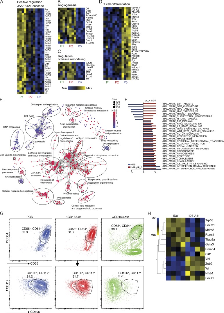
Figure S5. Transcriptomic analysis of ID8 ovarian cancer cells and TAMs from omentum. (A–D) Heatmaps showing expression of genes associated with the enriched pathways in Fig. 5 E: Positive regulation of JAK-STAT (A), angiogenesis (B), regulation of tissue remodeling (C), and T cell differentiation (D). (E) GO term enrichment analysis of RNAseq data comparing cultured ID8 cells (n = 4) and ID8 cells harvested from ascites 11 wk after transplantation in vivo (ID8-A11, n = 4). Identified GO terms are represented by nodes with a size according to the number of enriched genes. Nodes are colored according to the normalized enrichment score (NES). (F) GSEA comparing ID8 and ID8-A11 against the hallmark gene sets in MSigDB. (G) Flow cytometric analysis of CSC markers CD54, CD55, CD106, and CD117 on tumor cells from ascites of mice specifically depleted of Tim4+ CD163+ resident TAM (αCD163-dxr). Mice were injected i.p. with 1 mg/kg αCD163-dxr or controls (empty αCD163-LNP or PBS) on days 1, 3, and 5, before injection of ID8-Luc cells i.p. on day 8. 10 wk after ID8, tumor cells in ascites were harvested and analyzed for CSC marker expression by flow cytometry by gating on Live, CD45.2neg, and Linneg(CD11b, F4/80, CD5,CD19, Ly6G, NK1.1) cells. (H) Heatmap showing expression levels of TFs involved in EMT and MET in ID8 and ID8-A11 cells. All data are representative of three independent experiments.
Figure S5. Transcriptomic analysis of ID8 ovarian cancer cells and TAMs from omentum. (A–D) Heatmaps showing expression of genes associated with the enriched pathways in Fig. 5 E: Positive regulation of JAK-STAT (A), angiogenesis (B), regulation of tissue remodeling (C), and T cell differentiation (D). (E) GO term enrichment analysis of RNAseq data comparing cultured ID8 cells (n = 4) and ID8 cells harvested from ascites 11 wk after transplantation in vivo (ID8-A11, n = 4). Identified GO terms are represented by nodes with a size according to the number of enriched genes. Nodes are colored according to the normalized enrichment score (NES). (F) GSEA comparing ID8 and ID8-A11 against the hallmark gene sets in MSigDB. (G) Flow cytometric analysis of CSC markers CD54, CD55, CD106, and CD117 on tumor cells from ascites of mice specifically depleted of Tim4+ CD163+ resident TAM (αCD163-dxr). Mice were injected i.p. with 1 mg/kg αCD163-dxr or controls (empty αCD163-LNP or PBS) on days 1, 3, and 5, before injection of ID8-Luc cells i.p. on day 8. 10 wk after ID8, tumor cells in ascites were harvested and analyzed for CSC marker expression by flow cytometry by gating on Live, CD45.2neg, and Linneg(CD11b, F4/80, CD5,CD19, Ly6G, NK1.1) cells. (H) Heatmap showing expression levels of TFs involved in EMT and MET in ID8 and ID8-A11 cells. All data are representative of three independent experiments.CC BY-NC-SA · PubMed Central
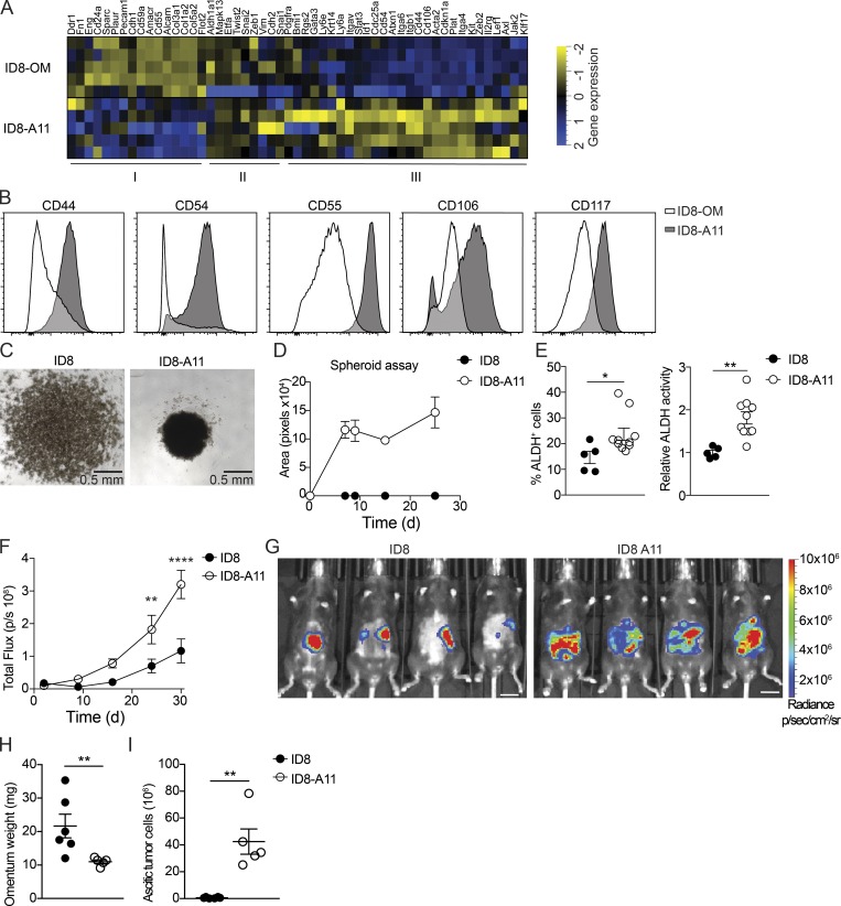
Figure 7. Ovarian cancer cells in ascites acquire CSC characteristics. (A) Heatmap showing expression of CSC markers in omental ID8 cells (ID8-OM) and ID8 cells from ascites (ID8-A11); see Table S1 for details. (B) Flow cytometry analysis of CSC surface markers on ascitic ID8-A11 cells compared with ID8 cells in omentum (ID8-OM). (C and D) Spheroid formation assay comparing cultured ID8 and ID8-A11 cells; 4 × 10⁴ cells were seeded in ultra-low-adherence 96-well plates, and formation of spheroids was assessed with a wide-field microscope. (E) Analysis of ALDH activity in ID8 and ID8-A11 cells by flow cytometry using the Aldefluor assay. ALDH+ cells were gated using DEAB-treated cells as negative control (left), and relative ALDH activity was calculated by measuring the median fluorescence intensity of Aldefluor. Data are represented as mean ± SEM of n = 5 (ID8) or n = 8 (ID8-A11), and statistically significant difference was calculated using Mann-Whitney U test; *, P < 0.05; **, P < 0.01. (F) Analysis of tumorigenic potential of ID8 and ID8-A11 cells in vivo; total tumor burden was monitored by in vivo bioluminescence imaging. Data are represented as mean ± SEM of n = 6, and statistically significant difference was calculated using two-way ANOVA followed by Tukey post hoc test; **, P < 0.01; ****, P < 0.0001. (G) Representative images of tumor localization at 30 d after injection of parental ID8-Luc and ID8-A11 cells; scale bar: 1 cm. (H and I) Ex vivo analysis of tumor burden in omentum (H) and ascites (I) at 30 d after transplantation of ID8-Luc or ID8-A11 cells. Data are represented as mean ± SEM of n = 6 (ID8) or n = 5 (ID8-A11), and statistically significant difference was calculated using Mann-Whitney U test; **, P < 0.01. All data are representative of two independent experiments.
Figure 7. Ovarian cancer cells in ascites acquire CSC characteristics. (A) Heatmap showing expression of CSC markers in omental ID8 cells (ID8-OM) and ID8 cells from ascites (ID8-A11); see Table S1 for details. (B) Flow cytometry analysis of CSC surface markers on ascitic ID8-A11 cells compared with ID8 cells in omentum (ID8-OM). (C and D) Spheroid formation assay comparing cultured ID8 and ID8-A11 cells; 4 × 10⁴ cells were seeded in ultra-low-adherence 96-well plates, and formation of spheroids was assessed with a wide-field microscope. (E) Analysis of ALDH activity in ID8 and ID8-A11 cells by flow cytometry using the Aldefluor assay. ALDH+ cells were gated using DEAB-treated cells as negative control (left), and relative ALDH activity was calculated by measuring the median fluorescence intensity of Aldefluor. Data are represented as mean ± SEM of n = 5 (ID8) or n = 8 (ID8-A11), and statistically significant difference was calculated using Mann-Whitney U test; *, P < 0.05; **, P < 0.01. (F) Analysis of tumorigenic potential of ID8 and ID8-A11 cells in vivo; total tumor burden was monitored by in vivo bioluminescence imaging. Data are represented as mean ± SEM of n = 6, and statistically significant difference was calculated using two-way ANOVA followed by Tukey post hoc test; **, P < 0.01; ****, P < 0.0001. (G) Representative images of tumor localization at 30 d after injection of parental ID8-Luc and ID8-A11 cells; scale bar: 1 cm. (H and I) Ex vivo analysis of tumor burden in omentum (H) and ascites (I) at 30 d after transplantation of ID8-Luc or ID8-A11 cells. Data are represented as mean ± SEM of n = 6 (ID8) or n = 5 (ID8-A11), and statistically significant difference was calculated using Mann-Whitney U test; **, P < 0.01. All data are representative of two independent experiments.CC BY-NC-SA · PubMed Central
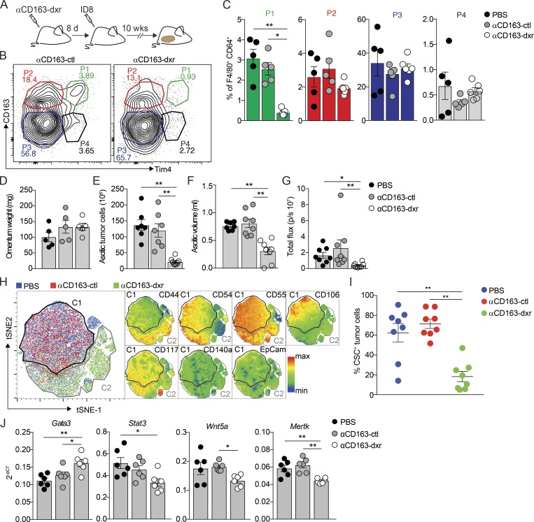
Figure 8. CD163+ Tim4+ tissue-resident macrophages promote the CSC-like phenotype of ovarian cancer cells. (A) Specific depletion of CD163+ Tim4+ (P4) macrophages using dxr-loaded αCD163-LNPs (αCD163-dxr); mice were injected i.p. with 1 mg/kg αCD163-dxr or controls (empty αCD163-LNPs or PBS) on days 1, 3, and 5, before injection of ID8-Luc cells on day 8. (B and C) Flow cytometry analysis of CD163hi Lyve-1+ macrophages in omentum at 10 wk after prophylactic treatment with αCD163-dxr. Data are represented as mean ± SEM of n = 5 (PBS, αCD163-LNP) or n = 6 (αCD163-dxr), and statistically significant difference was calculated using Kruskal-Wallis one-way ANOVA followed by Dunn’s multiple comparisons test; *, P < 0.05; **, P < 0.01. (D–F) Omentum weight (D), total tumor cells in ascites (E), and ascites volume (F) at 10 wk after prophylactic treatment with αCD163-dxr. (G) Ex vivo bioluminescence analysis of metastases on the diaphragm. Data are represented as mean ± SEM of n = 8, and statistically significant difference was calculated using Kruskal-Wallis one-way ANOVA followed by Dunn’s multiple comparisons test; *, P < 0.05; **, P < 0.01. (H) tSNE map of concatenated fcs files from flow cytometry analysis of tumor cells from ascites after prophylactic treatment with αCD163-dxr. tSNE parameters were computed on expression levels of CSC markers (CD44, CD54, CD55, CD106, CD117, CD140a, and EpCam), and individual sample files were color-coded according to treatment group (PBS, blue; αCD163-ctl, red; αCD163-dxr, green; n = 8). This analysis identified distinct clusters from control-treated mice (PBS or αCD163-LNP; C1, black) and αCD163-dxr–treated mice (C2, gray). Expression of the individual CSC markers was subsequently visualized by heatmap statistics in a tSNE map of the concatenated samples within C1 (PBS or αCD163-LNP) and C2 (αCD163-dxr; right). (I) Flow cytometry analysis identifying the proportion of malignant cells expressing specific CSC markers: CD54, CD55, CD106, and CD117. See Fig. S5 G for the gating strategy. Data are represented as mean ± SEM of n = 8, and statistically significant difference was calculated using Kruskal-Wallis one-way ANOVA followed by Dunn’s multiple comparisons test; **, P < 0.01. (J) Gene expression analysis of Gata3, Stat3, Wnt5a, and Mertk in tumor cells from omentum after specific depletion of CD163+ Tim4+ (P1) macrophages, as described in A. dCT, delta cycle threshold. Data are represented as mean ± SEM of n = 6, and statistically significant difference was calculated using Kruskal-Wallis one-way ANOVA followed by Dunn’s multiple comparisons test; *, P < 0.05; **, P < 0.01. A–G are representative of three independent experiments, whereas H–J are representative of two independent experiments.
Figure 8. CD163+ Tim4+ tissue-resident macrophages promote the CSC-like phenotype of ovarian cancer cells. (A) Specific depletion of CD163+ Tim4+ (P4) macrophages using dxr-loaded αCD163-LNPs (αCD163-dxr); mice were injected i.p. with 1 mg/kg αCD163-dxr or controls (empty αCD163-LNPs or PBS) on days 1, 3, and 5, before injection of ID8-Luc cells on day 8. (B and C) Flow cytometry analysis of CD163hi Lyve-1+ macrophages in omentum at 10 wk after prophylactic treatment with αCD163-dxr. Data are represented as mean ± SEM of n = 5 (PBS, αCD163-LNP) or n = 6 (αCD163-dxr), and statistically significant difference was calculated using Kruskal-Wallis one-way ANOVA followed by Dunn’s multiple comparisons test; *, P < 0.05; **, P < 0.01. (D–F) Omentum weight (D), total tumor cells in ascites (E), and ascites volume (F) at 10 wk after prophylactic treatment with αCD163-dxr. (G) Ex vivo bioluminescence analysis of metastases on the diaphragm. Data are represented as mean ± SEM of n = 8, and statistically significant difference was calculated using Kruskal-Wallis one-way ANOVA followed by Dunn’s multiple comparisons test; *, P < 0.05; **, P < 0.01. (H) tSNE map of concatenated fcs files from flow cytometry analysis of tumor cells from ascites after prophylactic treatment with αCD163-dxr. tSNE parameters were computed on expression levels of CSC markers (CD44, CD54, CD55, CD106, CD117, CD140a, and EpCam), and individual sample files were color-coded according to treatment group (PBS, blue; αCD163-ctl, red; αCD163-dxr, green; n = 8). This analysis identified distinct clusters from control-treated mice (PBS or αCD163-LNP; C1, black) and αCD163-dxr–treated mice (C2, gray). Expression of the individual CSC markers was subsequently visualized by heatmap statistics in a tSNE map of the concatenated samples within C1 (PBS or αCD163-LNP) and C2 (αCD163-dxr; right). (I) Flow cytometry analysis identifying the proportion of malignant cells expressing specific CSC markers: CD54, CD55, CD106, and CD117. See Fig. S5 G for the gating strategy. Data are represented as mean ± SEM of n = 8, and statistically significant difference was calculated using Kruskal-Wallis one-way ANOVA followed by Dunn’s multiple comparisons test; **, P < 0.01. (J) Gene expression analysis of Gata3, Stat3, Wnt5a, and Mertk in tumor cells from omentum after specific depletion of CD163+ Tim4+ (P1) macrophages, as described in A. dCT, delta cycle threshold. Data are represented as mean ± SEM of n = 6, and statistically significant difference was calculated using Kruskal-Wallis one-way ANOVA followed by Dunn’s multiple comparisons test; *, P < 0.05; **, P < 0.01. A–G are representative of three independent experiments, whereas H–J are representative of two independent experiments.CC BY-NC-SA · PubMed Central

Verification

Claims the paper makes

The claims and statistics of this paper haven’t been checked yet.

Run the check — free, no account needed. It reads the open-access text and checks the sources it cites.

A few free checks a day · papers already verified open instantly · results are public

Claims other papers make about this one

Each is checked against this paper’s own text: does it say what the citing paper says it does?

Self-check

Percentages, sums and reported differences re-computed from the paper’s own numbers.

The self-check needs the paper’s full text.

Citations

Cites

Research works printed but not linked: 79

  1. Abubaker, 2014, Inhibition of the JAK2/STAT3 pathway in ovarian cancer results in the loss of cancer stem cell-like characteristics and a reduced tumor burden, BMC Cancer., 14, 317, 10.1186/1471-2407-14-317 · DOI
  2. AlHossiny, 2016, Ly6E/K signaling to TGFβ promotes breast cancer progression, immune escape, and drug resistance, Cancer Res., 76, 3376, 10.1158/0008-5472.CAN-15-2654 · DOI
  3. Bapat, 2005, Stem and progenitor-like cells contribute to the aggressive behavior of human epithelial ovarian cancer, Cancer Res., 65, 3025, 10.1158/0008-5472.CAN-04-3931 · DOI
  4. Baratin, 2017, T Cell Zone Resident Macrophages Silently Dispose of Apoptotic Cells in the Lymph Node, Immunity, 47, 349, 10.1016/j.immuni.2017.07.019 · DOI
  5. Bénézech, 2015, Inflammation-induced formation of fat-associated lymphoid clusters, Nat. Immunol., 16, 819, 10.1038/ni.3215 · DOI
  6. Bushnell, 2014, BBMap: A Fast, Accurate, Splice-Aware Aligner.
  7. Chakrabarti, 2018, Notch ligand Dll1 mediates cross-talk between mammary stem cells and the macrophageal niche, Science., 360, 10.1126/science.aan4153 · DOI
  8. Charles, 2009, The tumor-promoting actions of TNF-α involve TNFR1 and IL-17 in ovarian cancer in mice and humans, J. Clin. Invest., 119, 3011, 10.1172/JCI39065 · DOI · Paper page
  9. Chen, 2017, CD59 regulation by SOX2 is required for epithelial cancer stem cells to evade complement surveillance, Stem Cell Reports., 8, 140, 10.1016/j.stemcr.2016.11.008 · DOI
  10. Chou, 2010, GATA3 in development and cancer differentiation: cells GATA have it!, J. Cell. Physiol., 222, 42, 10.1002/jcp.21943 · DOI
  11. Clark, 2013, Milky spots promote ovarian cancer metastatic colonization of peritoneal adipose in experimental models, Am. J. Pathol., 183, 576, 10.1016/j.ajpath.2013.04.023 · DOI
  12. De Schepper, 2018, Self-maintaining gut macrophages are essential for intestinal homeostasis, Cell., 175, 400, 10.1016/j.cell.2018.07.048 · DOI
  13. Dick, 2019, Self-renewing resident cardiac macrophages limit adverse remodeling following myocardial infarction, Nat. Immunol., 20, 29, 10.1038/s41590-018-0272-2 · DOI · Paper page
  14. Etzerodt, 2013, CD163 and inflammation: biological, diagnostic, and therapeutic aspects, Antioxid. Redox Signal., 18, 2352, 10.1089/ars.2012.4834 · DOI
  15. Etzerodt, 2012, Efficient intracellular drug-targeting of macrophages using stealth liposomes directed to the hemoglobin scavenger receptor CD163, J. Control. Release., 160, 72, 10.1016/j.jconrel.2012.01.034 · DOI
  16. Etzerodt, 2013, Plasma clearance of hemoglobin and haptoglobin in mice and effect of CD163 gene targeting disruption, Antioxid. Redox Signal., 18, 2254, 10.1089/ars.2012.4605 · DOI
  17. Etzerodt, 2019, Specific targeting of CD163+ TAMs mobilizes inflammatory monocytes and promotes T cell-mediated tumor regression, J. Exp. Med., 216, 2394, 10.1084/jem.20182124 · DOI
  18. Ferlay, 2018, Global Cancer Observatory: Cancer Today
  19. Ford, 2014, The non-canonical Wnt ligand, Wnt5a, is upregulated and associated with epithelial to mesenchymal transition in epithelial ovarian cancer, Gynecol. Oncol., 134, 338, 10.1016/j.ygyno.2014.06.004 · DOI
  20. Fritze, 2006, Remote loading of doxorubicin into liposomes driven by a transmembrane phosphate gradient, Biochimica et Biophysica Acta, 1758, 1633, 10.1016/j.bbamem.2006.05.028 · DOI
  21. Garson, 2015, Epithelial ovarian cancer stem cells: underlying complexity of a simple paradigm, Reproduction., 149, R59, 10.1530/REP-14-0234 · DOI
  22. Ginhoux, 2016, Tissue-resident macrophage ontogeny and homeostasis, Immunity., 44, 439, 10.1016/j.immuni.2016.02.024 · DOI · Paper page
  23. Ginhoux, 2010, Ontogeny and homeostasis of Langerhans cells, Immunol. Cell Biol., 88, 387, 10.1038/icb.2010.38 · DOI
  24. Goossens, 2019, Membrane cholesterol efflux drives tumor-associated macrophage reprogramming and tumor progression, Cell Metab., 29, 1376, 10.1016/j.cmet.2019.02.016 · DOI · Paper page
  25. Gray, 2012, Lymph node macrophages, J. Innate Immun., 4, 424, 10.1159/000337007 · DOI
  26. Hagemann, 2008, “Re-educating” tumor-associated macrophages by targeting NF-kappaB, J. Exp. Med., 205, 1261, 10.1084/jem.20080108 · DOI · Paper page
  27. Hagiwara, 1993, Milky spots as the implantation site for malignant cells in peritoneal dissemination in mice, Cancer Res., 53, 687
  28. Jäppinen, 2019, Fetal-derived macrophages dominate in adult mammary glands, Nat. Commun., 10, 281, 10.1038/s41467-018-08065-1 · DOI
  29. Jung, 2016, Endogenous GAS6 and Mer receptor signaling regulate prostate cancer stem cells in bone marrow, Oncotarget., 7, 25698, 10.18632/oncotarget.8365 · DOI
  30. Kim, 2018, High NRF2 level mediates cancer stem cell-like properties of aldehyde dehydrogenase (ALDH)-high ovarian cancer cells: inhibitory role of all-trans retinoic acid in ALDH/NRF2 signaling, Cell Death Dis., 9, 896, 10.1038/s41419-018-0903-4 · DOI
  31. Kipps, 2013, Meeting the challenge of ascites in ovarian cancer: new avenues for therapy and research, Nat. Rev. Cancer., 13, 273, 10.1038/nrc3432 · DOI
  32. Komohara, 2014, Clinical significance of macrophage heterogeneity in human malignant tumors, Cancer Sci., 105, 1, 10.1111/cas.12314 · DOI
  33. Kreso, 2014, Evolution of the cancer stem cell model, Cell Stem Cell., 14, 275, 10.1016/j.stem.2014.02.006 · DOI
  34. Krist, 1995, Cellular composition of milky spots in the human greater omentum: an immunochemical and ultrastructural study, Anat. Rec., 241, 163, 10.1002/ar.1092410204 · DOI
  35. Lahmar, 2016, Tissue-resident versus monocyte-derived macrophages in the tumor microenvironment, Biochim. Biophys. Acta., 1865, 23, 10.1016/j.bbcan.2015.06.009 · DOI
  36. Lee, 2012, The CD49d+/high subpopulation from isolated human breast sarcoma spheres possesses tumor-initiating ability, Int. J. Oncol., 40, 665, 10.3892/ijo.2011.1289 · DOI
  37. Lee, 2018, Macrophage-secreted interleukin-35 regulates cancer cell plasticity to facilitate metastatic colonization, Nat. Commun., 9, 3763, 10.1038/s41467-018-06268-0 · DOI
  38. Lee, 2019, Neutrophils facilitate ovarian cancer premetastatic niche formation in the omentum, J. Exp. Med., 216, 176, 10.1084/jem.20181170 · DOI · Paper page
  39. Lengyel, 2010, Ovarian cancer development and metastasis, Am. J. Pathol., 177, 1053, 10.2353/ajpath.2010.100105 · DOI
  40. Levin, 2010, Characterization of the intestinal cancer stem cell marker CD166 in the human and mouse gastrointestinal tract, Gastroenterology., 139, 2072, 10.1053/j.gastro.2010.08.053 · DOI
  41. Levina, 2009, Biological significance of prolactin in gynecologic cancers, Cancer Res., 69, 5226, 10.1158/0008-5472.CAN-08-4652 · DOI
  42. Lim, 2018, Hyaluronan receptor LYVE-1-expressing macrophages maintain arterial tone through hyaluronan-mediated regulation of smooth muscle cell collagen, Immunity., 49, 326, 10.1016/j.immuni.2018.06.008 · DOI · Paper page
  43. Loyher, 2018, Macrophages of distinct origins contribute to tumor development in the lung, J. Exp. Med., 215, 2536, 10.1084/jem.20180534 · DOI · Paper page
  44. Lupia, 2017, Ovarian cancer stem cells: still an elusive entity?, Mol. Cancer., 16, 64, 10.1186/s12943-017-0638-3 · DOI
  45. Meng, 2014, ALDH1A1 maintains ovarian cancer stem cell-like properties by altered regulation of cell cycle checkpoint and DNA repair network signaling, PLoS One., 9, 10.1371/journal.pone.0107142 · DOI
  46. Mossadegh-Keller, 2017, Developmental origin and maintenance of distinct testicular macrophage populations, J. Exp. Med., 214, 2829, 10.1084/jem.20170829 · DOI
  47. Motz, 2014, Tumor endothelium FasL establishes a selective immune barrier promoting tolerance in tumors, Nat. Med., 20, 607, 10.1038/nm.3541 · DOI
  48. Nieto, 2016, EMT: 2016, Cell., 166, 21, 10.1016/j.cell.2016.06.028 · DOI · Paper page
  49. Ojalvo, 2010, Gene expression analysis of macrophages that facilitate tumor invasion supports a role for Wnt-signaling in mediating their activity in primary mammary tumors, J. Immunol., 184, 702, 10.4049/jimmunol.0902360 · DOI
  50. Oktem, 2014, Cancer stem cell differentiation: TGFβ1 and versican may trigger molecules for the organization of tumor spheroids, Oncol. Rep., 32, 641, 10.3892/or.2014.3252 · DOI
  51. Parte, 2018, Characterization of stem cell and cancer stem cell populations in ovary and ovarian tumors, J. Ovarian Res., 11, 69, 10.1186/s13048-018-0439-3 · DOI
  52. Pattabiraman, 2014, Tackling the cancer stem cells - what challenges do they pose?, Nat. Rev. Drug Discov., 13, 497, 10.1038/nrd4253 · DOI
  53. Pearce, 2018, Deconstruction of a metastatic tumor microenvironment reveals a common matrix response in human cancers, Cancer Discov., 8, 304, 10.1158/2159-8290.CD-17-0284 · DOI
  54. Raggi, 2016, Cancer stem cells and tumor-associated macrophages: a roadmap for multitargeting strategies, Oncogene., 35, 671, 10.1038/onc.2015.132 · DOI
  55. Rangel-Moreno, 2009, Omental milky spots develop in the absence of lymphoid tissue-inducer cells and support B and T cell responses to peritoneal antigens, Immunity., 30, 731, 10.1016/j.immuni.2009.03.014 · DOI
  56. Roby, 2000, Development of a syngeneic mouse model for events related to ovarian cancer, Carcinogenesis., 21, 585, 10.1093/carcin/21.4.585 · DOI · Paper page
  57. Rosas, 2014, The transcription factor Gata6 links tissue macrophage phenotype and proliferative renewal, Science., 344, 645, 10.1126/science.1251414 · DOI
  58. Saygin, 2017, CD55 regulates self-renewal and cisplatin resistance in endometrioid tumors, J. Exp. Med., 214, 2715, 10.1084/jem.20170438 · DOI
  59. Schreiber, 2013, Intestinal monocytes and macrophages are required for T cell polarization in response to Citrobacter rodentium., J. Exp. Med., 210, 2025, 10.1084/jem.20130903 · DOI
  60. Scott, 2016, Bone marrow-derived monocytes give rise to self-renewing and fully differentiated Kupffer cells, Nat. Commun., 7, 10321, 10.1038/ncomms10321 · DOI · Paper page
  61. Sehouli, 2009, Intra-abdominal tumor dissemination pattern and surgical outcome in 214 patients with primary ovarian cancer, J. Surg. Oncol., 99, 424, 10.1002/jso.21288 · DOI
  62. Seo, 2016, Autotaxin regulates maintenance of ovarian cancer stem cells through lysophosphatidic acid-mediated autocrine mechanism, Stem Cells., 34, 551, 10.1002/stem.2279 · DOI
  63. Solar, 2008, Erythropoietin treatment of human ovarian cancer cells results in enhanced signaling and a paclitaxel-resistant phenotype, Int. J. Cancer., 122, 281, 10.1002/ijc.23071 · DOI
  64. Steg, 2012, Stem cell pathways contribute to clinical chemoresistance in ovarian cancer, Clin. Cancer Res., 18, 869, 10.1158/1078-0432.CCR-11-2188 · DOI
  65. Torchilin, 2001, p-Nitrophenylcarbonyl-PEG-PE-liposomes: fast and simple attachment of specific ligands, including monoclonal antibodies, to distal ends of PEG chains via p-nitrophenylcarbonyl groups, Biochim. Biophys. Acta., 1511, 397, 10.1016/S0005-2728(01)00165-7 · DOI
  66. Wang, 2015, DNA damage responses in cancer stem cells: Implications for cancer therapeutic strategies, World J. Biol. Chem., 6, 57, 10.4331/wjbc.v6.i3.57 · DOI
  67. Wang, 2011, Enhancing chemotherapy response with Bmi-1 silencing in ovarian cancer, PLoS One., 6, 10.1371/journal.pone.0017918 · DOI
  68. Wang, 2018, IL-6 mediates platinum-induced enrichment of ovarian cancer stem cells, JCI Insight., 3, 10.1172/jci.insight.122360 · DOI
  69. Wehrens, 2007, Self- and super-organizing maps in R: the kohonenpackage, J. Stat. Softw., 21, 1, 10.18637/jss.v021.i05 · DOI
  70. Wehrens, 2018, Flexible self-organizing maps in kohonen3.0, J. Stat. Softw., 87, 10.18637/jss.v087.i07 · DOI
  71. Wei, 2017, STROBE-compliant integrin through focal adhesion involve in cancer stem cell and multidrug resistance of ovarian cancer, Medicine (Baltimore)., 96, 10.1097/MD.0000000000006345 · DOI
  72. Yan, 2010, GATA3 inhibits breast cancer metastasis through the reversal of epithelial-mesenchymal transition, J. Biol. Chem., 285, 14042, 10.1074/jbc.M110.105262 · DOI
  73. Yang, 2017, The KMT1A-GATA3-STAT3 circuit is a novel self-renewal signaling of human bladder cancer stem cells, Clin. Cancer Res., 23, 6673, 10.1158/1078-0432.CCR-17-0882 · DOI
  74. Yang, 2018, Diverse functions of macrophages in different tumor microenvironments, Cancer Res., 78, 5492, 10.1158/0008-5472.CAN-18-1367 · DOI
  75. Yasuda, 2016, MAPK13 is preferentially expressed in gynecological cancer stem cells and has a role in the tumor-initiation, Biochem. Biophys. Res. Commun., 472, 643, 10.1016/j.bbrc.2016.03.004 · DOI
  76. Yates, 2016, Ensembl 2016, Nucleic Acids Res., 44, D710, 10.1093/nar/gkv1157 · DOI
  77. Yu, 2012, clusterProfiler: an R package for comparing biological themes among gene clusters, OMICS., 16, 284, 10.1089/omi.2011.0118 · DOI · Paper page
  78. Zhang, 2019, Human epithelial ovarian cancer cells expressing CD105, CD44 and CD106 surface markers exhibit increased invasive capacity and drug resistance, Oncol. Lett., 17, 5351, 10.3892/ol.2019.10221 · DOI
  79. Zhu, 2017, Tissue-resident macrophages in pancreatic ductal adenocarcinoma originate from embryonic hematopoiesis and promote tumor progression, Immunity., 47, 323, 10.1016/j.immuni.2017.07.014 · DOI

Cited by

Sorted by citations

See all 305

Metadata

Authors
Etzerodt, A., Moulin, M., Doktor, T. K., Delfini, M., Mossadegh-Keller, N., Bajenoff, M., Sieweke, M. H., Moestrup, S. K., Auphan-Anezin, N., & Lawrence, T.
Affiliations
  • Aix Marseille Univ, CNRS, INSERM, CIML, Marseille, France 1
  • Department of Biochemistry and Molecular Biology, University of Southern Denmark, Odense, Denmark 4
  • Department of Biomedicine, University of Aarhus, Aarhus, Denmark 2
Venue
Journal of Experimental Medicine
Published
2020-01-17
Volume
217
Issue
4
ISSN
  • 0022-1007
  • 1540-9538
Cited by
305
References
12
Language
en
Funders
  • Novo Nordisk Foundation (NNF14OC0008781)
  • Agence Nationale de la Recherche (ANR-09-MIEN-029-01, ANR-10-BLAN-1302-01)
  • FP7 Ideas: European Research Council (260753)
  • Institut national de la santé et de la recherche médicale
  • Centre national de la recherche scientifique
  • Aix Marseille Université
  • France Bio Imaging (ANR-10-INBS-04-01)

About these checks

Registry, retraction, Cites and statistics results are computed, not judged, with one exception: a printed reference that no index lists is read once by a model before it counts as a citation error. Claims are extracted by a model and kept only when their quote appears verbatim in the text; each cited source is then checked, with a model’s help, for whether it says what the claim relies on. Those verdicts can be wrong — read the claim’s quote and the cited source before relying on one.

Statistics and arithmetic are recomputed, not judged.

Confidence bands are shown only for fields calibrated on our annotated test set. None is calibrated yet, so no band is shown.

What counts as a claim ›How we measure confidence ›How we check this ›

Are you an author, or do you see a problem with this page? Request a review