

The DOI matches its Crossref record; 50 of the 50 references it cites were found in a registry or an index.
Mitochondrial defects in acute multiple sclerosis lesions
Open accessAbstract
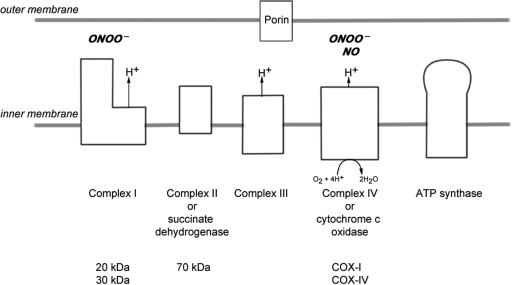
Multiple sclerosis is a chronic inflammatory disease, which leads to focal plaques of demyelination and tissue injury in the CNS. The structural and immunopathological patterns of demyelination suggest that different immune mechanisms may be involved in tissue damage. In a subtype of lesions, which are mainly found in patients with acute fulminant multiple sclerosis with Balo's type concentric sclerosis and in a subset of early relapsing remitting multiple sclerosis, the initial myelin changes closely resemble those seen in white matter stroke (WMS), suggesting a hypoxia-like tissue injury. Since mitochondrial injury may be involved in the pathogenesis of such lesions, we analysed a number of mitochondrial respiratory chain proteins in active lesions from acute multiple sclerosis and from WMS using immunohistochemistry. Functionally important defects of mitochondrial respiratory chain complex IV [cytochrome c oxidase (COX)] including its catalytic component (COX-I) are present in Pattern III but not in Pattern II multiple sclerosis lesions. The lack of immunohistochemically detected COX-I is apparent in oligodendrocytes, hypertrophied astrocytes and axons, but not in microglia. The profile of immunohistochemically detected mitochondrial respiratory chain complex subunits differs between multiple sclerosis and WMS. The findings suggest that hypoxia-like tissue injury in Pattern III multiple sclerosis lesions may be due to mitochondrial impairment.
Overview
The claims and statistics of this paper haven’t been checked yet.
Free: it reads the open-access text and checks the first 5 claims that cite a source we can check. Create a free account to check every claim.
Integrity signals
Registry
Cites
Statistics
Figures


Verification
Claims the paper makes
The claims and statistics of this paper haven’t been checked yet.
Free: it reads the open-access text and checks the first 5 claims that cite a source we can check. Create a free account to check every claim.
Claims other papers make about this one
Each is checked against this paper’s own text: does it say what the citing paper says it does?
The source backs part of the claim, not all of it — or backs it for a different population, condition or comparison.
In the citing paper
“Oxidative damage and mitochondrial dysfunction are key features of MS pathology [277,278,279,280].”
From Ageing, Cellular Senescence and Neurodegenerative Disease (2018) · cites this paper as [279]
Experimental — accuracy not yet measuredThe source backs part of the claim, not all of it — or backs it for a different population, condition or comparison.
In the citing paper
“Other features common to both cuprizone-induced and type 3 MS lesions are mitochondrial stress, selective loss of myelin-associated glycoprotein (MAG), lesions with indistinct borders that are not perivascular, pronounced glial activation, and lymphocyte infiltration [43, 46, 65, 69–71].”
From Oligodendrocyte death and myelin loss in the cuprizone model: an updated overview of the intrinsic and extrinsic causes of cuprizone demyelination (2022) · cites this paper as [69]
Experimental — accuracy not yet measured
Self-check
Percentages, sums and reported differences re-computed from the paper’s own numbers.
The self-check needs the paper’s full text.
Citations
Cites
Mitochondrial DNA mutations in human disease
Taylor, R. W., & Turnbull, D. M. (2005) · Nature Reviews Genetics · doi:10.1038/nrg1606 · Cited by 1,852 · cited 5×
“…c oxidase Abbreviations: CNPase=cyclic nucleotide phosphodiesterase; COX=cytochrome c oxidase; LFB=Luxol fast blue; MAG=myelin associated glycoprotein; MBP=myelin basic protein; mtDNA=mitochondrial DNA; NO=nitric oxide; NWM=normal white matter; PLP=proteolipid protein; WMS=white matter stroke”
BackgroundMethodInfluentialSelf-citationMultiple Sclerosis — The Plaque and Its Pathogenesis
Frohman, E. M., Racke, M. K., & Raine, C. S. (2006) · New England Journal of Medicine · doi:10.1056/nejmra052130 · Cited by 1,740 · cited 1×
“Multiple sclerosis is an inflammatory demyelinating disease of the central nervous system with axonal degeneration and astrogliosis (Frohman et al., 2006).”
BackgroundMitochondrial Respiratory-Chain Diseases
DiMauro, S., & Schon, E. A. (2003) · New England Journal of Medicine · doi:10.1056/nejmra022567 · Cited by 1,661 · cited 5×
“Keywords: multiple sclerosis; Pattern III lesion; mitochondria; cytochrome c oxidase Abbreviations: CNPase=cyclic nucleotide phosphodiesterase; COX=cytochrome c oxidase; LFB=Luxol fast blue; MAG=myelin associated glycoprotein; MBP=myelin basic protein; mtDNA=mitochondrial DNA; NO=nitric oxide;…”
BackgroundInfluentialvon Büdingen, H.-C., Palanichamy, A., Lehmann-Horn, K., Michel, B. A., & Zamvil, S. S. (2015) · European Neurology · doi:10.1159/000377675 · Cited by 1,463
Cleeter, M. W. J., Cooper, J. M., Darley-Usmar, V. M., Moncada, S., & Schapira, A. H. V. (1994) · FEBS Letters · doi:10.1016/0014-5793(94)00424-2 · Cited by 1,241 · cited 1×
“NO competitively inhibits the binding of oxygen to complex IV (Brown and Cooper, 1994; Cleeter et al., 1994).”
BackgroundHIF-1 Regulates Cytochrome Oxidase Subunits to Optimize Efficiency of Respiration in Hypoxic Cells
Fukuda, R., Zhang, H., Kim, J.-W., Shimoda, L., Dang, C. V., & Semenza, G. L. (2007) · Cell · doi:10.1016/j.cell.2007.01.047 · Cited by 1,225 · cited 5×
“Widespread defects of mitochondrial respiratory chain complexes inWMS lesions The transcription of mtDNA and nuclear DNA encoded subunits of the mitochondrial respiratory chain complexes are modulated by hypoxia (Vijayasarathy et al., 2003; Piruat and Lopez-Barneo, 2005; Fukuda et al., 2007).”
BackgroundInfluentialMicrodomains of Intracellular Ca2+: Molecular Determinants and Functional Consequences
Rizzuto, R., & Pozzan, T. (2006) · Physiological Reviews · doi:10.1152/physrev.00004.2005 · Cited by 1,206 · cited 1×
“Mitochondria also play an important role in apoptosis and calcium homeostasis (Rizzuto and Pozzan, 2006; Kalman et al., 2007).”
BackgroundRelapsing and remitting multiple sclerosis: Pathology of the newly forming lesion
Barnett, M. H., & Prineas, J. W. (2004) · Annals of Neurology · doi:10.1002/ana.20016 · Cited by 1,185 · cited 5×
“Mitochondrial defects in acute multiple sclerosis lesions Don Mahad,1 Iryna Ziabreva,1Hans Lassmann2 and DouglasTurnbull1 1The Mitochondrial Research Group, University of Newcastle uponTyne, UK and 2Centre for Brain Research, Medical University of Vienna, Austria Correspondence to: Prof. Dr Hans Lassmann, Center for B…”
BackgroundInfluentialBrown, G. C., & Cooper, C. E. (1994) · FEBS Letters · doi:10.1016/0014-5793(94)01290-3 · Cited by 1,055 · cited 2×
“…c oxidase Abbreviations: CNPase=cyclic nucleotide phosphodiesterase; COX=cytochrome c oxidase; LFB=Luxol fast blue; MAG=myelin associated glycoprotein; MBP=myelin basic protein; mtDNA=mitochondrial DNA; NO=nitric oxide; NWM=normal white matter; PLP=proteolipid protein; WMS=white matter stroke”
BackgroundSystemic infections and inflammation affect chronic neurodegeneration
Perry, V. H., Cunningham, C., & Holmes, C. (2007) · Nature Reviews Immunology · doi:10.1038/nri2015 · Cited by 1,027 · cited 1×
“These activated microglia, found in close proximity to apoptotic oligodendrocytes, may release a range of mediators (Barnett and Prineas, 2004; Perry et al., 2007).”
Background
Research works printed but not linked: 1
- Lucchinetti, 2000, Heterogeneity of multiple sclerosis lesions: implications for the pathogenesis of demyelination, Ann Neurol, 47, 707, 10.1002/1531-8249(200006)47:6<707::aid-ana3>3.0.co;2-q · DOI
Cited by
Compston, A., & Coles, A. (2008) · The Lancet · doi:10.1016/s0140-6736(08)61620-7 · Cited by 4,372
Pathological mechanisms in progressive multiple sclerosis
Mahad, D. H., Trapp, B. D., & Lassmann, H. (2015) · The Lancet Neurology · doi:10.1016/s1474-4422(14)70256-x · Cited by 1,137
Self-citationProgressive multiple sclerosis: pathology and pathogenesis
Lassmann, H., van Horssen, J., & Mahad, D. (2012) · Nature Reviews Neurology · doi:10.1038/nrneurol.2012.168 · Cited by 1,033
Self-citationLassmann, H. (2018) · Cold Spring Harbor Perspectives in Medicine · doi:10.1101/cshperspect.a028936 · Cited by 937
Self-citationOligodendrocytes: biology and pathology
Bradl, M., & Lassmann, H. (2010) · Acta Neuropathologica · doi:10.1007/s00401-009-0601-5 · Cited by 933
Self-citation
Metadata
- Authors
- Mahad, D., Ziabreva, I., Lassmann, H., & Turnbull, D.
- Affiliations
- Medical University of Vienna (AT)
- Corresponding author
- Lassmann, Hans
- Venue
- Brain
- Publisher
- Oxford University Press (OUP)
- Published
- 2008-05-30
- Volume
- 131
- Issue
- 7
- Pages
- 1722-1735
- ISSN
- 0006-8950 (ISSN-L)
- 1460-2156
- PMC
- PMC2442422
- OpenAlex
- W2149715738
- Cited by
- 406
- References
- 50
- Citations by year
- 2026: 8 · 2025: 8 · 2024: 22 · 2023: 18 · 2022: 33 · 2021: 25 · 2020: 30 · 2019: 26 · 2018: 13 · 2017: 23 · 2016: 25 · 2015: 28 · 2014: 28 · 2013: 30 · 2012: 24
- FWCI
- 15.42
- Type
- journal article
- Language
- en
- Open access
- hybrid
- doi.org
- Licence
- cc-by-nc
- Locations
- Brain (publishedVersion, cc-by-nc)
- Europe PMC (PubMed Central) (publishedVersion, cc-by-nc)
- PubMed Central (publishedVersion, cc-by-nc)
- PubMed (publishedVersion)
- Edinburgh Research Explorer (University of Edinburgh) (submittedVersion, cc-by-nc)
- Newcastle University ePrints (Newcastle Univesity)
- Funders
- Wellcome Trust (078415)
- Austrian Science Fund (P 19854)
- Topics
- Health Sciences › Medicine › Clinical Neurology › Multiple Sclerosis Research Studies
- Life Sciences › Biochemistry, Genetics and Molecular Biology › Molecular Biology › Mitochondrial Function and Pathology
- Health Sciences › Medicine › Clinical Neurology › Parkinson's Disease Mechanisms and Treatments
- Keywords
- multiple sclerosis, mitochondrial dysfunction, cytochrome c oxidase, coxI, mitochondrial respiratory chain, Balo's concentric sclerosis, demyelination, oligodendrocytes, astrocytes, microglia, relapsing-remitting multiple sclerosis
- MeSH terms
- Acute Disease, Adult, Aged, Aged, 80 and over, Astrocytes, Axons, Diffuse Cerebral Sclerosis of Schilder, Electron Transport Complex IV, Electron Transport, Female, Humans, Male, Middle Aged, Mitochondria, Multiple Sclerosis, Oligodendroglia, Mitochondrial Encephalomyopathies, Mitochondrial Proteins, Mitochondrial Diseases
About these checks
Registry, retraction, Cites and statistics results are computed, not judged, with one exception: a printed reference that no index lists is read once by a model before it counts as a citation error. Claims are extracted by a model and kept only when their quote appears verbatim in the text; each cited source is then checked, with a model’s help, for whether it says what the claim relies on. Those verdicts can be wrong — read the claim’s quote and the cited source before relying on one.
Statistics and arithmetic are recomputed, not judged.
Confidence bands are shown only for fields calibrated on our annotated test set. None is calibrated yet, so no band is shown.
What counts as a claim ›How we measure confidence ›How we check this ›
Are you an author, or do you see a problem with this page? Request a review
How to cite
Cite CoChat for the checks on this page, and the paper for its own findings.
- This page
- CoChat. (2026). Citation check: Mitochondrial defects in acute multiple sclerosis lesions. https://app.cochat.ai/papers/10.1093/brain/awn105
- The paper
- Mahad, D., Ziabreva, I., Lassmann, H., & Turnbull, D. (2008). Mitochondrial defects in acute multiple sclerosis lesions. Brain. https://doi.org/10.1093/brain/awn105
Data sources
Bibliographic data: Semantic Scholar, OpenAlex, Crossref.
Contains information from the Semantic Scholar Academic Graph Datasets, made available under the ODC Attribution License (ODC-BY 1.0).
Semantic Scholar’s data is described in: Kinney, R., et al. (2023). The Semantic Scholar Open Data Platform. arXiv:2301.10140.
