

The DOI matches its Crossref record; 27 of the 28 references it cites were found in a registry or an index.
Endothelial-specific Nox2 overexpression increases vascular superoxide and macrophage recruitment in ApoE−/− mice
Open accessAbstract
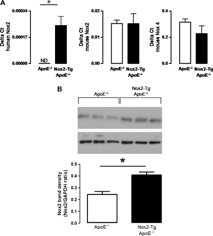
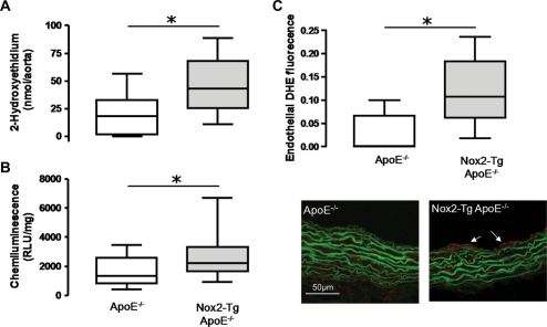
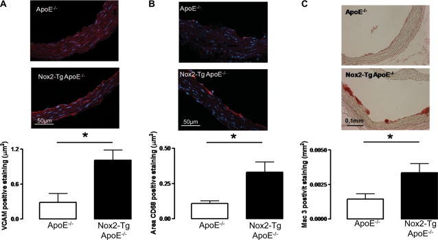
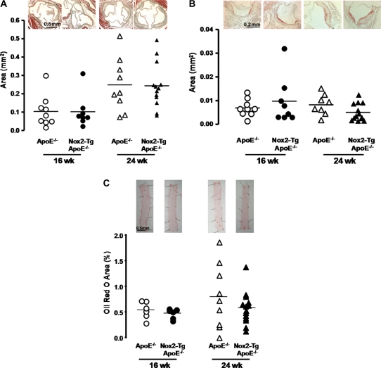
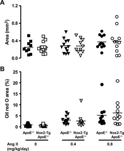
AIMS: Vascular disease states are associated with endothelial dysfunction and increased production of reactive oxygen species derived from NADPH oxidases. However, it remains unclear whether a primary increase in superoxide production specifically in the endothelium alters the initiation or progression of atherosclerosis. METHODS AND RESULTS: Mice overexpressing Nox2 specifically in the endothelium (Nox2-Tg) were crossed with ApoE(-/-) mice to produce Nox2-Tg ApoE(-/-) mice and ApoE(-/-) littermates. Endothelial overexpression of Nox2 in ApoE(-/-) mice did not alter blood pressure, but significantly increased vascular superoxide production compared with ApoE(-/-) littermates, measured using both lucigenin chemiluminescence and 2-hydroxyethidium production (ApoE(-/-), 19.9 ± 6.3 vs. Nox2-Tg ApoE(-/-), 47.0 ± 7.0 nmol 2-hydroxyethidium/aorta, P< 0.05). Increased endothelial superoxide production increased endothelial levels of vascular cell adhesion protein 1 and enhanced macrophage recruitment in early lesions in the aortic roots of 9-week-old mice, indicating increased atherosclerotic plaque initiation. However, endothelial-specific Nox2 overexpression did not alter native or angiotensin II-driven atherosclerosis in either the aortic root or the descending aorta. CONCLUSION: Endothelial-targeted Nox2 overexpression in ApoE(-/-) mice is sufficient to increase vascular superoxide production and increase macrophage recruitment possible via activation of endothelial cells. However, this initial increase in macrophage recruitment did not alter the progression of atherosclerosis. These results indicate that Nox-mediated reactive oxygen species signalling has important cell-specific and distinct temporal roles in the initiation and progression of atherosclerosis.
Overview
The claims and statistics of this paper haven’t been checked yet.
Run the check — free, no account needed. It reads the open-access text and checks the sources it cites.
Integrity signals
Registry
Cites
Statistics
Figures
Verification
Claims the paper makes
The claims and statistics of this paper haven’t been checked yet.
Run the check — free, no account needed. It reads the open-access text and checks the sources it cites.
Claims other papers make about this one
Each is checked against this paper’s own text: does it say what the citing paper says it does?
In the citing paper
“However, in other cases, an increased expression of endothelial NOX2 has not been linked to pathological conditions even though an increased production of superoxide and macrophage recruitment was observed (243, 443).”
From Sources of Vascular Nitric Oxide and Reactive Oxygen Species and Their Regulation (2019) · cites this paper as [243]
In the citing paper
“The current paradigm indicates that macrophage NOX2 is more detrimental in the development of cardiovascular disease (395, 876), with endothelial NOX2 probably involved in the initiation, but not so much in the development of the disease, and a subsequent progress of the atherosclerotic lesion dependent on immune cells (243, 573, 976).”
From Sources of Vascular Nitric Oxide and Reactive Oxygen Species and Their Regulation (2019) · cites this paper as [243]
In the citing paper
“The current paradigm indicates that macrophage NOX2 is more detrimental in the development of cardiovascular disease (395, 876), with endothelial NOX2 probably involved in the initiation, but not so much in the development of the disease, and a subsequent progress of the atherosclerotic lesion dependent on immune cells (243, 573, 976).”
From Sources of Vascular Nitric Oxide and Reactive Oxygen Species and Their Regulation (2019) · cites this paper as [243]
Self-check
Percentages, sums and reported differences re-computed from the paper’s own numbers.
The self-check needs the paper’s full text.
Citations
Cites
Endothelial dysfunction in cardiovascular diseases: the role of oxidant stress
Cai, H., & Harrison, D. G. (2000) · Circulation Research · doi:10.1161/01.res.87.10.840 · Cited by 3,839
ApoE-deficient mice develop lesions of all phases of atherosclerosis throughout the arterial tree.
Nakashima, Y., Plump, A. S., Raines, E. W., Breslow, J. L., & Ross, R. (1994) · Arteriosclerosis and Thrombosis: A Journal of Vascular Biology · doi:10.1161/01.atv.14.1.133 · Cited by 1,693
Mechanisms of Increased Vascular Superoxide Production in Human Diabetes Mellitus
Guzik, T. J., Mussa, S., Gastaldi, D., Sadowski, J., Ratnatunga, C., Pillai, R. G., & Channon, K. M. (2002) · Circulation · doi:10.1161/01.cir.0000012748.58444.08 · Cited by 979
Self-citationSuperoxide Production and Expression of Nox Family Proteins in Human Atherosclerosis
Sorescu, D., Weiss, D., Lassègue, B., Clempus, R. E., Szöcs, K., Sorescu, G. P., Valppu, L., Quinn, M. T., Lambeth, J. D., Vega, J. D., et al. (2002) · Circulation · doi:10.1161/01.cir.0000012917.74432.66 · Cited by 847
Lassègue, B., Sorescu, D., Szöcs, K., Yin, Q., Akers, M., Zhang, Y., Grant, S. L., Lambeth, J. D., & Griendling, K. K. (2001) · Circulation Research · doi:10.1161/hh0901.090299 · Cited by 839
NADPH Oxidases in Cardiovascular Health and Disease
Cave, A. C., Brewer, A. C., Narayanapanicker, A., Ray, R., Grieve, D. J., Walker, S., & Shah, A. M. (2006) · Antioxidants & Redox Signaling · doi:10.1089/ars.2006.8.691 · Cited by 630
Görlach, A., Brandes, R. P., Nguyen, K., Amidi, M., Dehghani, F., & Busse, R. (2000) · Circulation Research · doi:10.1161/01.res.87.1.26 · Cited by 598
NADPH Oxidases: Functions and Pathologies in the Vasculature
Lassègue, B., & Griendling, K. K. (2010) · Arteriosclerosis, Thrombosis, and Vascular Biology · doi:10.1161/atvbaha.108.181610 · Cited by 592
Vascular Superoxide Production by NAD(P)H Oxidase
Guzik, T. J., West, N. E. J., Black, E., McDonald, D., Ratnatunga, C., Pillai, R. G., & Channon, K. M. (2000) · Circulation Research · doi:10.1161/01.res.86.9.e85 · Cited by 584
Self-citationPueyo, M. E., Gonzalez, W., Nicoletti, A., Savoie, F., Arnal, J., & Michel, J. (2000) · Arteriosclerosis Thrombosis and Vascular Biology · doi:10.1161/01.atv.20.3.645 · Cited by 523
Nox4 as the Major Catalytic Component of an Endothelial NAD(P)H Oxidase
Ago, T., Kitazono, T., Ooboshi, H., Iyama, T., Han, Y. H., Takada, J., Wakisaka, M., Ibayashi, S., Utsumi, H., & Iida, M. (2004) · Circulation · doi:10.1161/01.cir.0000105680.92873.70 · Cited by 497
p47phox is required for atherosclerotic lesion progression in ApoE–/– mice
Barry-Lane, P. A., Patterson, C., van der Merwe, M., Hu, Z., Holland, S. M., Yeh, E. T. H., & Runge, M. S. (2001) · Journal of Clinical Investigation · doi:10.1172/jci200111927 · Cited by 489
Dikalov, S., Dikalova, A., Bikineyeva, A. T., Schmidt, H. H. H. W., Harrison, D. G., & Griendling, K. K. (2008) · Free Radical Biology and Medicine · doi:10.1016/j.freeradbiomed.2008.08.013 · Cited by 391
Judkins, C. P., Diep, H., Broughton, B. R. S., Mast, A. E., Hooker, E. U., Miller, A. A., Selemidis, S., Dusting, G. J., Sobey, C. G., & Drummond, G. R. (2010) · American Journal of Physiology-Heart and Circulatory Physiology · doi:10.1152/ajpheart.00799.2009 · Cited by 299
Coronary Artery Superoxide Production and Nox Isoform Expression in Human Coronary Artery Disease
Guzik, T. J., Sadowski, J., Guzik, B., Jopek, A., Kapelak, B., Przybyłowski, P., Wierzbicki, K., Korbut, R., Harrison, D. G., & Channon, K. M. (2006) · Arteriosclerosis, Thrombosis, and Vascular Biology · doi:10.1161/01.atv.0000196651.64776.51 · Cited by 278
Self-citationYang, H., Roberts, L. J., Shi, M., Zhou, L. C., Ballard, B. R., Richardson, A. G., & Guo, Z. M. (2004) · Circulation Research · doi:10.1161/01.res.0000149564.49410.0d · Cited by 230
Dansky, H. M., Barlow, C. B., Lominska, C. E., Sikes, J. L., Kao, C., Weinsaft, J. W., Cybulsky, M. I., & Smith, J. D. (2001) · Arteriosclerosis Thrombosis and Vascular Biology · doi:10.1161/hq1001.096625 · Cited by 219
The contribution of Nox4 to NADPH oxidase activity in mouse vascular smooth muscle
ELLMARK, S., DUSTING, G., NGTANGFUI, M., GUZZOPERNELL, N., & DRUMMOND, G. (2005) · Cardiovascular Research · doi:10.1016/j.cardiores.2004.10.026 · Cited by 213
Bendall, J. K., Alp, N. J., Warrick, N., Cai, S., Adlam, D., Rockett, K. A., Yokoyama, M., Kawashima, S., & Channon, K. M. (2005) · Circulation Research · doi:10.1161/01.res.0000187447.03525.72 · Cited by 209
Self-citationCrabtree, M. J., Tatham, A. L., Al-Wakeel, Y., Warrick, N., Hale, A. B., Cai, S., Channon, K. M., & Alp, N. J. (2009) · Journal of Biological Chemistry · doi:10.1074/jbc.m805403200 · Cited by 200
Self-citationAnilkumar, N. P., Weber, R., Zhang, M., Brewer, A. C., & Shah, A. M. (2008) · Arteriosclerosis Thrombosis and Vascular Biology · doi:10.1161/atvbaha.108.164277 · Cited by 195
Kirk, E. A., Dinauer, M. C., Rosen, H., Chait, A., Heinecke, J. W., & LeBoeuf, R. C. (2000) · Arteriosclerosis, Thrombosis, and Vascular Biology · doi:10.1161/01.atv.20.6.1529 · Cited by 171
Hsich, E., Segal, B. H., Pagano, P. J., Rey, F. E., Paigen, B., Deleonardis, J., Hoyt, R. F., Holland, S. M., & Finkel, T. (2000) · Circulation · doi:10.1161/01.cir.101.11.1234 · Cited by 158
Bendall, J. K., Rinze, R., Adlam, D., Tatham, A. L., de Bono, J., & Channon, K. M. (2007) · Circulation Research · doi:10.1161/01.res.0000263381.83835.7b · Cited by 157
Self-citationPendyala, S., Usatyuk, P. V., Gorshkova, I. A., Garcia, J. G. N., & Natarajan, V. (2008) · Antioxidants and Redox Signaling · doi:10.1089/ars.2008.2231 · Cited by 124
Angiotensin II-Mediated Development of Vascular Diseases
Daugherty, A. (2004) · Trends in Cardiovascular Medicine · doi:10.1016/j.tcm.2004.01.002 · Cited by 121
Coleman, R., Hayek, T., Keidar, S., & Aviram, M. (2006) · Acta Histochemica · doi:10.1016/j.acthis.2006.07.002 · Cited by 104
Cited by
Sorted by citations
Reactive Oxygen Species in Metabolic and Inflammatory Signaling
Forrester, S. J., Kikuchi, D. S., Hernandes, M. S., Xu, Q., & Griendling, K. K. (2018) · Circulation Research · doi:10.1161/circresaha.117.311401 · Cited by 2,081
Roles of vascular oxidative stress and nitric oxide in the pathogenesis of atherosclerosis
Förstermann, U., Xia, N., & Li, H. (2017) · Circulation Research · doi:10.1161/circresaha.116.309326 · Cited by 1,519
Angiotensin II Signal Transduction: An Update on Mechanisms of Physiology and Pathophysiology
Forrester, S. J., Booz, G. W., Sigmund, C. D., Coffman, T. M., Kawai, T., Rizzo, V., Scalia, R. G., & Eguchi, S. (2018) · Physiological Reviews · doi:10.1152/physrev.00038.2017 · Cited by 1,135
Vascular oxidative stress, nitric oxide and atherosclerosis
Li, H., Horke, S., & Förstermann, U. (2014) · Atherosclerosis · doi:10.1016/j.atherosclerosis.2014.09.001 · Cited by 678
Sources of Vascular Nitric Oxide and Reactive Oxygen Species and Their Regulation
Tejero, J., Shiva, S., & Gladwin, M. T. (2019) · Physiological Reviews · doi:10.1152/physrev.00036.2017 · Cited by 541
Metadata
- Authors
- Douglas, G., Bendall, J. K., Crabtree, M. J., Tatham, A. L., Carter, E. E., Hale, A. B., & Channon, K. M.
- Affiliations
- John Radcliffe Hospital (GB)
- University of Oxford (GB)
- Corresponding author
- Douglas, Gillian
- Venue
- Cardiovascular Research
- Publisher
- Oxford University Press (OUP)
- Published
- 2012-01-27
- Volume
- 94
- Issue
- 1
- Pages
- 20-29
- ISSN
- 0008-6363 (ISSN-L)
- 1755-3245
- PMC
- PMC3307381
- OpenAlex
- W1988097865
- Cited by
- 111
- References
- 28
- Citations by year
- 2026: 5 · 2025: 5 · 2024: 8 · 2023: 7 · 2022: 12 · 2021: 5 · 2020: 1 · 2019: 6 · 2018: 8 · 2017: 13 · 2016: 10 · 2015: 6 · 2014: 10 · 2013: 10 · 2012: 5
- FWCI
- 6.65
- Type
- journal article
- Language
- en
- Open access
- hybrid
- doi.org
- Licence
- other-oa
- Locations
- Cardiovascular Research (publishedVersion, other-oa)
- PubMed Central (publishedVersion, cc-by-nc)
- PubMed (publishedVersion)
- Funders
- Wellcome Trust (090532)
- British Heart Foundation (PG/05/141/20098, RG/07/003/23133, RG/12/5/29576)
- Topics
- Health Sciences › Medicine › Physiology (medical) › Nitric Oxide and Endothelin Effects
- Life Sciences › Immunology and Microbiology › Immunology › Atherosclerosis and Cardiovascular Diseases
- Life Sciences › Immunology and Microbiology › Immunology › Neutrophil, Myeloperoxidase and Oxidative Mechanisms
- Keywords
- NOX2, NADPH oxidase, atherosclerosis initiation, macrophage recruitment, ApoE-deficient mice, VCAM-1, reactive oxygen species, endothelial dysfunction, angiotensin II, aortic root lesions
- MeSH terms
- NADPH Oxidase 2, Angiotensin II, Animals, Aortic Diseases, Apolipoproteins E, Blood Pressure, Chemotaxis, Disease Models, Animal, Humans, Macrophages, Membrane Glycoproteins, Mice, Transgenic, Superoxides, Time Factors, Signal Transduction, Up-Regulation, Mice, Knockout, Disease Progression, Vascular Cell Adhesion Molecule-1, NADPH Oxidases, Endothelial Cells, Atherosclerosis, Mice
About these checks
Registry, retraction, Cites and statistics results are computed, not judged, with one exception: a printed reference that no index lists is read once by a model before it counts as a citation error. Claims are extracted by a model and kept only when their quote appears verbatim in the text; each cited source is then checked, with a model’s help, for whether it says what the claim relies on. Those verdicts can be wrong — read the claim’s quote and the cited source before relying on one.
Statistics and arithmetic are recomputed, not judged.
Confidence bands are shown only for fields calibrated on our annotated test set. None is calibrated yet, so no band is shown.
What counts as a claim ›How we measure confidence ›How we check this ›
Are you an author, or do you see a problem with this page? Request a review
