

The DOI matches its Crossref record; 59 of the 68 references it cites were found in a registry or an index.
Maternal diet-induced obesity programmes cardiac dysfunction in male mice independently of post-weaning diet
Open accessAbstract
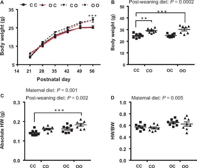
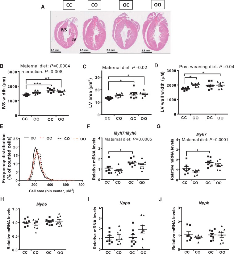
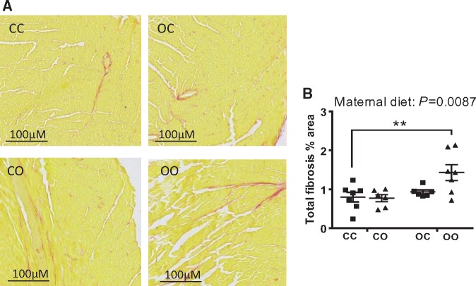
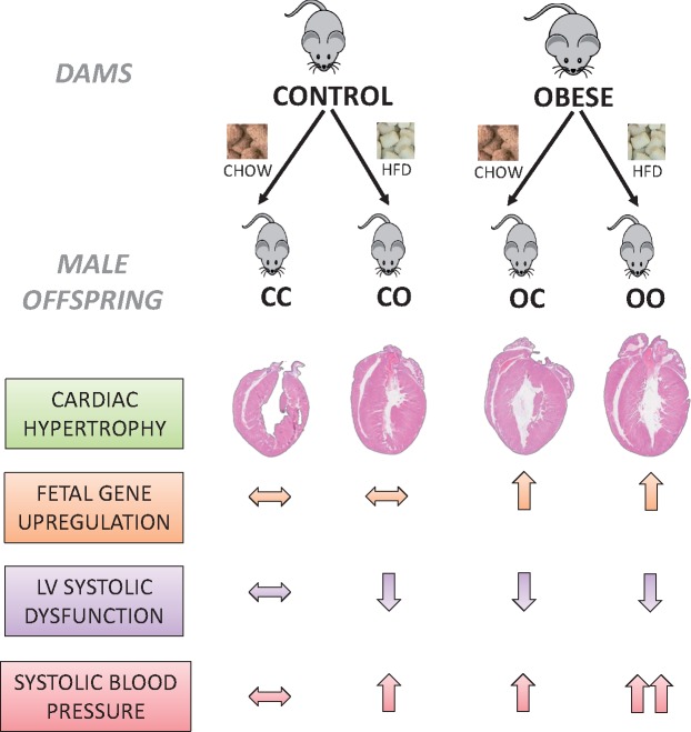
Aims: Obesity during pregnancy increases risk of cardiovascular disease (CVD) in the offspring and individuals exposed to over-nutrition during fetal life are likely to be exposed to a calorie-rich environment postnatally. Here, we established the consequences of combined exposure to a maternal and post-weaning obesogenic diet on offspring cardiac structure and function using an established mouse model of maternal diet-induced obesity. Methods and results: The impact of the maternal and postnatal environment on the offspring metabolic profile, arterial blood pressure, cardiac structure, and function was assessed in 8-week-old C57BL/6 male mice. Measurement of cardiomyocyte cell area, the transcriptional re-activation of cardiac fetal genes as well as genes involved in the regulation of contractile function and matrix remodelling in the adult heart were determined as potential mediators of effects on cardiac function. In the adult offspring: a post-weaning obesogenic diet coupled with exposure to maternal obesity increased serum insulin (P < 0.0001) and leptin levels (P < 0.0001); maternal obesity (P = 0.001) and a post-weaning obesogenic diet (P = 0.002) increased absolute heart weight; maternal obesity (P = 0.01) and offspring obesity (P = 0.01) caused cardiac dysfunction but effects were not additive; cardiac dysfunction resulting from maternal obesity was associated with re-expression of cardiac fetal genes (Myh7: Myh6 ratio; P = 0.0004), however, these genes were not affected by offspring diet; maternal obesity (P = 0.02); and offspring obesity (P = 0.05) caused hypertension and effects were additive. Conclusions: Maternal diet-induced obesity and offspring obesity independently promote cardiac dysfunction and hypertension in adult male progeny. Exposure to maternal obesity alone programmed cardiac dysfunction, associated with hallmarks of pathological left ventricular hypertrophy, including increased cardiomyocyte area, upregulation of fetal genes, and remodelling of cardiac structure. These data highlight that the perinatal period is just as important as adult-onset obesity in predicting CVD risk. Therefore, early developmental periods are key intervention windows to reduce the prevalence of CVD.
Overview
The claims and statistics of this paper haven’t been checked yet.
Run the check — free, no account needed. It reads the open-access text and checks the sources it cites.
Integrity signals
Registry
Cites
Statistics
Figures
Verification
Claims the paper makes
The claims and statistics of this paper haven’t been checked yet.
Run the check — free, no account needed. It reads the open-access text and checks the sources it cites.
Claims other papers make about this one
Each is checked against this paper’s own text: does it say what the citing paper says it does?
The cited source says what the claim attributes to it.
In the citing paper
“High-fat diet feeding had similar effects on increasing body weight in offspring from obese dams as well as control dams [114].”
From Recent Experimental Studies of Maternal Obesity, Diabetes during Pregnancy and the Developmental Origins of Cardiovascular Disease (2022) · cites this paper as [114]
The cited source says what the claim attributes to it.
In the citing paper
“The combination of exposure to maternal obesity and a postnatal high-fat diet also markedly increased systolic blood pressure in mouse offspring [114].”
From Recent Experimental Studies of Maternal Obesity, Diabetes during Pregnancy and the Developmental Origins of Cardiovascular Disease (2022) · cites this paper as [114]
Self-check
Percentages, sums and reported differences re-computed from the paper’s own numbers.
The self-check needs the paper’s full text.
Citations
Cites
Analyzing real-time PCR data by the comparative CT method
Schmittgen, T. D., & Livak, K. J. (2008) · Nature Protocols · doi:10.1038/nprot.2008.73 · Cited by 26,190
Effect of In Utero and Early-Life Conditions on Adult Health and Disease
Gluckman, P. D., Hanson, M. A., Cooper, C., & Thornburg, K. L. (2008) · New England Journal of Medicine · doi:10.1056/nejmra0708473 · Cited by 3,898
Body Fatness and Cancer — Viewpoint of the IARC Working Group
Lauby-Secretan, B., Scoccianti, C., Loomis, D., Grosse, Y., Bianchini, F., & Straif, K. (2016) · New England Journal of Medicine · doi:10.1056/nejmsr1606602 · Cited by 3,596
Regulation of cardiac hypertrophy by intracellular signalling pathways
Heineke, J., & Molkentin, J. D. (2006) · Nature Reviews Molecular Cell Biology · doi:10.1038/nrm1983 · Cited by 1,890
Cardiovascular disease in Europe: epidemiological update 2016
Townsend, N. P., Wilson, L., Bhatnagar, P., Wickramasinghe, K. K., Rayner, M., & Nichols, M. S. (2016) · European Heart Journal · doi:10.1093/eurheartj/ehw334 · Cited by 1,579
Preconceptional and maternal obesity: epidemiology and health consequences
Poston, L., Caleyachetty, R., Cnattingius, S., Corvalán, C., Uauy, R., Herring, S., & Gillman, M. W. (2016) · The Lancet Diabetes & Endocrinology · doi:10.1016/s2213-8587(16)30217-0 · Cited by 1,111
Influence of Maternal Obesity on the Long-Term Health of Offspring
Godfrey, K. M., Reynolds, R. M., Prescott, S. L., Nyirenda, M., Jaddoe, V. W. V., Eriksson, J. G., & Broekman, B. F. P. (2017) · The Lancet Diabetes & Endocrinology · doi:10.1016/s2213-8587(16)30107-3 · Cited by 1,059
Samuelsson, A.-M., Matthews, P. A., Argenton, M., Christie, M. R., McConnell, J. M., Jansen, E. H. J. M., Piersma, A. H., Ozanne, S. E., Twinn, D. F., Remacle, C., et al. (2008) · Hypertension · doi:10.1161/hypertensionaha.107.101477 · Cited by 923
Self-citationPhysiological and pathological cardiac hypertrophy
Shimizu, I., & Minamino, T. (2016) · Journal of Molecular and Cellular Cardiology · doi:10.1016/j.yjmcc.2016.06.001 · Cited by 919
Obesity-Associated Hypertension
Rahmouni, K., Correia, M., Haynes, W. G., & Mark, A. L. (2004) · Hypertension · doi:10.1161/01.hyp.0000151325.83008.b4 · Cited by 788
Fetuses of obese mothers develop insulin resistance in utero
Catalano, P. M., Presley, L., Minium, J., & Hauguel-de Mouzon, S. (2009) · Diabetes Care · doi:10.2337/dc08-2077 · Cited by 655
BMP8B Increases Brown Adipose Tissue Thermogenesis through Both Central and Peripheral Actions
Whittle, A. J., Carobbio, S., Martins, L., Slawik, M., Hondares, E., Vázquez, M. J., Morgan, D., Csikasz, R. I., Gallego, R., Rodriguez-Cuenca, S., et al. (2012) · Cell · doi:10.1016/j.cell.2012.02.066 · Cited by 573
Reynolds, R. M., Allan, K. M., Raja, E. A., Bhattacharya, S., McNeill, G., Hannaford, P. C., Sarwar, N., Lee, A. J., Bhattacharya, S., & Norman, J. E. (2013) · BMJ · doi:10.1136/bmj.f4539 · Cited by 571
Maternal nutritional history predicts obesity in adult offspring independent of postnatal diet
Howie, G. J., Sloboda, D., Kamal, T., & Vickers, M. (2009) · Journal of Physiology · doi:10.1113/jphysiol.2008.163477 · Cited by 449
Role of resistin in diet-induced hepatic insulin resistance
Muse, E. D., Obici, S., Bhanot, S., Monia, B. P., McKay, R. A., Rajala, M. W., Scherer, P. E., & Rossetti, L. (2004) · Journal of Clinical Investigation · doi:10.1172/jci21270 · Cited by 375
Regulation of Resistin Expression and Circulating Levels in Obesity, Diabetes, and Fasting
Rajala, M. W., Qi, Y., Patel, H. R., Takahashi, N., Banerjee, R. R., Pajvani, U. B., Sinha, M. K., Gingerich, R. L., Scherer, P. E., & Ahima, R. S. (2004) · Diabetes · doi:10.2337/diabetes.53.7.1671 · Cited by 351
Taylor, P. D., McConnell, J., Khan, I. Y., Holemans, K., Lawrence, K. M., Asare-Anane, H., Persaud, S. J., Jones, P. M., Petrie, L., Hanson, M. A., et al. (2005) · American Journal of Physiology-Regulatory, Integrative and Comparative Physiology · doi:10.1152/ajpregu.00355.2004 · Cited by 297
Regulation of fetal gene expression in heart failure
Dirkx, E., Martins, P. A. D. C., & Windt, L. J. D. (2013) · Biochimica et Biophysica Acta (BBA) - Molecular Basis of Disease · doi:10.1016/j.bbadis.2013.07.023 · Cited by 292
Developmental Programming of Cardiovascular Dysfunction by Prenatal Hypoxia and Oxidative Stress
Giussani, D. A., Camm, E. J., Niu, Y., Richter, H. G., Blanco, C. E., Gottschalk, R., Blake, E. Z., Horder, K. A., Thakor, A. S., Hansell, J. A., et al. (2012) · PLoS ONE · doi:10.1371/journal.pone.0031017 · Cited by 279
Self-citationElahi, M. M., Cagampang, F. R. A., Mukhtar, D., Anthony, F. W., Ohri, S. K., & Hanson, M. A. (2009) · British Journal Of Nutrition · doi:10.1017/s000711450820749x · Cited by 259
Periasamy, M., Bhupathy, P., & Babu, G. J. (2007) · Cardiovascular Research · doi:10.1093/cvr/cvm056 · Cited by 256
Catalano, P., & deMouzon, S. H. (2015) · International Journal of Obesity · doi:10.1038/ijo.2015.15 · Cited by 243
Guénard, F., Deshaies, Y., Cianflone, K., Kral, J. G., Marceau, P., & Vohl, M.-C. (2013) · Proceedings of the National Academy of Sciences · doi:10.1073/pnas.1216959110 · Cited by 236
Animal models of in utero exposure to a high fat diet: A review
Williams, L., Seki, Y., Vuguin, P. M., & Charron, M. J. (2013) · Biochimica et Biophysica Acta (BBA) - Molecular Basis of Disease · doi:10.1016/j.bbadis.2013.07.006 · Cited by 234
Eriksson, J. G., Sandboge, S., Salonen, M. K., Kajantie, E., & Osmond, C. (2014) · Annals of Medicine · doi:10.3109/07853890.2014.919728 · Cited by 213
Left ventricular hypertrophy in hypertension: its arrhythmogenic potential
Kahan, T., & Bergfeldt, L. (2005) · Heart · doi:10.1136/hrt.2004.042473 · Cited by 208
Myocardial Collagen Turnover in Hypertrophic Cardiomyopathy
Lombardi, R., Betocchi, S., Losi, M. A., Tocchetti, C. G., Aversa, M., Miranda, M., D’Alessandro, G., Cacace, A., Ciampi, Q., & Massimo Chiariello, A. (2003) · Circulation · doi:10.1161/01.cir.0000090687.97972.10 · Cited by 205
Armitage, J. A., Lakasing, L., Taylor, P. D., Balachandran, A. A., Jensen, R. I., Dekou, V., Ashton, N., Nyengaard, J. R., & Poston, L. (2005) · The Journal of Physiology · doi:10.1113/jphysiol.2005.084947 · Cited by 195
Developmental programming of cardiovascular disease by prenatal hypoxia
Giussani, D. A., & Davidge, S. T. (2013) · Journal of Developmental Origins of Health and Disease · doi:10.1017/s204017441300010x · Cited by 185
Self-citationCardiovascular disease in Europe 2016: an epidemiological update
Townsend, N. P., Wilson, L., Bhatnagar, P., Wickramasinghe, K., Rayner, M., & Nichols, M. (2016) · European Heart Journal · doi:10.1093/eurheartj/ehw468 · Cited by 153
Fernandez-Twinn, D. S., Blackmore, H. L., Siggens, L., Giussani, D. A., Cross, C. M., Foo, R., & Ozanne, S. E. (2012) · Endocrinology · doi:10.1210/en.2012-1508 · Cited by 141
Self-citationTardiff, J. C., Hewett, T. E., Factor, S. M., Vikstrom, K. L., Robbins, J. M., & Leinwand, L. A. (2000) · American Journal of Physiology-Heart and Circulatory Physiology · doi:10.1152/ajpheart.2000.278.2.h412 · Cited by 141
Blackmore, H. L., Niu, Y., Fernandez-Twinn, D. S., Tarry-Adkins, J. L., Giussani, D. A., & Ozanne, S. E. (2014) · Endocrinology · doi:10.1210/en.2014-1383 · Cited by 127
Self-citationLiang, C., Oest, M. E., & Prater, M. R. (2009) · Birth Defects Research Part B Developmental and Reproductive Toxicology · doi:10.1002/bdrb.20206 · Cited by 121
Fernandez-Twinn, D. S., Gascoin, G., Musial, B., Carr, S., Duque-Guimaraes, D., Blackmore, H. L., Alfaradhi, M. Z., Loche, E., Sferruzzi-Perri, A. N., Fowden, A. L., et al. (2017) · Scientific Reports · doi:10.1038/srep44650 · Cited by 115
Self-citationWang, J., Ma, H., Tong, C., Zhang, H., Lawlis, G. B., Li, Y., Zang, M., Ren, J. T., Nijland, M. J., Ford, S. P., et al. (2010) · The FASEB Journal · doi:10.1096/fj.09-142315 · Cited by 109
Zambrano, E., Ibáñez, C. A., Martínez‐Samayoa, P. M., Lomas‐Soria, C., Durand-Carbajal, M., & Rodríguez-González, G. L. (2016) · Archives of Medical Research · doi:10.1016/j.arcmed.2016.01.004 · Cited by 107
Maternal high-fat diet impacts endothelial function in nonhuman primate offspring
Fan, L., Lindsley, S. R., Comstock, S. M., Takahashi, D. L., Evans, A. E., He, G.-W., Thornburg, K. L., & Grove, K. L. (2013) · International Journal of Obesity · doi:10.1038/ijo.2012.42 · Cited by 98
Liang, X., Yang, Q., Zhang, L., Maricelli, J. W., Rodgers, B. D., Zhu, M. J., & Du, . (2016) · Scientific Reports · doi:10.1038/srep34345 · Cited by 98
Mechanisms underlying the developmental origins of disease
Martin‐Gronert, M. S., & Ozanne, S. E. (2012) · Reviews in Endocrine and Metabolic Disorders · doi:10.1007/s11154-012-9210-z · Cited by 97
Self-citationISGANAITIS, E. M., Woo, M., Ma, H., Chen, M., Kong, W., Lytras, A., Sales, V., DeCoste-Lopez, J., Lee, K.-J., Leatherwood, C. L., et al. (2013) · Diabetes · doi:10.2337/db13-0558 · Cited by 89
Turdi, S., Ge, W., Hu, N., Bradley, K. M., Wang, X., & Ren, J. (2013) · Journal of Molecular and Cellular Cardiology · doi:10.1016/j.yjmcc.2012.12.007 · Cited by 88
Maternal obesity induces fibrosis in fetal myocardium of sheep
Huang, Y., Yan, X., Zhao, J. X., Zhu, M. J., McCormick, R. J., Ford, S. P., Nathanielsz, P. W., Ren, J. T., & Du, . (2010) · American Journal of Physiology-Endocrinology and Metabolism · doi:10.1152/ajpendo.00434.2010 · Cited by 79
Fletcher, A. J. W., McGARRIGLE, H. H. G., Edwards, C. M. B., Fowden, A. L., & Giussani, D. A. (2002) · The Journal of Physiology · doi:10.1113/jphysiol.2001.015693 · Cited by 75
Self-citationMaternal High-Fat Diet Programs for Metabolic Disturbances in Offspring despite Leptin Sensitivity
Volpato, A. M., Schultz, A., Magalhães-da-Costa, E., Correia, M., Águila, M. B., & Mandarim‐de‐Lacerda, C. A. (2012) · Neuroendocrinology · doi:10.1159/000336377 · Cited by 71
Torrens, C., Ethirajan, P., Bruce, K., Cagampang, F., Siow, R., Hanson, M., Byrne, C., Mann, G., & Clough, G. (2012) · PLoS ONE · doi:10.1371/journal.pone.0050671 · Cited by 67
Altered adipose tissue metabolism in offspring of dietary obese rat dams
Benkalfat, N. B., Merzouk, H., Bouanane, S., Merzouk, S.-A., Bellenger, J., Gresti, J., Tessier, C., & Narce, M. (2011) · Clinical Science · doi:10.1042/cs20100534 · Cited by 66
Resistin Impairs Insulin-Evoked Vasodilation
Gentile, M. T., Vecchione, C., Marino, G., Aretini, A., Pardo, A. D., Antenucci, G., Maffei, A., Cifelli, G., Iorio, L., Landolfi, A., et al. (2007) · Diabetes · doi:10.2337/db07-0557 · Cited by 65
Guberman, C., Jellyman, J. K., Han, G., Ross, M. G., & Desai, M. (2013) · American Journal of Obstetrics and Gynecology · doi:10.1016/j.ajog.2013.05.023 · Cited by 61
Maternal obesity affects fetal myocardial function as early as in the first trimester
Ingul, C. B., Loraas, L., Tegnander, E., Eik‐Nes, S. H., & Brantberg, A. (2015) · Ultrasound in Obstetrics and Gynecology · doi:10.1002/uog.14841 · Cited by 59
Fan, X., Turdi, S., Ford, S. P., Hua, Y., Nijland, M. J., Zhu, M. J., Nathanielsz, P. W., & Ren, J. T. (2010) · The Journal of Nutritional Biochemistry · doi:10.1016/j.jnutbio.2009.11.006 · Cited by 41
Central role for melanocortin-4 receptors in offspring hypertension arising from maternal obesity
Samuelsson, A., Mullier, A., Maicas, N., Oosterhuis, N. R., Bae, S., Novoselova, T. V., Chan, L. F., Pombo, J., Taylor, P. D., Joles, J. A., et al. (2016) · Proceedings of the National Academy of Sciences · doi:10.1073/pnas.1607464113 · Cited by 40
de Almeida-Faria, J., Duque-Guimaraes, D. E., Carpenter, A. A. M., Loche, E., & Ozanne, S. E. (2017) · Scientific Reports · doi:10.1038/srep44949 · Cited by 32
Self-citationChen, H., Simar, D., Ting, J., Erkelens, J. R. S., & Morris, M. J. (2012) · Journal of Neuroendocrinology · doi:10.1111/j.1365-2826.2012.02339.x · Cited by 32
Vieira, A. K. G., Soares, V. M., Bernardo, A. F., Neves, F. A., de Mattos, A. B. M., Guedes, R. M., Cortez, E. A. C., de Andrade, D. C., Miranda, G. L., Garcia‐Souza, É. P., et al. (2015) · Nutrition Metabolism and Cardiovascular Diseases · doi:10.1016/j.numecd.2015.07.009 · Cited by 30
Janoschek, R., Bae‐Gartz, I., Vohlen, C., Alcazar, M. A. A., Dinger, K., Appel, S., Dötsch, J., & Hucklenbruch‐Rother, E. (2016) · Obesity · doi:10.1002/oby.21486 · Cited by 29
Xie, P. L., McDowell, T. S., Chapleau, M. W., Hajduczok, G., & Abboud, F. M. (1991) · Hypertension · doi:10.1161/01.hyp.17.1.72 · Cited by 28
Schrempft, S., van Jaarsveld, C. H. M., Fisher, A., Fildes, A., & Wardle, J. (2016) · Appetite · doi:10.1016/j.appet.2016.08.108 · Cited by 23
Fetal Hyperinsulinemia and Protein Turnover in Fetal Rat Tissues
Johnson, J. D., Dunham, T., Wogenrich, F. J., Greenberg, R. E., Loftfield, R. B., & SKIPPER, B. J. (1990) · Diabetes · doi:10.2337/diab.39.5.541 · Cited by 11
Research works printed but not linked: 1
- Ingul, 2016, Maternal obesity affects foetal myocardial function already in first trimester, Ultrasound Obstet Gynecol, 47, 433
Cited by
Sorted by citations
Developmental origins of metabolic diseases
Hoffman, D. J., Powell, T. L., Barrett, E. S., & Hardy, D. B. (2020) · Physiological Reviews · doi:10.1152/physrev.00002.2020 · Cited by 504
Morgan, H. L., Paganopoulou, P., Akhtar, S., Urquhart, N., Philomin, R., Dickinson, Y., & Watkins, A. J. (2019) · The Journal of Physiology · doi:10.1113/jp278270 · Cited by 79
Beeson, J. H., Blackmore, H. L., Carr, S. K., Dearden, L., Duque-Guimarães, D. E., Kusinski, L. C., Pantaleão, L. C., Pinnock, A. G., Aiken, C. E., Giussani, D. A., et al. (2018) · Molecular Metabolism · doi:10.1016/j.molmet.2018.06.009 · Cited by 73
Self-citationFerey, J. L. A., Boudoures, A. L., Reid, M., Drury, A. M., Scheaffer, S. M., Modi, Z. N., Kovács, A. D., Pietka, T., DeBosch, B. J., Thompson, M. D., et al. (2019) · American Journal of Physiology-Heart and Circulatory Physiology · doi:10.1152/ajpheart.00013.2019 · Cited by 67
Programming of cardiometabolic health: the role of maternal and fetal hyperinsulinaemia
Hufnagel, A., Dearden, L., Fernandez‐Twinn, D. S., & Ozanne, S. E. (2022) · Journal of Endocrinology · doi:10.1530/joe-21-0332 · Cited by 78
Self-citation
Metadata
- Authors
- Loche, E., Blackmore, H. L., Carpenter, A. A., Beeson, J. H., Pinnock, A., Ashmore, T. J., Aiken, C. E., de Almeida-Faria, J., Schoonejans, J. M., Giussani, D. A., Fernandez-Twinn, D. S., & Ozanne, S. E.
- Affiliations
- University of Cambridge (GB)
- Universidade Estadual de Campinas (UNICAMP) (BR)
- University of Cambridge Metabolic Research Laboratories and MRC Metabolic Diseases Unit, Wellcome Trust-MRC Institute of Metabolic Science, Level 4, Box 289, Addenbrookes' Treatment Centre, Addenbrookes' Hospital, Hills Road, Cambridge, CB2 OQQ, UK
- Corresponding author
- Loche, Elena
- ORCID
- Venue
- Cardiovascular Research
- Publisher
- Oxford University Press (OUP)
- Published
- 2018-04-04
- Volume
- 114
- Issue
- 10
- Pages
- 1372-1384
- ISSN
- 0008-6363 (ISSN-L)
- 1755-3245
- PMC
- PMC6054211
- OpenAlex
- W2795824202
- Cited by
- 105
- References
- 68
- Citations by year
- 2026: 6 · 2025: 12 · 2024: 10 · 2023: 9 · 2022: 17 · 2021: 14 · 2020: 13 · 2019: 18 · 2018: 6
- FWCI
- 14.08
- Type
- journal article
- Language
- en
- Open access
- hybrid
- doi.org
- Licence
- Locations
- Cardiovascular Research (publishedVersion, cc-by)
- Europe PMC (PubMed Central) (publishedVersion, cc-by)
- PubMed Central (publishedVersion, cc-by)
- PubMed (publishedVersion)
- Apollo (University of Cambridge) (submittedVersion, cc-by)
- Apollo (cc-by)
- Funders
- Medical Research Council (MRC_MC_UU_12012/4)
- British Heart Foundation (FS/12/64/30001, PG/14/20/30769, RG/17/12/33167)
- São Paulo Research Foundation (2017/03525-8)
- Topics
- Health Sciences › Medicine › Pediatrics, Perinatology and Child Health › Birth, Development, and Health
- Health Sciences › Medicine › Cardiology and Cardiovascular Medicine › Cardiovascular Function and Risk Factors
- Health Sciences › Medicine › Cardiology and Cardiovascular Medicine › Cardiac Fibrosis and Remodeling
- Keywords
- maternal obesity, diet-induced obesity, cardiac dysfunction, fetal programming, hypertension, left ventricular hypertrophy, cardiomyocyte size, MYH7, MYH6, cardiac remodeling, insulin, leptin, cardiovascular risk, perinatal development
- MeSH terms
- Gestational Weight Gain, Age Factors, Animals, Animal Nutritional Physiological Phenomena, Disease Models, Animal, Female, Heart Diseases, Hypertension, Male, Mice, Inbred C57BL, Myocardial Contraction, Nutritional Status, Obesity, Pregnancy, Prenatal Exposure Delayed Effects, Ventricular Function, Left, Ventricular Remodeling, Myocytes, Cardiac, Maternal Nutritional Physiological Phenomena
About these checks
Registry, retraction, Cites and statistics results are computed, not judged, with one exception: a printed reference that no index lists is read once by a model before it counts as a citation error. Claims are extracted by a model and kept only when their quote appears verbatim in the text; each cited source is then checked, with a model’s help, for whether it says what the claim relies on. Those verdicts can be wrong — read the claim’s quote and the cited source before relying on one.
Statistics and arithmetic are recomputed, not judged.
Confidence bands are shown only for fields calibrated on our annotated test set. None is calibrated yet, so no band is shown.
What counts as a claim ›How we measure confidence ›How we check this ›
Are you an author, or do you see a problem with this page? Request a review
