

The DOI matches its Crossref record; 33 of the 34 references it cites were found in a registry or an index.
Inherited and de novo SHANK2 variants associated with autism spectrum disorder impair neuronal morphogenesis and physiology
Open accessAbstract
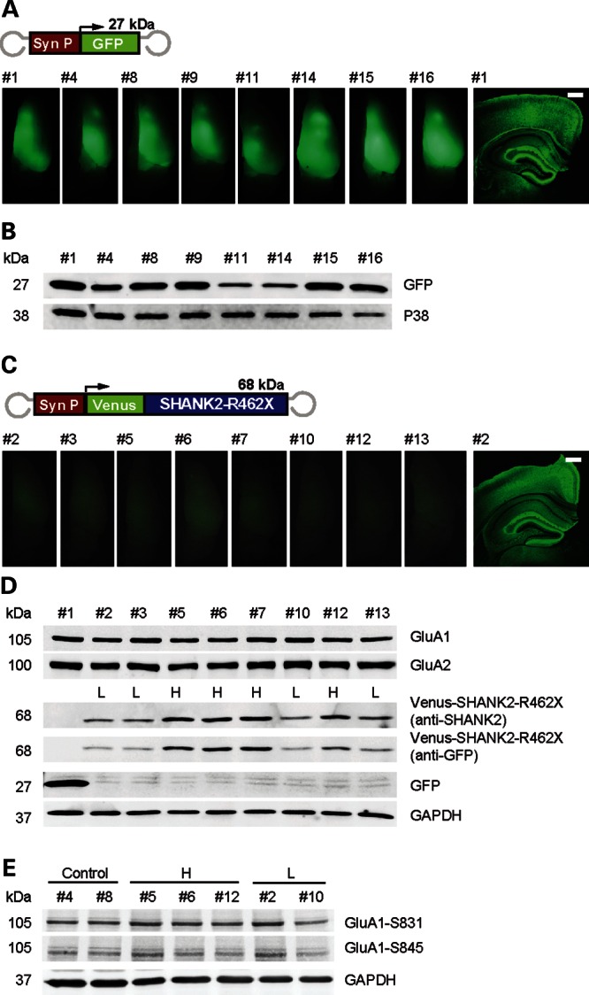
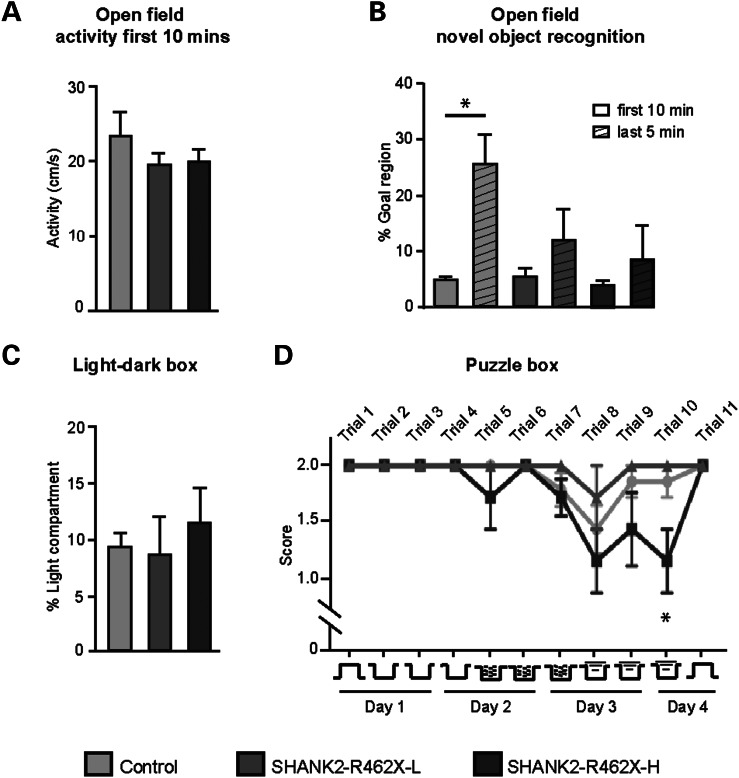
Mutations in the postsynaptic scaffolding gene SHANK2 have recently been identified in individuals with autism spectrum disorder (ASD) and intellectual disability. However, the cellular and physiological consequences of these mutations in neurons remain unknown. We have analyzed the functional impact caused by two inherited and one de novo SHANK2 mutations from ASD individuals (L1008_P1009dup, T1127M, R462X). Although all three variants affect spine volume and have smaller SHANK2 cluster sizes, T1127M additionally fails to rescue spine volume in Shank2 knock-down neurons. R462X is not able to rescue spine volume and dendritic branching and lacks postsynaptic clustering, indicating the most severe dysfunction. To demonstrate that R462X when expressed in mouse can be linked to physiological effects, we analyzed synaptic transmission and behavior. Principal neurons of mice expressing rAAV-transduced SHANK2-R462X present a specific, long-lasting reduction in miniature postsynaptic AMPA receptor currents. This dominant negative effect translates into dose-dependent altered cognitive behavior of SHANK2-R462X-expressing mice, with an impact on the penetrance of ASD.
Overview
The claims and statistics of this paper haven’t been checked yet.
Run the check — free, no account needed. It reads the open-access text and checks the sources it cites.
Integrity signals
Registry
Cites
Statistics
Figures
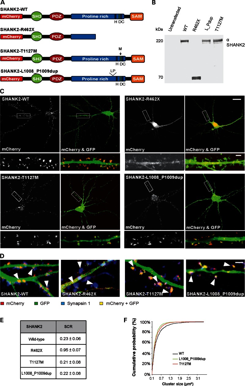
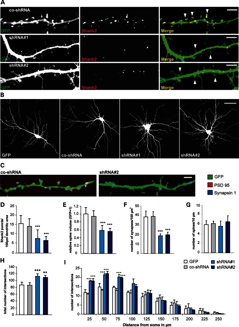
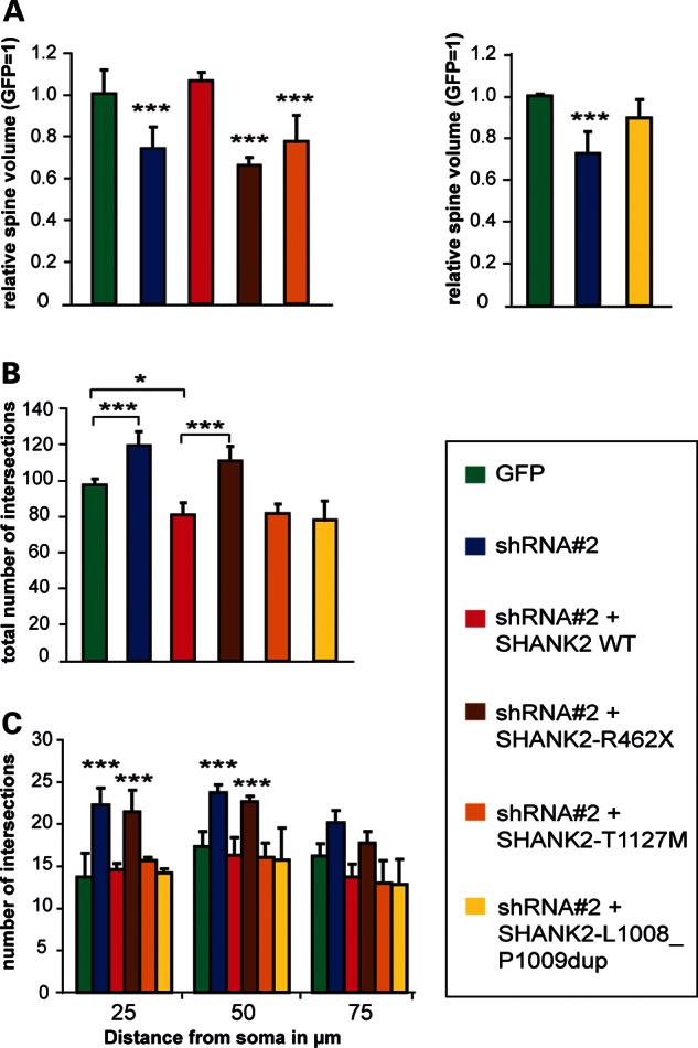
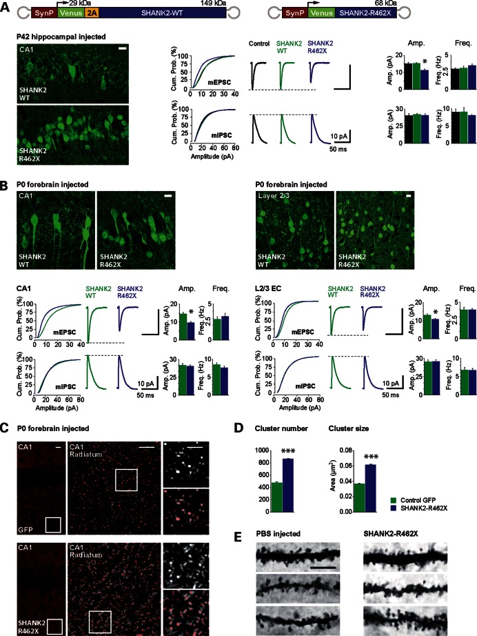
Verification
Claims the paper makes
The claims and statistics of this paper haven’t been checked yet.
Run the check — free, no account needed. It reads the open-access text and checks the sources it cites.
Claims other papers make about this one
Each is checked against this paper’s own text: does it say what the citing paper says it does?
In the citing paper
“Recent human genetic studies indicate that SHANK family genes (SHANK1, SHANK2, and SHANK3) are causative genes for idiopathic autism spectrum disorders (ASD).”
From Modeling Autism by SHANK Gene Mutations in Mice (2013) · cites this paper as Berkel et al., 2012
In the citing paper
“SHANK/ProSAP family proteins (SHANK1, SHANK2, SHANK3) have emerged as promising candidates for modeling ASD in mice due to strong genetic evidence showing molecular defects of SHANK in patients with ASD (Berkel et al., 2010; Berkel et al., 2012; Durand et al., 2007; Gauthier et al., 2010; Marshall et al., 2008; Pinto et al., 2010; Sato et al., 2012).”
From Modeling Autism by SHANK Gene Mutations in Mice (2013) · cites this paper as Berkel et al., 2012
In the citing paper
“Recently, mutations in SHANK1 and SHANK2 have also been associated with ASD (Berkel et al., 2010; Berkel et al., 2012; Sato et al., 2012), supporting a general function for this gene family in common molecular pathways associated with ASD.”
From Modeling Autism by SHANK Gene Mutations in Mice (2013) · cites this paper as Berkel et al., 2012
Self-check
Percentages, sums and reported differences re-computed from the paper’s own numbers.
The self-check needs the paper’s full text.
Citations
Cites
Functional impact of global rare copy number variation in autism spectrum disorders
Pinto, D., Pagnamenta, A. T., Klei, L., Anney, R., Merico, D., Regan, R., Conroy, J., Magalhaes, T. R., Correia, C., Abrahams, B. S., et al. (2010) · Nature · doi:10.1038/nature09146 · Cited by 2,013 · cited 1×
“In previous studies, mutations and copy number variations in the scaffolding gene SHANK2 have been reported in individuals with ASD and ID (6,7).”
Neuroligins and neurexins link synaptic function to cognitive disease
Südhof, T. C. (2008) · Nature · doi:10.1038/nature07456 · Cited by 1,849
Durand, C. M., Betancur, C., Boeckers, T. M., Bockmann, J., Chaste, P., Fauchereau, F., Nygren, G., Rastam, M., Gillberg, I. C., Anckarsäter, H., et al. (2007) · Nature Genetics · doi:10.1038/ng1933 · Cited by 1,602
Shank3 mutant mice display autistic-like behaviours and striatal dysfunction
Peça, J., Feliciano, C., Ting, J. T., Wang, W., Wells, M. F., Venkatraman, T. N., Lascola, C. D., Fu, Z., & Feng, G. (2011) · Nature · doi:10.1038/nature09965 · Cited by 1,570
Dendritic spine pathology in neuropsychiatric disorders
Penzes, P., Cahill, M. E., Jones, K. A., VanLeeuwen, J.-E., & Woolfrey, K. M. (2011) · Nature Neuroscience · doi:10.1038/nn.2741 · Cited by 1,523
Naisbitt, S., Kim, E., Tu, J. C., Xiao, B., Sala, C., Valtschanoff, J., Weinberg, R. J., Worley, P. F., & Sheng, M. (1999) · Neuron · doi:10.1016/s0896-6273(00)80809-0 · Cited by 1,009
T-maze alternation in the rodent
Deacon, R. M. J., & Rawlins, J. N. P. (2006) · Nature Protocols · doi:10.1038/nprot.2006.2 · Cited by 903
Increased dendritic spine densities on cortical projection neurons in autism spectrum disorders
Hutsler, J. J., & Zhang, H. (2010) · Brain Research · doi:10.1016/j.brainres.2009.09.120 · Cited by 765
The Autistic Neuron: Troubled Translation?
Kelleher, R. J., & Bear, M. F. (2008) · Cell · doi:10.1016/j.cell.2008.10.017 · Cited by 598
The Shank family of scaffold proteins
Sheng, M., & Kim, E. (2000) · Journal of Cell Science · doi:10.1242/jcs.113.11.1851 · Cited by 578
Cited by
Sorted by citations
Genetics of autism spectrum disorders
Geschwind, D. H. (2011) · Trends in Cognitive Sciences · doi:10.1016/j.tics.2011.07.003 · Cited by 696
Autistic-like social behaviour in Shank2-mutant mice improved by restoring NMDA receptor function
Won, H., Lee, H.-R., Gee, H. Y., Mah, W., Kim, J.-I., Lee, J., Ha, S., Chung, C., Jung, E. S., Cho, Y. S., et al. (2012) · Nature · doi:10.1038/nature11208 · Cited by 695
Leblond, C. S., Nava, C., Polge, A., Gauthier, J., Huguet, G., Lumbroso, S., Giuliano, F., Stordeur, C., Depienne, C., Mouzat, K., et al. (2014) · PLoS Genetics · doi:10.1371/journal.pgen.1004580 · Cited by 630
“Previous functional studies have shown that inherited variants are associated with a statistically significant reduction in the density of synapses, although not as severe as the reduction caused by the de novo or truncating mutations [6,18,42–44].”
BackgroundSelf-citationAutistic-like behaviours and hyperactivity in mice lacking ProSAP1/Shank2
Schmeisser, M. J., Ey, E., Wegener, S., Bockmann, J., Stempel, A. V., Kuebler, A., Janssen, A.-L., Udvardi, P. T., Shiban, E., Spilker, C., et al. (2012) · Nature · doi:10.1038/nature11015 · Cited by 638
Genetic architecture in autism spectrum disorder
Devlin, B., & Scherer, S. W. (2012) · Current Opinion in Genetics & Development · doi:10.1016/j.gde.2012.03.002 · Cited by 545
Self-citationModeling Autism by SHANK Gene Mutations in Mice
Jiang, Y.-H., & Ehlers, M. D. (2013) · Neuron · doi:10.1016/j.neuron.2013.03.016 · Cited by 488
The Genetic Landscapes of Autism Spectrum Disorders
Huguet, G., Ey, E., & Bourgeron, T. (2013) · Annual Review of Genomics and Human Genetics · doi:10.1146/annurev-genom-091212-153431 · Cited by 486
Leblond, C. S., Heinrich, J., Delorme, R., Proepper, C., Betancur, C., Huguet, G., Konyukh, M., Chaste, P., Ey, E., Rastam, M., et al. (2012) · PLoS Genetics · doi:10.1371/journal.pgen.1002521 · Cited by 429
Self-citationSHANK1 Deletions in Males with Autism Spectrum Disorder
Sato, D., Lionel, A. C., Leblond, C. S., Prasad, A., Pinto, D., Walker, S., O'Connor, I., Russell, C., Drmic, I. E., Hamdan, F. F., et al. (2012) · The American Journal of Human Genetics · doi:10.1016/j.ajhg.2012.03.017 · Cited by 318
Self-citationGenetics of autism spectrum disorder
Ramaswami, G., & Geschwind, D. H. (2018) · Handbook of clinical neurology · doi:10.1016/b978-0-444-63233-3.00021-x · Cited by 233
Autism spectrum disorder causes, mechanisms, and treatments: focus on neuronal synapses
Won, H., Mah, W., & Kim, E. (2013) · Frontiers in Molecular Neuroscience · doi:10.3389/fnmol.2013.00019 · Cited by 217
Behavioral phenotypes of genetic mouse models of autism
Kazdoba, T. M., Leach, P. T., & Crawley, J. N. (2015) · Genes Brain & Behavior · doi:10.1111/gbb.12256 · Cited by 187
Sala, C., Vicidomini, C., Bigi, I., Mossa, A., & Verpelli, C. (2015) · Journal of Neurochemistry · doi:10.1111/jnc.13232 · Cited by 182
SHANK2 mutations associated with autism spectrum disorder cause hyperconnectivity of human neurons
Zaslavsky, K., Zhang, W.-B., McCready, F. P., Rodrigues, D. C., Deneault, E., Loo, C., Zhao, M., Ross, P. J., El Hajjar, J., Romm, A., et al. (2019) · Nature Neuroscience · doi:10.1038/s41593-019-0365-8 · Cited by 174
Self-citationNMDA and AMPA receptors dysregulation in Alzheimer's disease
Babaei, P. (2021) · European Journal of Pharmacology · doi:10.1016/j.ejphar.2021.174310 · Cited by 155
Regulation of neuronal migration, an emerging topic in autism spectrum disorders
Reiner, O., Karzbrun, E., Kshirsagar, A., & Kaibuchi, K. (2015) · Journal of Neurochemistry · doi:10.1111/jnc.13403 · Cited by 129
Role of Mesolimbic Brain-Derived Neurotrophic Factor in Depression
Koo, J. W., Chaudhury, D., Han, M.-H., & Nestler, E. J. (2019) · Biological Psychiatry · doi:10.1016/j.biopsych.2019.05.020 · Cited by 112
Lee, E., Lee, H., Huang, T., Chung, C., Shin, W., Kim, K., Koh, J., Hsueh, Y., & Kim, E. (2015) · Nature Communications · doi:10.1038/ncomms8168 · Cited by 110
Ha, S., Lee, D., Cho, Y. S., Chung, C., Yoo, Y.-E., Kim, J., Lee, J., Kim, W., Kim, H., Bae, Y. C., et al. (2016) · Journal of Neuroscience · doi:10.1523/jneurosci.1849-16.2016 · Cited by 110
Homann, O. R., Misura, K. M. S., Lamas, E., Sandrock, R. W., Nelson, P., McDonough, S. I., & DeLisi, L. E. (2016) · Molecular Psychiatry · doi:10.1038/mp.2016.24 · Cited by 105
AMPA Receptors as Therapeutic Targets for Neurological Disorders
Lee, K., Goodman, L., Fourie, C., Schenk, S., Leitch, B., & Montgomery, J. M. (2015) · Advances in protein chemistry and structural biology · doi:10.1016/bs.apcsb.2015.10.004 · Cited by 106
Risk factors diagnosis prognosis and treatment of autism
Al-Dewik, N. I. E. (2020) · Frontiers in bioscience · doi:10.2741/4873 · Cited by 123
Identification and functional characterization of rare SHANK2 variants in schizophrenia
Peykov, S. Z., Berkel, S., Schoen, M., Weiss, K., Degenhardt, F., Strohmaier, J., Weiss, B., Proepper, C., Schratt, G. M., Nöthen, M. M., et al. (2015) · Molecular Psychiatry · doi:10.1038/mp.2014.172 · Cited by 100
Self-citationA Subset of Autism-Associated Genes Regulate the Structural Stability of Neurons
Lin, Y., Frei, J. A., Kilander, M. B. C., Shen, W., & Blatt, G. J. (2016) · Frontiers in Cellular Neuroscience · doi:10.3389/fncel.2016.00263 · Cited by 97
Molecular basis of dendritic atrophy and activity in stress susceptibility
Francis, T. C., Chandra, R., Gaynor, A., Konkalmatt, P., Metzbower, S. R., Evans, B., Engeln, M., Blanpied, T. A., & Lobo, M. K. (2017) · Molecular Psychiatry · doi:10.1038/mp.2017.178 · Cited by 89
Iasevoli, F., Tomasetti, C., & de Bartolomeis, A. (2012) · Neurochemical Research · doi:10.1007/s11064-012-0886-y · Cited by 91
Vinuesa, Á., Bentivegna, M., Calfa, G. D., Filipello, F., Pomilio, C. J., Bonaventura, M. M., Lux-Lantos, V. A., Matzkin, M. E., Gregosa, A., Presa, J., et al. (2018) · Molecular Neurobiology · doi:10.1007/s12035-018-1435-8 · Cited by 86
Harony‐Nicolas, H., Rubeis, S. D., Kolevzon, A. L., & Buxbaum, J. D. (2015) · Journal of Child Neurology · doi:10.1177/0883073815600872 · Cited by 83
Cell-Type-SpecificShank2Deletion in Mice Leads to Differential Synaptic and Behavioral Phenotypes
Kim, R., Kim, J., Chung, C., Ha, S., Lee, S., Lee, E., Yoo, Y.-E., Kim, W., Shin, W., & Kim, E. (2018) · Journal of Neuroscience · doi:10.1523/jneurosci.2684-17.2018 · Cited by 80
Neurobiology of autism gene products: towards pathogenesis and drug targets
Kleijer, K. T. E., Schmeißer, M. J., Krueger‐Burg, D., Boeckers, T. M., Scheiffele, P., Bourgeron, T., Brose, N., & Burbach, J. P. H. (2014) · Psychopharmacology · doi:10.1007/s00213-013-3403-3 · Cited by 80
Chung, C., Ha, S., Kang, H., Lee, J., Um, S. M., Yan, H., Yoo, Y.-E., Yoo, T., Jung, H., Lee, D., et al. (2018) · Biological Psychiatry · doi:10.1016/j.biopsych.2018.09.025 · Cited by 75
Autism Spectrum Disorder Genetics: Diverse Genes with Diverse Clinical Outcomes
Talkowski, M. E., Minikel, E. V., & Gusella, J. F. (2014) · Harvard Review of Psychiatry · doi:10.1097/hrp.0000000000000002 · Cited by 72
Mao, W., Watanabe, T., Cho, S., Frost, J. L., Truong, T., Zhao, X., & Futai, K. (2015) · European Journal of Neuroscience · doi:10.1111/ejn.12877 · Cited by 59
Deficiency of Shank2 causes mania-like behavior that responds to mood stabilizers
Pappas, A. L., Bey, A. L., Wang, X., Rossi, M. A., Kim, Y. H., Yan, H., Porkka, F., Duffney, L. J., Phillips, S. M., Cao, X., et al. (2017) · JCI Insight · doi:10.1172/jci.insight.92052 · Cited by 76
Shank and Zinc Mediate an AMPA Receptor Subunit Switch in Developing Neurons
Ha, H., Leal‐Ortiz, S., Lalwani, K., Kiyonaka, S., Hamachi, I., Mysore, S. P., Montgomery, J. M., Garner, C. C., Huguenard, J. R., & Kim, S. A. (2018) · Frontiers in Molecular Neuroscience · doi:10.3389/fnmol.2018.00405 · Cited by 69
Eltokhi, A., Rappold, G., & Sprengel, R. (2018) · Frontiers in Molecular Neuroscience · doi:10.3389/fnmol.2018.00240 · Cited by 63
Self-citationMacGillavry, H. D., Kerr, J. M., Kassner, J. D., Frost, N. A., & Blanpied, T. A. (2015) · European Journal of Neuroscience · doi:10.1111/ejn.13129 · Cited by 63
de Bartolomeis, A., & Tomasetti, C. (2012) · Molecular Neurobiology · doi:10.1007/s12035-012-8293-6 · Cited by 58
Lee, S., Lee, E., Kim, R., Kim, J., Lee, S., Park, H., Yang, E., Kim, H., & Kim, E. (2018) · Frontiers in Molecular Neuroscience · doi:10.3389/fnmol.2018.00209 · Cited by 54
ProSAP1 and membrane nanodomain-associated syndapin I promote postsynapse formation and function
Schneider, K., Seemann, E. A., Liebmann, L., Ahuja, R., Koch, D. C., Westermann, M., Hübner, C. A., Kessels, M. M., & Qualmann, B. (2014) · The Journal of Cell Biology · doi:10.1083/jcb.201307088 · Cited by 48
Oelsner, K. T., Guo, Y. P., To, S. B., Non, A. L., & Barkin, S. L. (2017) · BMC Genomics · doi:10.1186/s12864-016-3473-9 · Cited by 47
Ohashi, R., Takao, K., Miyakawa, T., & Shiina, N. (2016) · Scientific Reports · doi:10.1038/srep20775 · Cited by 45
Clinical and Neurobiological Relevance of Current Animal Models of Autism Spectrum Disorders
Kim, K. C., Gonzales, E. L., Lázaro, M. T., Choi, C. S., Bahn, G. H., Yoo, H. J., & Shin, C. Y. (2016) · Biomolecules & Therapeutics · doi:10.4062/biomolther.2016.061 · Cited by 44
O’Connor, E. C., Bariselli, S., & Bellone, C. (2014) · European Journal of Neuroscience · doi:10.1111/ejn.12510 · Cited by 40
Eltokhi, A., Gonzalez-Lozano, M. A., Oettl, L.-L., Rozov, A., Pitzer, C., Röth, R., Berkel, S., Hüser, M., Harten, A., Kelsch, W., et al. (2021) · Molecular Psychiatry · doi:10.1038/s41380-021-01140-y · Cited by 38
Self-citationIdentifying Genes Responsible for Intellectual Disability in Consanguineous Families
Iqbal, Z., & van Bokhoven, H. (2014) · Human Heredity · doi:10.1159/000360539 · Cited by 39
Genetic and neuroendocrine regulation of the postpartum brain
Gammie, S. C., Driessen, T. M., Zhao, C., Saul, M. C., & Eisinger, B. E. (2016) · Frontiers in Neuroendocrinology · doi:10.1016/j.yfrne.2016.05.002 · Cited by 36
USP8 Deubiquitinates SHANK3 to Control Synapse Density and SHANK3 Activity-Dependent Protein Levels
Campbell, M. K., & Sheng, M. (2018) · Journal of Neuroscience · doi:10.1523/jneurosci.3305-17.2018 · Cited by 35
Minocycline does not evoke anxiolytic and antidepressant-like effects in C57BL/6 mice
Vogt, M. A., Mallien, A. S., Pfeiffer, N., Inta, I. M., Gass, P., & Inta, D. (2015) · Behavioural Brain Research · doi:10.1016/j.bbr.2015.12.015 · Cited by 34
Self-citationOrnoy, A., Echefu, B. E., & Becker, M. (2024) · International Journal of Molecular Sciences · doi:10.3390/ijms251910469 · Cited by 36
Modi, M. E., Brooks, J. M., Guilmette, E. R., Beyna, M., Graf, R., Reim, D., Schmeißer, M. J., Boeckers, T. M., O’Donnell, P., & Buhl, D. L. (2018) · Frontiers in Molecular Neuroscience · doi:10.3389/fnmol.2018.00107 · Cited by 36
Schmidt, M., Braun, K., Brandwein, C., Rossetti, A. C., Ciurana, S. G., Riva, M. A., Deuschle, M., Bock, J., Gass, P., & Gröger, N. (2018) · Behavioural Brain Research · doi:10.1016/j.bbr.2018.06.027 · Cited by 33
Self-citationScheefhals, N., Catsburg, L. A. E., Westerveld, M. L., Blanpied, T. A., Hoogenraad, C. C., & MacGillavry, H. D. (2019) · Cell Reports · doi:10.1016/j.celrep.2019.08.102 · Cited by 32
Matsuoka, K., Makinodan, M., Kitamura, S., Takahashi, M., Yoshikawa, H., Yasuno, F., Ishida, R., Kishimoto, N., Yasuda, Y., Hashimoto, R., et al. (2020) · Cerebral Cortex · doi:10.1093/cercor/bhaa121 · Cited by 29
Grabrucker, S., Pagano, J., Schweizer, J., Urrutia‐Ruiz, C., Schön, M., Thome, K., Ehret, G., Grabrucker, A. M., Zhang, R., Hengerer, B., et al. (2021) · The EMBO Journal · doi:10.15252/embj.2019104267 · Cited by 28
Sungur, A. Ö., Jochner, M. C. E., Harb, H., Kılıç, A., Garn, H., Schwarting, R. K. W., & Wöhr, M. (2017) · Hippocampus · doi:10.1002/hipo.22741 · Cited by 30
A Presynaptic Function of Shank Protein inDrosophila
Wu, S. H., Gan, G., Zhang, Z., Sun, J. Y., Wang, Q., Gao, Z., Li, M., Jin, S., Huang, J., Thomas, U., et al. (2017) · Journal of Neuroscience · doi:10.1523/jneurosci.0893-17.2017 · Cited by 27
Adaptive Choice Biases in Mice and Humans
Treviño, M., y León, R. M., & Haro, B. (2020) · Frontiers in Behavioral Neuroscience · doi:10.3389/fnbeh.2020.00099 · Cited by 24
Self-citationSungur, A. Ö., Schwarting, R. K. W., & Wöhr, M. (2017) · Behavioural Brain Research · doi:10.1016/j.bbr.2017.09.038 · Cited by 24
Wang, L., Li, J., Shuang, M., Lu, T., Wang, Z., Zhang, T., Yue, W., Jia, M., Ruan, Y., Liu, J., et al. (2018) · Translational Psychiatry · doi:10.1038/s41398-018-0197-4 · Cited by 27
Sasaki, K., Kojitani, N., Hirose, H., Yoshihama, Y., Suzuki, H., Shimada, M., Takayanagi, A., Yamashita, A., Nakaya, M., Hirano, H., et al. (2020) · Cell Reports · doi:10.1016/j.celrep.2020.02.088 · Cited by 23
Cortabitarte, A. D. S., Berkel, S., Cristian, F.-B., Fischer, C., & Rappold, G. (2018) · Journal of Neurodevelopmental Disorders · doi:10.1186/s11689-018-9233-1 · Cited by 23
Self-citationAltered nociceptive behavior and emotional contagion of pain in mouse models of autism
Martin, L. J., Poulson, S. J., Mannan, E., Sivaselvachandran, S., Cho, M., Setak, F., & Chan, C. (2021) · Genes Brain & Behavior · doi:10.1111/gbb.12778 · Cited by 19
Therapeutic approaches for shankopathies
Wang, X., Bey, A. L., Chung, L., Krystal, A. D., & Jiang, Y. (2013) · Developmental Neurobiology · doi:10.1002/dneu.22084 · Cited by 23
Effects of p75 NTR deficiency on cholinergic innervation of the amygdala and anxiety‐like behavior
Busch, R., Baldus, M., Vogt, M. A., Berger, S. M., Bartsch, D., Gass, P., & Halbach, O. V. B. U. (2017) · Journal of Neurochemistry · doi:10.1111/jnc.14006 · Cited by 22
Self-citationEmerging Role of Non-coding RNAs in Autism Spectrum Disorder
Ghafouri‐Fard, S., Noroozi, R., Brand, S., Hussen, B. M., Eghtedarian, R., Taheri, M. N., & Ebrahimzadeh, K. (2021) · Journal of Molecular Neuroscience · doi:10.1007/s12031-021-01934-3 · Cited by 23
POSH regulates assembly of the NMDAR/PSD-95/Shank complex and synaptic function
Yao, M., Meng, M., Yang, X., Wang, S., Zhang, H., Zhang, F., Shi, L., Zhang, Y., Zhang, X., & Xu, Z. (2022) · Cell Reports · doi:10.1016/j.celrep.2022.110642 · Cited by 21
Morc1 knockout evokes a depression-like phenotype in mice
Schmidt, M., Brandwein, C., Luoni, A., Sandrini, P., Calzoni, T., Deuschle, M., Cirulli, F., Riva, M. A., & Gass, P. (2015) · Behavioural Brain Research · doi:10.1016/j.bbr.2015.08.005 · Cited by 21
Self-citationLight-induced modification of silver nanoparticles with functional polymers
Stolzer, L., Ahmed, I., Rodriguez‐Emmenegger, C., Trouillet, V., Bockstaller, P., Barner‐Kowollik, C., & Fruk, L. (2014) · Chemical Communications · doi:10.1039/c4cc00960f · Cited by 20
SHANK family on stem cell fate and development
Liu, X., Yuan, M., Lau, B. W.-M., & Li, Y. (2022) · Cell Death and Disease · doi:10.1038/s41419-022-05325-3 · Cited by 21
Phenotypic spectrum of SHANK2-related neurodevelopmental disorder
Caumes, R., Smol, T., Thuillier, C., Balerdi, M., Roche‐Lestienne, C., Manouvrier‐Hanu, S., & Ghoumid, J. (2020) · European Journal of Medical Genetics · doi:10.1016/j.ejmg.2020.104072 · Cited by 19
Unsicker, C., Cristian, F.-B., von Hahn, M., Eckstein, V., Rappold, G., & Berkel, S. (2021) · Scientific Reports · doi:10.1038/s41598-021-81241-4 · Cited by 21
Self-citationDhaliwal, J., Qiao, Y., Calli, K., Martell, S., Race, S., Chijiwa, C., Glodjo, A., Jones, S. J. M., Rajcan‐Separovic, E., Scherer, S. W., et al. (2021) · Genes · doi:10.3390/genes12071053 · Cited by 19
Self-citationVogt, M. A., Inta, D., Luoni, A., Elkin, H., Pfeiffer, N., Riva, M. A., & Gass, P. (2014) · Frontiers in Behavioral Neuroscience · doi:10.3389/fnbeh.2014.00407 · Cited by 18
Self-citationNia, F. H., Woike, D., Bento, I., Niebling, S., Tibbe, D., Schulz, K., Hirnet, D., Skiba, M., Hönck, H., Veith, K., et al. (2022) · Molecular Psychiatry · doi:10.1038/s41380-022-01882-3 · Cited by 18
Li, N., Ren, C., Li, S., Yu, W., Jin, K., & Ji, X. (2023) · Progress in Neurobiology · doi:10.1016/j.pneurobio.2023.102514 · Cited by 17
Distinct roles of Bdnf I and Bdnf IV transcript variant expression in hippocampal neurons
Bach, S. V., Bauman, A. J., Hosein, D., Tuscher, J. J., Ianov, L., Greathouse, K. M., Henderson, B. W., Herskowitz, J. H., Martinowich, K., & Day, J. J. (2024) · Hippocampus · doi:10.1002/hipo.23600 · Cited by 18
Genetic association between SHANK2 polymorphisms and susceptibility to autism spectrum disorder
Bai, Y., Qiu, S., Li, Y., Li, Y., Zhong, W., Shi, M., Zhu, X., Jiang, H., Yu, Y., Cheng, Y., et al. (2018) · IUBMB Life · doi:10.1002/iub.1876 · Cited by 17
Integrin β1/FAK/SRC signal pathway is involved in autism spectrum disorder inTspan7knockout rats
Pang, S., Luo, Z., Dong, W., Gao, S., Chen, W., Liu, N., Zhang, X., Gao, X., Li, J., Gao, K., et al. (2022) · Life Science Alliance · doi:10.26508/lsa.202201616 · Cited by 17
Mallien, A. S., Soukup, S. T., Pfeiffer, N., Brandwein, C., Kulling, S. E., Chourbaji, S., & Gass, P. (2019) · Journal of the American Association for Laboratory Animal Science · doi:10.30802/aalas-jaalas-18-000129 · Cited by 16
Self-citationBennett, M. R., & Lagopoulos, J. (2015) · International Journal of Developmental Neuroscience · doi:10.1016/j.ijdevneu.2015.02.007 · Cited by 16
Goikolea-Vives, A., & Stolp, H. B. (2021) · International Journal of Molecular Sciences · doi:10.3390/ijms22158220 · Cited by 15
Chen, S., Lai, W.-J., Zhang, W., Chen, Q., Zhou, L., So, K., & Shi, L.-L. (2020) · Neural Regeneration Research · doi:10.4103/1673-5374.285002 · Cited by 14
New Horizons for Molecular Genetics Diagnostic and Research in Autism Spectrum Disorder
Al-Dewik, N. I. E., & Alsharshani, M. (2020) · Advances in neurobiology · doi:10.1007/978-3-030-30402-7_2 · Cited by 13
Early Chronic Memantine Treatment-Induced Transcriptomic Changes in Wild-Type and Shank2-Mutant Mice
Yoo, Y.-E., Lee, S., Kim, W., Kim, H., Chung, C., Ha, S., Park, J., Chung, Y., Kang, H., & Kim, E. (2021) · Frontiers in Molecular Neuroscience · doi:10.3389/fnmol.2021.712576 · Cited by 12
Baum, P., Vogt, M. A., Gass, P., Unsicker, K., & Halbach, O. V. B. U. (2016) · International Journal of Developmental Neuroscience · doi:10.1016/j.ijdevneu.2016.03.002 · Cited by 14
Self-citationEnhanced fear limits behavioral flexibility in Shank2-deficient mice
Yun, M., Kim, E., & Jung, M. W. (2022) · Molecular Autism · doi:10.1186/s13229-022-00518-1 · Cited by 13
Expression profiles of the autism-related SHANK proteins in the human brain
Woelfle, S., Pedro, M. T., Wagner, J., Schön, M., & Boeckers, T. M. (2023) · BMC Biology · doi:10.1186/s12915-023-01712-0 · Cited by 15
A blueprint for research on Shankopathies: A view from research on autism spectrum disorder
Carbonetto, S. (2013) · Developmental Neurobiology · doi:10.1002/dneu.22150 · Cited by 13
Fakhro, K. A. (2020) · Advances in neurobiology · doi:10.1007/978-3-030-30402-7_3 · Cited by 12
Identification of a Novel SHANK2 Pathogenic Variant in a Patient with a Neurodevelopmental Disorder
Doddato, G., Fabbiani, A., Scandurra, V., Canitano, R., Mencarelli, M. A., Renieri, A., & Ariani, F. (2022) · Genes · doi:10.3390/genes13040688 · Cited by 11
Role of the nitric oxide donor sodium nitroprusside in the antidepressant effect of ketamine in mice
Vogt, M. A., Vogel, A. S., Pfeiffer, N., Gass, P., & Inta, D. (2015) · European Neuropsychopharmacology · doi:10.1016/j.euroneuro.2015.06.012 · Cited by 10
Self-citationObenhaus, H. A., Rozov, A. V., Bertocchi, I., Tang, W., Kirsch, J., Betz, H., & Sprengel, R. (2016) · Frontiers in Molecular Neuroscience · doi:10.3389/fnmol.2016.00075 · Cited by 10
Self-citationBrain region and gene dosage-differential transcriptomic changes in Shank2-mutant mice
Yoo, Y.-E., Yoo, T., Kang, H., & Kim, E. (2022) · Frontiers in Molecular Neuroscience · doi:10.3389/fnmol.2022.977305 · Cited by 10
Richter, S. H., Wollmann, E., Schmidt, M., Zillmann, U., Hellweg, R., Sprengel, R., & Gass, P. (2014) · Behavioural Brain Research · doi:10.1016/j.bbr.2014.05.002 · Cited by 9
Self-citationDe novo 11q deletion including SHANK2 in a patient with global developmental delay
Marcou, C. A., Jones, A. L. S., Murphree, M. I., Kirmani, S., & Hoppman, N. L. (2017) · American Journal of Medical Genetics Part A · doi:10.1002/ajmg.a.38075 · Cited by 9
Lima‐Ojeda, J. M., Vogt, M. A., Richter, S. H., Dormann, C., Schneider, M., Gass, P., & Inta, D. (2014) · Neuroscience Letters · doi:10.1016/j.neulet.2014.06.022 · Cited by 9
Self-citationLee, S., Kang, H., Jung, H., Kim, E., & Lee, E. (2021) · Frontiers in Molecular Neuroscience · doi:10.3389/fnmol.2021.683196 · Cited by 9
Farra, N., Manickaraj, A. K., Ellis, J., & Mital, S. R. (2012) · Future Cardiology · doi:10.2217/fca.12.3 · Cited by 8
Chow, F. E., Gong, Y., & Lippa, C. F. (2014) · American Journal of Alzheimer s Disease & Other Dementias® · doi:10.1177/1533317513518645 · Cited by 8
Metadata
- Authors
- Berkel, S., Tang, W., Treviño, M., Vogt, M., Obenhaus, H. A., Gass, P., Scherer, S. W., Sprengel, R., Schratt, G., & Rappold, G. A.
- Affiliations
- Heidelberg University (DE)
- Max Planck Institute for Medical Research (DE)
- University of Toronto (CA)
- Philipps University of Marburg (DE)
- Corresponding author
- Rappold, Gudrun Anna
- ORCID
- Venue
- Human Molecular Genetics
- Publisher
- Oxford University Press (OUP)
- Published
- 2011-10-12
- Volume
- 21
- Issue
- 2
- Pages
- 344-357
- ISSN
- 0964-6906 (ISSN-L)
- 1460-2083
- PMC
- PMC3276277
- OpenAlex
- W2120195741
- Cited by
- 162
- References
- 34
- Citations by year
- 2026: 6 · 2025: 4 · 2024: 23 · 2023: 7 · 2022: 10 · 2021: 12 · 2020: 11 · 2019: 8 · 2018: 14 · 2017: 9 · 2016: 9 · 2015: 14 · 2014: 14 · 2013: 9 · 2012: 11
- FWCI
- 6.98
- Type
- journal article
- Language
- en
- Open access
- hybrid
- doi.org
- Licence
- cc-by-nc
- Locations
- Human Molecular Genetics (publishedVersion, cc-by-nc)
- PubMed Central (publishedVersion, cc-by-nc)
- PubMed (publishedVersion)
- Max Planck Digital Library (submittedVersion)
- MPG.PuRe (Max Planck Society) (submittedVersion)
- Funders
- GlaxoSmithKline
- Deutsche Forschungsgemeinschaft (EXC 81, SFB636.A4, SFB 636, SFB 488)
- Bundesministerium für Bildung und Forschung (01GS08168, EXC81)
- University of Toronto
- Hospital for Sick Children
- Canadian Institutes of Health Research
- Topics
- Life Sciences › Biochemistry, Genetics and Molecular Biology › Genetics › Genetics and Neurodevelopmental Disorders
- Life Sciences › Neuroscience › Cognitive Neuroscience › Autism Spectrum Disorder Research
- Life Sciences › Neuroscience › Cellular and Molecular Neuroscience › Neuroscience and Neuropharmacology Research
- Keywords
- Shank2, autism spectrum disorder, de novo mutations, postsynaptic scaffolding, dendritic spine morphology, synaptic transmission, intellectual disability, cognitive behavior, dominant negative effect, penetrance, neuronal morphogenesis
- MeSH terms
- Animals, Child, Child Development Disorders, Pervasive, Hippocampus, Humans, Morphogenesis, Mutation, Nerve Tissue Proteins, Neurons, Mice, Gene Knockdown Techniques
About these checks
Registry, retraction, Cites and statistics results are computed, not judged, with one exception: a printed reference that no index lists is read once by a model before it counts as a citation error. Claims are extracted by a model and kept only when their quote appears verbatim in the text; each cited source is then checked, with a model’s help, for whether it says what the claim relies on. Those verdicts can be wrong — read the claim’s quote and the cited source before relying on one.
Statistics and arithmetic are recomputed, not judged.
Confidence bands are shown only for fields calibrated on our annotated test set. None is calibrated yet, so no band is shown.
What counts as a claim ›How we measure confidence ›How we check this ›
Are you an author, or do you see a problem with this page? Request a review
