

The DOI matches its Crossref record; 127 of the 128 references it cites were found in a registry or an index.
RNA circularization strategies in vivo and in vitro
Open accessAbstract
In the plenitude of naturally occurring RNAs, circular RNAs (circRNAs) and their biological role were underestimated for years. However, circRNAs are ubiquitous in all domains of life, including eukaryotes, archaea, bacteria and viruses, where they can fulfill diverse biological functions. Some of those functions, as for example playing a role in the life cycle of viral and viroid genomes or in the maturation of tRNA genes, have been elucidated; other putative functions still remain elusive. Due to the resistance to exonucleases, circRNAs are promising tools for in vivo application as aptamers, trans-cleaving ribozymes or siRNAs. How are circRNAs generated in vivo and what approaches do exist to produce ring-shaped RNAs in vitro? In this review we illustrate the occurrence and mechanisms of RNA circularization in vivo, survey methods for the generation of circRNA in vitro and provide appropriate protocols.
Overview
The claims and statistics of this paper haven’t been checked yet.
Run the check — free, no account needed. It reads the open-access text and checks the sources it cites.
Integrity signals
Registry
Cites
Statistics
Figures
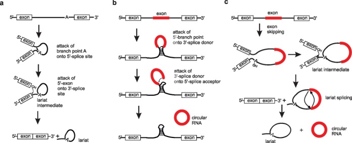
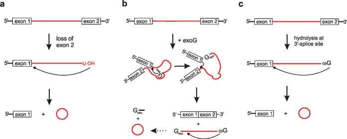
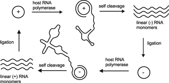
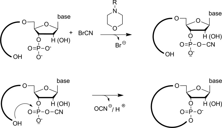
Verification
Claims the paper makes
The claims and statistics of this paper haven’t been checked yet.
Run the check — free, no account needed. It reads the open-access text and checks the sources it cites.
Claims other papers make about this one
Each is checked against this paper’s own text: does it say what the citing paper says it does?
The source backs part of the claim, not all of it — or backs it for a different population, condition or comparison.
In the citing paper
“A ribozymatic method utilizing a permuted group I catalytic intron has been reported to be more applicable to long RNA circularization and requires only the addition of GTP and Mg2+ as cofactors17.”
From Engineering circular RNA for potent and stable translation in eukaryotic cells (2018) · cites this paper as [17]
Experimental — accuracy not yet measuredThe cited source says what the claim attributes to it.
In the citing paper
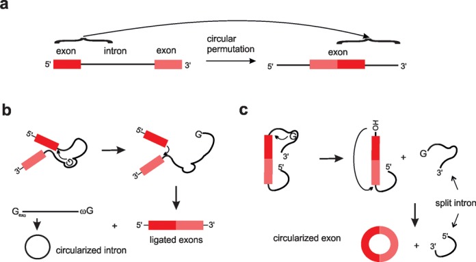
“In vitro, these constructs undergo the double transesterification reactions characteristic of group I catalytic introns, but because the exons are already fused they are excised as covalently 5′ to 3′ linked circles17 (Fig. 1a).”
From Engineering circular RNA for potent and stable translation in eukaryotic cells (2018) · cites this paper as [17]
Self-check
Percentages, sums and reported differences re-computed from the paper’s own numbers.
The self-check needs the paper’s full text.
Citations
Cites
2 retracted
Liu, Y., Cui, H., Wang, W., Li, L., Wang, Z., Yang, S., & Zhang, X. (2013) · The International Journal of Biochemistry & Cell Biology · doi:10.1016/j.biocel.2013.09.003 · Cited by 44
RetractedLiu, Y., Cui, H., Wang, W., Li, L., Wang, Z., Yang, S., & Zhang, X. (2014) · The International Journal of Biochemistry & Cell Biology · doi:10.1016/j.biocel.2014.03.013 · Cited by 42
RetractedCircular RNAs are a large class of animal RNAs with regulatory potency
Memczak, S., Jens, M., Elefsinioti, A., Torti, F., Krueger, J., Rybak, A., Maier, L., Mackowiak, S. D., Gregersen, L. H., Munschauer, M., et al. (2013) · Nature · doi:10.1038/nature11928 · Cited by 7,990 · cited 2×
“Based on the results of a computational approach for identification and analysis of circRNAs, the authors suggest that apart from CDR1as, which indeed appears to function as miR-7 sponge (11,12), a large majority of circRNAs might be just inconsequential side products of pre-mRNA splicing (13).”
BackgroundNatural RNA circles function as efficient microRNA sponges
Hansen, T. B., Jensen, T. I., Clausen, B. H., Bramsen, J. B., Finsen, B., Damgaard, C. K., & Kjems, J. (2013) · Nature · doi:10.1038/nature11993 · Cited by 7,985 · cited 2×
“Based on the results of a computational approach for identification and analysis of circRNAs, the authors suggest that apart from CDR1as, which indeed appears to function as miR-7 sponge (11,12), a large majority of circRNAs might be just inconsequential side products of pre-mRNA splicing (13).”
BackgroundCircular RNAs are abundant, conserved, and associated with ALU repeats
Jeck, W. R., Sorrentino, J. A., Wang, K., Slevin, M. K., Burd, C. E., Liu, J., Marzluff, W. F., & Sharpless, N. E. (2013) · RNA · doi:10.1261/rna.035667.112 · Cited by 4,388 · cited 1×
“The functions of circRNAs in eukaryotes still remain more or less elusive, although it has been suggested that circRNAs may act as transcription regulators (10) or as competing endogenous RNAs to bind miRNAs (RNA sponges) or RNA binding proteins (protein sponges), and thus to modulate their local free concentration (1…”
BackgroundcircRNA biogenesis competes with pre-mRNA splicing
Ashwal-Fluss, R., Meyer, M., Pamudurti, N. R., Ivanov, A., Bartok, O., Hanan, M., Evantal, N., Memczak, S., Rajewsky, N., & Kadener, S. (2014) · Molecular Cell · doi:10.1016/j.molcel.2014.08.019 · Cited by 3,083 · cited 1×
“Furthermore, it was demonstrated that circularization and linear splicing compete against each other, assigning circRNAs a functional role in gene regulation (15,34).”
BackgroundDetecting and characterizing circular RNAs
Jeck, W. R., & Sharpless, N. E. (2014) · Nature Biotechnology · doi:10.1038/nbt.2890 · Cited by 2,710 · cited 2×
“Both mechanisms are plausible in vivo, although direct backsplicing is favored as the more frequently used pathway (25).”
BackgroundSalzman, J., Gawad, C., Wang, P. L., Lacayo, N., & Brown, P. O. (2012) · PLoS ONE · doi:10.1371/journal.pone.0030733 · Cited by 2,239
Complementary Sequence-Mediated Exon Circularization
Zhang, X.-O., Wang, H.-B., Zhang, Y., Lu, X., Chen, L.-L., & Yang, L. (2014) · Cell · doi:10.1016/j.cell.2014.09.001 · Cited by 1,999
Expanded identification and characterization of mammalian circular RNAs
Guo, J. U., Agarwal, V., Guo, H., & Bartel, D. P. (2014) · Genome biology · doi:10.1186/s13059-014-0409-z · Cited by 1,591 · cited 1×
“Based on the results of a computational approach for identification and analysis of circRNAs, the authors suggest that apart from CDR1as, which indeed appears to function as miR-7 sponge (11,12), a large majority of circRNAs might be just inconsequential side products of pre-mRNA splicing (13).”
Background
Cited by
Sorted by citations
Circular RNA: a new star of noncoding RNAs
Qu, S., Yang, X., Li, X., Wang, J., Gao, Y., Shang, R., Sun, W., Dou, K., & Li, H. (2015) · Cancer Letters · doi:10.1016/j.canlet.2015.06.003 · Cited by 1,808
“Furthermore, several researchers have confirmed that reversed complementary sequences including inverted repeated Alu pairs (IRAlus) and exon skipping are essential to circRNA formation [17–25].”
BackgroundEngineering circular RNA for potent and stable translation in eukaryotic cells
Wesselhoeft, R. A., Kowalski, P. S., & Anderson, D. G. (2018) · Nature Communications · doi:10.1038/s41467-018-05096-6 · Cited by 853
Translation and functional roles of circular RNAs in human cancer
Lei, M., Zheng, G., Ning, Q., Zheng, J., & Dong, D. (2020) · Molecular Cancer · doi:10.1186/s12943-020-1135-7 · Cited by 814
Detecting circular RNAs: bioinformatic and experimental challenges
Szabo, L. A., & Salzman, J. (2016) · Nature Reviews Genetics · doi:10.1038/nrg.2016.114 · Cited by 684
CircHIPK3 sponges miR‐558 to suppress heparanase expression in bladder cancer cells
Li, Y., Zheng, F., Xiao, X., Xie, F., Tao, D., Huang, C., Liu, D., Wang, M., Wang, L., Zeng, F., et al. (2017) · EMBO Reports · doi:10.15252/embr.201643581 · Cited by 562
“Two main mechanisms both involving the canonical spliceosome have been proposed for the biogenesis of exonic circRNAs [37,38].”
BackgroundKramer, M. C., Liang, D., Tatomer, D. C., Gold, B., March, Z. M., Cherry, S. R., & Wilusz, J. E. (2015) · Genes & Development · doi:10.1101/gad.270421.115 · Cited by 498
“However, the lack of efficientmethods formodulating circular RNA levels or ectopically expressing circular RNAs (for review, see Petkovic and Muller 2015) has limited our ability to define functions for these transcripts.”
BackgroundRNA Circularization Diminishes Immunogenicity and Can Extend Translation Duration In Vivo
Wesselhoeft, R. A., Kowalski, P. S., Parker-Hale, F. C., Huang, Y., Bisaria, N., & Anderson, D. G. (2019) · Molecular Cell · doi:10.1016/j.molcel.2019.02.015 · Cited by 482
“We turned to an alternative method of circRNA preparation using T4 RNA ligase I and splint oligonucleotides designed to bring the ends of the precursor RNA into proximity for ligation (Figure S3A) (Petkovic & Müller 2015).”
MethodInsights into circular RNA biology
Ebbesen, K. K., Hansen, T. B., & Kjems, J. (2016) · RNA Biology · doi:10.1080/15476286.2016.1271524 · Cited by 458
A 360° view of circular RNAs: From biogenesis to functions
Wilusz, J. E. (2018) · Wiley Interdisciplinary Reviews - RNA · doi:10.1002/wrna.1478 · Cited by 446
“Binding to near-perfect target sites (similar to the miR-671 site in CDR1as) can cause tailing and degradation of microRNAs (Ameres et al., 2010), and thus the observed increase in miR-671 levels is likely directly due to the lack of CDR1as expression in the knockout mice (Piwecka et al., 2017).”
BackgroundInfluentialChen, Y., Yang, F., Fang, E., Xiao, W., Mei, H., Li, H., Li, D., Song, H., Wang, J., Mei, H., et al. (2018) · Cell Death and Differentiation · doi:10.1038/s41418-018-0220-6 · Cited by 322
Circular RNA circSATB2 promotes progression of non-small cell lung cancer cells
Zhang, N., Nan, A., Chen, L., Li, X., Jia, Y., Qiu, M., Dai, X., Zhou, H., Zhu, J., Zhang, H., et al. (2020) · Molecular Cancer · doi:10.1186/s12943-020-01221-6 · Cited by 327
Lessons from non-canonical splicing
Sibley, C. R., Blázquez, L., & Ule, J. (2016) · Nature Reviews Genetics · doi:10.1038/nrg.2016.46 · Cited by 327
Biogenesis and Functions of Circular RNAs Come into Focus
Xiao, M., Ai, Y., & Wilusz, J. E. (2020) · Trends in Cell Biology · doi:10.1016/j.tcb.2019.12.004 · Cited by 316
A Circular RNA Protects Dormant Hematopoietic Stem Cells from DNA Sensor cGAS-Mediated Exhaustion
Xia, P., Wang, S., Ye, B., Du, Y., Li, C., Xiong, Z., Qu, Y., & Fan, Z. (2018) · Immunity · doi:10.1016/j.immuni.2018.03.016 · Cited by 285
Circular RNAs: A Novel Class of Functional RNA Molecules with a Therapeutic Perspective
Santer, L., Bär, C., & Thum, T. (2019) · Molecular Therapy · doi:10.1016/j.ymthe.2019.07.001 · Cited by 279
“Moreover, adding a short oligonucleotide splint (complementary to the circRNA backsplice site) can help to increase intramolecular circularization.(122) Finally, the desired circular product can be purified from site products by PAGE or HPLC.”
BackgroundGlobal circular RNA expression profile of human gastric cancer and its clinical significance
Shao, Y., Li, J., Lu, R., Li, T., Yang, Y., Xiao, B., & Guo, J. (2017) · Cancer Medicine · doi:10.1002/cam4.1055 · Cited by 268
“Circular RNAs (circRNAs) are special class of endogenous noncoding RNAs that are formed by back- splicing events via exon or intron circularization [5].”
BackgroundCircular RNA vaccine in disease prevention and treatment
Niu, D., Wu, Y., & Lian, J. (2023) · Signal Transduction and Targeted Therapy · doi:10.1038/s41392-023-01561-x · Cited by 281
“59,124 Group I intron catalysis of cyclic ribonucleotide ligation is a commonly used PIE cyclization strategy that assembles the pre-tRNA leu and thymidylate synthase (td) genes of the cyanobacteria Anabaena and T4 bacteriophage, respectively, and only requires the addition of GTP and Mg 2 + as…”
MethodIRES-mediated cap-independent translation, a path leading to hidden proteome
Yang, Y., & Wang, Z. (2019) · Journal of Molecular Cell Biology · doi:10.1093/jmcb/mjz091 · Cited by 260
“The translatable circRNAs can be generated by the circularization of in vitro synthesized RNA using chemical or enzymatic ligation, or through a self-splicing Group I intron system (Petkovic and Muller, 2015).”
MethodCircular RNAs as potential biomarkers for cancer diagnosis and therapy.
Wang, F., Nazarali, A. J., & Ji, S. (2016) · PubMed Central · Cited by 254
Circular RNA cancer vaccines drive immunity in hard-to-treat malignancies
Li, H., Peng, K., Yang, K., Ma, W., Qi, S., Yu, X., He, J., Lin, X., & Yu, G. (2022) · Theranostics · doi:10.7150/thno.77350 · Cited by 256
“Different from previous studies which used chemical reaction or T4 ligase to generate circRNA molecule with stringent size restriction [40], we adopted a novel circRNA synthesis method that used a permuted intron-exon (PIE) element to generate circRNA via in vitro back-splicing [31].”
MethodBiogenesis, functions and clinical significance of circRNAs in gastric cancer
Shan, C., Zhang, Y., Hao, X., Gao, J., Chen, X., & Wang, K. (2019) · Molecular Cancer · doi:10.1186/s12943-019-1069-0 · Cited by 232
“ageing in Drosophila [21] and detecting diseases from human body fluid [22, 23].”
BackgroundBest practice standards for circular RNA research
Nielsen, A. F., Bindereif, A., Bozzoni, I., Hanan, M., Hansen, T. B., Irimia, M., Kadener, S., Kristensen, L. S., Legnini, I., Morlando, M., et al. (2022) · Nature Methods · doi:10.1038/s41592-022-01487-2 · Cited by 238
The complexity of the translation ability of circRNAs
Granados-Riveron, J. T., & Aquino‐Jarquín, G. (2016) · Biochimica et Biophysica Acta (BBA) - Gene Regulatory Mechanisms · doi:10.1016/j.bbagrm.2016.07.009 · Cited by 218
Yang, F., Fang, E., Mei, H., Chen, Y., Li, H., Li, D., Song, H., Wang, J., Mei, H., Xiao, W., et al. (2018) · Cancer Research · doi:10.1158/0008-5472.can-18-1559 · Cited by 213
Synthetic circular RNA functions as a miR-21 sponge to suppress gastric carcinoma cell proliferation
Liu, X., Abraham, J. M., Cheng, Y., Wang, Z., Wang, Z., Zhang, G., Ashktorab, H., Smoot, D. T., Cole, R. N., Boronina, T. N., et al. (2018) · Molecular Therapy Nucleic Acids · doi:10.1016/j.omtn.2018.09.010 · Cited by 212
Metazoan tRNA introns generate stable circular RNAs in vivo
Lu, Z., Filonov, G. S., Noto, J. J., Schmidt, C. A., Hatkevich, T., Wen, Y., Jaffrey, S. R., & Matera, A. G. (2015) · RNA · doi:10.1261/rna.052944.115 · Cited by 209
Ma, C., Wang, X., Yang, F., Zang, Y., Liu, J., Wang, X., Xu, X., Li, W., Jia, J., & Liu, Z. (2020) · Molecular Cancer · doi:10.1186/s12943-020-01268-5 · Cited by 208
Regulatory Role of Circular RNAs and Neurological Disorders
Floris, G., Zhang, L., Follesa, P., & Sun, T. (2016) · Molecular Neurobiology · doi:10.1007/s12035-016-0055-4 · Cited by 208
“Currently, the following two mechanisms involved in back-splicing are proposed: Bdirect back-splicing^ and Bexon skipping^ [38].”
BackgroundCircular RNAs: a new frontier for cancer diagnosis and therapy
Zhang, M., & Xin, Y. (2018) · Journal of Hematology & Oncology · doi:10.1186/s13045-018-0569-5 · Cited by 199
“Hence, to further research circRNAs, minigene construct design should be improved and alternative methods should be developed to facilitate circRNA overexpression [71].”
BackgroundLong non‐coding RNA regulation of reproduction and development
Taylor, D. H., Chu, E. T., Spektor, R., & Soloway, P. D. (2015) · Molecular Reproduction and Development · doi:10.1002/mrd.22581 · Cited by 191
“Finally, (6) some lnRNAs are circular in structure (Hansen et al., 2013; Memczak et al., 2013; Zhang et al., 2013; Petkovic and Muller, 2015).”
BackgroundEger, N., Schoppe, L., Schuster, S., Laufs, U., & Boeckel, J. (2018) · Advances in experimental medicine and biology · doi:10.1007/978-981-13-1426-1_4 · Cited by 184
Tracing the expression of circular RNAs in human pre-implantation embryos
Dang, Y., Yan, L., Hu, B., Fan, X., Ren, Y., Li, R., Lian, Y., Yan, J., Li, Q., Zhang, Y., et al. (2016) · Genome biology · doi:10.1186/s13059-016-0991-3 · Cited by 177
The design and synthesis of circular RNAs
Obi, P., & Chen, Y. G. (2021) · Methods · doi:10.1016/j.ymeth.2021.02.020 · Cited by 171
“Beyond RNA secondary structure, the terminal nucleotides to be joined also affect the yield of a T4 RNA ligase 1 reaction, with cytidine and adenosine preferred at the 5′ and 3′ ends, respectively [41,55].”
BackgroundMethodInfluentialFeng, Y., Yang, Y., Zhao, X., Fan, Y., Zhou, L., Rong, J., & Yu, Y. (2019) · Cell Death and Disease · doi:10.1038/s41419-019-2028-9 · Cited by 168
Circular RNAs: Expression, localization, and therapeutic potentials
Yang, Q., Li, F., He, A. T., & Yang, B. B. (2021) · Molecular Therapy · doi:10.1016/j.ymthe.2021.01.018 · Cited by 166
Therapeutic targeting of circ‐CUX1/EWSR1/MAZ axis inhibits glycolysis and neuroblastoma progression
Li, H., Yang, F., Hu, A., Wang, X., Fang, E., Chen, Y., Li, D., Song, H., Wang, J., Guo, Y., et al. (2019) · EMBO Molecular Medicine · doi:10.15252/emmm.201910835 · Cited by 149
“…directly interacts with EWSR1 protein in NB cells To explore the protein partner of circ-CUX1, RNA pull-down was performed using biotin-labeled probes generated by ligation of linear transcript in vitro (Petkovic & Muller, 2015) or synthesized as oligonucleotides targeting junction site (Fig 4A).”
MethodNanomedicine-Based Approaches for mRNA Delivery
Uchida, S., Perche, F., Pichon, C., & Cabral, H. (2020) · Molecular Pharmaceutics · doi:10.1021/acs.molpharmaceut.0c00618 · Cited by 139
Insights into the biogenesis and potential functions of exonic circular RNA
Ragan, C., Goodall, G. J., Shirokikh, N. E., & Preiss, T. (2019) · Scientific Reports · doi:10.1038/s41598-018-37037-0 · Cited by 137
CircRNAs: biogenesis, functions, and role in drug-resistant Tumours
Ma, S., Kong, S., Wang, F., & Ju, S. (2020) · Molecular Cancer · doi:10.1186/s12943-020-01231-4 · Cited by 140
Emerging landscape of circular RNAs in lung cancer
Hu, W., Bi, Z., Chen, Z., Liu, C., Li, L., Zhang, F., Zhou, Q. Y., Zhu, W., Song, Y., Zhan, B.-T., et al. (2018) · Cancer Letters · doi:10.1016/j.canlet.2018.04.006 · Cited by 135
Clinical delivery of circular RNA: Lessons learned from RNA drug development
Young, T. L., Wang, K. C., Varley, A., & Li, B. (2023) · Advanced Drug Delivery Reviews · doi:10.1016/j.addr.2023.114826 · Cited by 130
Circular RNA: A promising new star of vaccine
Xie, J., Ye, F., Deng, X., Tang, Y., Liang, J., Huang, X., Sun, Y., Tang, H., Lei, J., Zheng, S., et al. (2023) · Journal of Translational Internal Medicine · doi:10.2478/jtim-2023-0122 · Cited by 129
“[87] Ribozymatic strategies Ribozymatic strategies are the most commonly used methods for RNA circularization, particularly when the production of large circular RNAs is required.”
BackgroundZhang, Y., Yang, H., Long, Y., & Li, W. (2016) · BJOG An International Journal of Obstetrics & Gynaecology · doi:10.1111/1471-0528.13897 · Cited by 119
The therapeutic potential of circular RNAs
O’Leary, E. T., Jiang, Y., Kristensen, L. S., Hansen, T. B., & Kjems, J. (2025) · Nature Reviews Genetics · doi:10.1038/s41576-024-00806-x · Cited by 131
Circular RNAs: Unexpected outputs of many protein-coding genes
Wilusz, J. E. (2016) · RNA Biology · doi:10.1080/15476286.2016.1227905 · Cited by 116
“Methods, such as splint ligation or the use of selfcleaving ribozymes, can also be used to generate circular RNAs in vitro (For a review see ref.(95)).”
BackgroundNew insight into circRNAs: characterization, strategies, and biomedical applications
Feng, X., Zhu, S.-X., Pu, K., Huang, H., Chen, Y., & Wang, W. (2023) · Experimental Hematology and Oncology · doi:10.1186/s40164-023-00451-w · Cited by 119
“Several methods based on chemical synthesis and enzymatic ligation have been used to generate circRNAs in vitro; however, circRNA production in vivo has only recently been delineated [47, 128, 147, 148].”
MethodIn vitro circularization of RNA
Müller, S., & Appel, B. (2016) · RNA Biology · doi:10.1080/15476286.2016.1239009 · Cited by 113
Self-citationLu, D., Chatterjee, S., Xiao, K., Riedel, I., Huang, C.-K., Costa, A., Cushman, S., Neufeldt, D., Rode, L., Schmidt, A., et al. (2022) · European Heart Journal · doi:10.1093/eurheartj/ehac337 · Cited by 112
Circular RNA Cwc27 contributes to Alzheimer’s disease pathogenesis by repressing Pur-α activity
Song, C., Zhang, Y., Huang, W., Shi, J., Huang, Q., Jiang, M., Qiu, Y. L., Wang, T., Chen, H., & Wang, H. (2021) · Cell Death and Differentiation · doi:10.1038/s41418-021-00865-1 · Cited by 109
Engineering and expressing circular RNAs via tRNA splicing
Noto, J. J., Schmidt, C. A., & Matera, A. G. (2017) · RNA Biology · doi:10.1080/15476286.2017.1317911 · Cited by 108
Hu, X., Ao, J., Li, X., Zhang, H., Wu, J., & Cheng, W. (2018) · Clinical Epigenetics · doi:10.1186/s13148-018-0482-3 · Cited by 103
“This structure helps circRNA to avoid the action of RNA exonuclease, which allows it to maintain stable expression and makes it a more suitable biomarker than other RNA types [19, 20].”
BackgroundUsing plasma circRNA_002453 as a novel biomarker in the diagnosis of lupus nephritis
Ouyang, Q., Huang, Q., Jiang, Z., Zhao, J., Shi, G., & Yang, M. (2018) · Molecular Immunology · doi:10.1016/j.molimm.2018.07.029 · Cited by 97
Guan, Y., Ma, J.-Y., & Song, W. (2019) · Cancer Cell International · doi:10.1186/s12935-019-0905-z · Cited by 99
Circular RNAs: a rising star in respiratory diseases
Wang, J., Zhu, M., Pan, J., Chen, C., Xia, S., & Song, Y. (2019) · Respiratory Research · doi:10.1186/s12931-018-0962-1 · Cited by 94
“So far, two well-known models of EcircRNA formation have been proposed, namely the 'direct back-splicing' model and the 'exon skipping' model [7, [20] [21] [22] .”
MethodCircular RNA Regulation of Myogenesis
Zhang, P., Chao, Z., Zhang, R., Ding, R., Wang, Y., Wu, W., Qiu, H., Li, C., Xu, H., Wang, L., et al. (2019) · Cells · doi:10.3390/cells8080885 · Cited by 89
Deep Insights in Circular RNAs: from biogenesis to therapeutics
Mumtaz, P. T., Taban, Q., Dar, M. A., Mir, S., Haq, Z. U., Zargar, S. M., Shah, R. A., & Ahmad, S. M. (2020) · Biological Procedures Online · doi:10.1186/s12575-020-00122-8 · Cited by 94
RNA chemistry and therapeutics
Wang, S., Weissman, D., & Dong, Y. (2025) · Nature Reviews Drug Discovery · doi:10.1038/s41573-025-01237-x · Cited by 109
“The permuted intron–exon (PIE) splicing approach incorporates partial exons fused together and surrounded by half-intron sequences 173 .”
InfluentialDecreased expression of hsa_circ_0001895 in human gastric cancer and its clinical significances
Shao, Y., Chen, L., Lu, R., Zhang, X., Xiao, B., Ye, G., & Guo, J. (2017) · Tumor Biology · doi:10.1177/1010428317699125 · Cited by 89
A guide to large-scale RNA sample preparation
Baronti, L., Karlsson, H., Marušič, M., & Petzold, K. (2018) · Analytical and Bioanalytical Chemistry · doi:10.1007/s00216-018-0943-8 · Cited by 87
An inducible circular RNA circKcnt2 inhibits ILC3 activation to facilitate colitis resolution
Liu, B., Ye, B., Zhu, X., Yang, L., Li, H., Liu, N., Zhu, P., Lu, T., He, L., Tian, Y., et al. (2020) · Nature Communications · doi:10.1038/s41467-020-17944-5 · Cited by 84
Best practices to ensure robust investigation of circular RNAs: pitfalls and tips
Dodbele, S., Mutlu, N., & Wilusz, J. E. (2021) · EMBO Reports · doi:10.15252/embr.202052072 · Cited by 85
“A number of approaches have been described, including ones based on ribozymes (Ford & Ares, 1994; Wesselhoeft et al , 2018; Litke & Jaffrey, 2019), splint ligation (Moore, 1999), and chemical methods (Petkovic & Muller, 2015; M € uller & Appel, 2017).”
Synthetic circular RNA switches and circuits that control protein expression in mammalian cells
Kameda, S., Ohno, H., & Saito, H. (2023) · Nucleic Acids Research · doi:10.1093/nar/gkac1252 · Cited by 88
“Several strategies for in vitro mRNA cyclization have been reported (39).”
BackgroundLiang, X., Li, L., Tang, J., Komiyama, M., & Ariga, K. (2020) · Bulletin of the Chemical Society of Japan · doi:10.1246/bcsj.20200012 · Cited by 79
mRNA vaccines: the most recent clinical applications of synthetic mRNA
Kwon, S., Kwon, M., Im, S., Lee, K., & Lee, H. (2022) · Archives of Pharmacal Research · doi:10.1007/s12272-022-01381-7 · Cited by 85
“In the permuted group 1 catalytic intron-based system, the exon sequence is flanked by split introns, causing a double transesterification reaction in the presence of GTP and Mg2+ cofactors (Petkovic and Müller 2015).”
BackgroundMethodCircular RNA: Biosynthesis in vitro
Chen, X., & Lu, Y. (2021) · Frontiers in Bioengineering and Biotechnology · doi:10.3389/fbioe.2021.787881 · Cited by 74
“In this way, as little as 6 to 8 nucleotides of circRNAs can be synthesized (Kaufmann et al., 1974; Petkovic and Müller, 2015).”
BackgroundMethodInfluentialCui, Y., Wu, X., Jin, J., Man, W., Li, J., Li, X., Li, Y., He, Y., Liu, M., Zhong, R., et al. (2023) · Molecular Cancer · doi:10.1186/s12943-023-01888-7 · Cited by 76
“In addition, circRNAs can interact with RNA binding proteins and participate in the development of different cancers [8, 13, 18–20].”
BackgroundModified nucleic acid aptamers: development, characterization, and biological applications
Ji, D., Feng, H., Liew, S. W., & Kwok, C. K. (2023) · Trends in biotechnology · doi:10.1016/j.tibtech.2023.05.005 · Cited by 75
Noncoding RNAs: New Players in Cancers
Chen, X., Fan, S., & Song, E. (2016) · Advances in experimental medicine and biology · doi:10.1007/978-981-10-1498-7_1 · Cited by 73
CircRNA—Protein Interactions in Muscle Development and Diseases
Zheng, S., Zhang, X., Odame, E., Xu, X., Chen, Y., Ye, J., Zhou, H., Dai, D., Kyei, B., Zhan, S., et al. (2021) · International Journal of Molecular Sciences · doi:10.3390/ijms22063262 · Cited by 72
“The differences are that co-IP systems were launched depending on a biotin-labeled in vitro synthesized circRNA instead of a probe [102-104].”
Wu, S., Liu, H., Huang, P.-J., Chang, I. Y., Lee, C., Yang, C.-Y., Tsai, W., & Tan, B. C. (2017) · GigaScience · doi:10.1093/gigascience/gix118 · Cited by 72
“Circular RNAs are a more recently discovered class of noncoding RNAs that are defined not by length but rather the unique structure of covalently closed circularity [5, 6].”
The understanding of circular RNAs as special triggers in carcinogenesis
Xin, Z., Ma, Q., Ren, S., Wang, G., & Fan, L. (2016) · Briefings in Functional Genomics · doi:10.1093/bfgp/elw001 · Cited by 69
Circular RNAs: A Novel Player in Development and Disease of the Central Nervous System
Xie, L., Mao, M., Xiong, K., & Jiang, B. (2017) · Frontiers in Cellular Neuroscience · doi:10.3389/fncel.2017.00354 · Cited by 70
“Although the exact details on how the splicesome carry out exon circularization is not known, the canonical splice signals and canonical spliceosomal machinery are important in the processing of circRNAs biogenesis (Chen and Yang, 2015; Petkovic and Müller, 2015).”
BackgroundFeng, Z., Zhang, X., Zhou, J., Li, Q., Chu, L., Di, G., Xu, Z., Chen, Q., Wang, M., Jiang, X., et al. (2023) · Nature Cancer · doi:10.1038/s43018-023-00650-8 · Cited by 70
“We used the permuted intron-exon (PIE) splicing strategy to construct circRNAs 36 .”
Wu, Q., Wang, H., Liu, L., Zhu, K., Yu, W., & Guo, J. (2019) · Biochemical and Biophysical Research Communications · doi:10.1016/j.bbrc.2019.10.117 · Cited by 67
Novel Molecular Markers in Glioblastoma—Benefits of Liquid Biopsy
Birkó, Z., Nagy, B., Klekner, Á., & Virga, J. (2020) · International Journal of Molecular Sciences · doi:10.3390/ijms21207522 · Cited by 67
“circRNAs can be involved in cancer development as oncogenes or tumor suppressors on the base of several different mechanisms: acting as miRNA sponges [63], regulating gene expression [64], interacting with RNA-binding proteins (RBPs), or to be involved in RNA and protein transfer and storage [65].”
BackgroundRole of Circular RNAs in Preeclampsia
Jia, N., & Li, J. (2019) · Disease Markers · doi:10.1155/2019/7237495 · Cited by 65
“The structure of circRNAs helps them escape degradation, thereby allowing for stable expression and adding to their potential as suitable biomarkers, compared with other ncRNAs [58, 59].”
BackgroundSharma, A. R., Bhattacharya, M., Bhakta, S., Saha, A., Lee, S., & Chakraborty, C. (2021) · Molecular Therapy — Nucleic Acids · doi:10.1016/j.omtn.2021.05.022 · Cited by 66
Template-jumping prime editing enables large insertion and exon rewriting in vivo
Zheng, C., Liu, B., Dong, X., Gaston, N., Sontheimer, E. J., & Xue, W. (2023) · Nature Communications · doi:10.1038/s41467-023-39137-6 · Cited by 70
Lee, K. H., Kim, S., Song, J., Han, S. R., Kim, J. H., & Lee, S. (2023) · Molecular Therapy — Nucleic Acids · doi:10.1016/j.omtn.2023.07.034 · Cited by 64
Circular RNA‐based neoantigen vaccine for hepatocellular carcinoma immunotherapy
Wang, F., Guang, C., Wang, Y., Zhuang, Q., Cai, Z., Li, Y., Gao, S., Li, F., Zhang, C., Zhao, B., et al. (2024) · MedComm · doi:10.1002/mco2.667 · Cited by 64
“18 In our study, the PIE strategy was used to generate circRNAs, an approach that allowed us to generate circRNAs of sufficient length to insert efficient IRES elements and antigen coding sequences.”
MethodCircRNAs: A new perspective of biomarkers in the nervous system
Ma, Y., Liu, Y., & Jiang, Z. (2020) · Biomedicine & Pharmacotherapy · doi:10.1016/j.biopha.2020.110251 · Cited by 61
Zhou, W., Jiang, L., Liao, S., Wu, F., Yang, G., Hou, L., Liu, L., Pan, X., Jia, W. W.-G., & Zhang, Y. (2023) · Viruses · doi:10.3390/v15081760 · Cited by 64
“(3) T4 RNA ligase 2, also used successfully for RNA cyclization, is compatible with both single-stranded and double-stranded RNA ligation [37].”
BackgroundHe, L., Zhang, A., Xiong, L., Li, Y., Huang, R., Liao, L., Zhu, Z., & Wang, A. Y. (2017) · International Journal of Molecular Sciences · doi:10.3390/ijms18091977 · Cited by 59
“A number of studies have led to circRNAs being divided into four types: circular exonic RNAs (ecircRNAs), circular intronic RNAs (ciRNAs), exon-intron circRNAs (eiciRNAs), and intergenic circRNAs [2,10,11].”
BackgroundEnzyme-free ligation of dimers and trimers to RNA primers
Sosson, M., Pfeffer, D., & Richert, C. (2019) · Nucleic Acids Research · doi:10.1093/nar/gkz160 · Cited by 60
“Further, a methodology for ligating RNA strands would also be useful for practical applications, including the synthesis of circular RNA (39), labeled siRNA (40), modified mRNA (41) and functional nanostructures (42,43).”
Methods for analysis of circular RNAs
Pandey, P. R., Munk, R. B., Kundu, G., De, S., Abdelmohsen, K., & Gorospe, M. (2019) · Wiley Interdisciplinary Reviews - RNA · doi:10.1002/wrna.1566 · Cited by 61
“They can be synthesized chemically using cyanogen bromide or enzymatically using T4 RNA ligases as well as by using an RNA cyclase ribozyme (L. L. Chen & Yang, 2015; Petkovic & Muller, 2015).”
MethodSystematic characterization of non‐coding RNAs in triple‐negative breast cancer
Mei, J., Hao, L., Wang, H., Xu, R., Liu, Y., Zhu, Y., & Liu, C. (2020) · Cell Proliferation · doi:10.1111/cpr.12801 · Cited by 57
circROBO1 promotes prostate cancer growth and enzalutamide resistance via accelerating glycolysis
Zhou, Z., Qin, J., Song, C., Wu, T., Quan, Q., Zhang, Y., Zou, Y., Liu, L., Tang, H., & Zhao, J. (2023) · Journal of Cancer · doi:10.7150/jca.86940 · Cited by 62
“Additionally, circRNAs are highly conserved and tissue-specific, and their expression levels are regulated in a cell-and developmental stage-specific manner [14].”
BackgroundCircRNA as an Achilles heel of cancer: characterization, biomarker and therapeutic modalities
Zhang, J., Luo, Z., Yang, Z. K., Duan, M., Qiu, Z., & Huang, C. (2024) · Journal of Translational Medicine · doi:10.1186/s12967-024-05562-4 · Cited by 58
“The enzymatic strategy for RNA circularization utilizes various nucleotide ligases to form phosphodiester bonds at both ends of linear RNA, thereby achieve cyclization [126, 127].”
BackgroundA flexible, efficient, and scalable platform to produce circular RNAs as new therapeuticsPreprint
Wei, H. Y., Chen, C., Zhang, K., Li, Z., Wei, T., Tang, C., Yang, Y., & Wang, Z. (2022) · bioRxiv (Cold Spring Harbor Laboratory) · doi:10.1101/2022.05.31.494115 · Cited by 60
Chen, H., Cheng, K., Liu, X., An, R., Komiyama, M., & Liang, X. (2020) · Nucleic Acids Research · doi:10.1093/nar/gkaa181 · Cited by 58
Sun, Y., Chen, W., Liu, J., Li, J., Zhang, Y., Cai, W., Liu, L., Tang, X., Hou, J., Wang, M., et al. (2022) · Angewandte Chemie International Edition · doi:10.1002/anie.202212413 · Cited by 55
Jiang, F., & Shen, X. (2019) · Cellular & Molecular Biology Letters · doi:10.1186/s11658-019-0178-5 · Cited by 54
“CircRNAs are a special class of endogenous noncoding RNAs usually resulting from splicing events or back-splicing events via exon or intron circularization in vitro and vivo experiments [5].”
BackgroundRegulation of microRNAs function by circular RNAs in human cancer
Han, C., Seebacher, N. A., Hornicek, F. J., Kan, Q., & Duan, Z. (2017) · Oncotarget · doi:10.18632/oncotarget.19930 · Cited by 55
“More recent studies have further verified the model of intron-pairing-driven circularization [17, 33, 41, 42].”
Circular L-RNA aptamer promotes target recognition and controls gene activity
Ji, D., Lyu, K., Zhao, H., & Kwok, C. K. (2021) · Nucleic Acids Research · doi:10.1093/nar/gkab593 · Cited by 56
“Studies on cyclization of natural nucleic acid have revealed that circular structures possess better thermal stability and increased conformational stability owing to the lack of free ends and reduction in misfolding (35–37).”
The emerging roles of exosomal circRNAs in diseases
Guo, X., Tan, W., & Wang, C. (2020) · Clinical & Translational Oncology · doi:10.1007/s12094-020-02485-6 · Cited by 51
Therapeutic mRNA Engineering from Head to Tail
Jia, L., & Qian, S. (2021) · Accounts of Chemical Research · doi:10.1021/acs.accounts.1c00541 · Cited by 51
Macrocyclic polymers: Synthesis, purification, properties and applications
Ochs, J., Pagnacco, C. A., & Barroso‐Bujans, F. (2022) · Progress in Polymer Science · doi:10.1016/j.progpolymsci.2022.101606 · Cited by 50
Efficient circular RNA synthesis for potent rolling circle translation
Du, Y., Zuber, P. K., Xiao, H., Li, X., Gordiyenko, Y., & Ramakrishnan, V. (2024) · Nature Biomedical Engineering · doi:10.1038/s41551-024-01306-3 · Cited by 51
“CircRNAs can be synthesised in vitro by either chemical or enzymatic ligation or the group I intron-based permuted intron–exon (PIE) method 12 .”
Circular RNAs: Functions and Prospects in Glioma
Zheng, H., Hu, S., Liu, Z., Wei-xin, S., Zhao, Y., & Li, M. (2018) · Journal of Molecular Neuroscience · doi:10.1007/s12031-018-1211-2 · Cited by 49
Wang, G., Sun, D., Li, W., & Yan, X. (2021) · Cancer Cell International · doi:10.1186/s12935-021-01964-2 · Cited by 48
Metadata
- Authors
- Petkovic, S., & Müller, S.
- Affiliations
- Universität Greifswald (DE)
- Corresponding author
- Müller, Sabine
- Venue
- Nucleic Acids Research
- Publisher
- Oxford University Press (OUP)
- Published
- 2015-02-06
- Volume
- 43
- Issue
- 4
- Pages
- 2454-2465
- ISSN
- 0305-1048 (ISSN-L)
- 1362-4962
- PMC
- PMC4344496
- OpenAlex
- W2121626677
- Cited by
- 386
- References
- 128
- Citations by year
- 2026: 21 · 2025: 42 · 2024: 51 · 2023: 39 · 2022: 40 · 2021: 51 · 2020: 46 · 2019: 31 · 2018: 26 · 2017: 18 · 2016: 15 · 2015: 5
- FWCI
- 2.07
- Type
- journal article
- Language
- en
- Open access
- gold
- doi.org
- Locations
- Nucleic Acids Research (publishedVersion, cc-by)
- Europe PMC (PubMed Central) (publishedVersion, cc-by)
- PubMed Central (publishedVersion, cc-by)
- PubMed (publishedVersion)
- Funders
- University of Oxford
- Topics
- Life Sciences › Biochemistry, Genetics and Molecular Biology › Molecular Biology › Circular RNAs in diseases
- Life Sciences › Biochemistry, Genetics and Molecular Biology › Molecular Biology › RNA Research and Splicing
- Life Sciences › Biochemistry, Genetics and Molecular Biology › Molecular Biology › RNA and protein synthesis mechanisms
- Keywords
- circular RNA, RNA circularization, aptamers, siRNA, tRNA maturation, RNA biogenesis
- MeSH terms
- RNA, Circular, Exons, Introns, RNA Ligase (ATP), RNA, RNA, Viral, RNA, Archaeal
About these checks
Registry, retraction, Cites and statistics results are computed, not judged, with one exception: a printed reference that no index lists is read once by a model before it counts as a citation error. Claims are extracted by a model and kept only when their quote appears verbatim in the text; each cited source is then checked, with a model’s help, for whether it says what the claim relies on. Those verdicts can be wrong — read the claim’s quote and the cited source before relying on one.
Statistics and arithmetic are recomputed, not judged.
Confidence bands are shown only for fields calibrated on our annotated test set. None is calibrated yet, so no band is shown.
What counts as a claim ›How we measure confidence ›How we check this ›
Are you an author, or do you see a problem with this page? Request a review
