

The DOI matches its Crossref record; 37 of the 37 references it cites were found in a registry or an index.
Pathogenic T-cells and inflammatory monocytes incite inflammatory storms in severe COVID-19 patients
Open accessPublished version of a preprint
Abstract
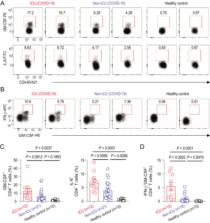
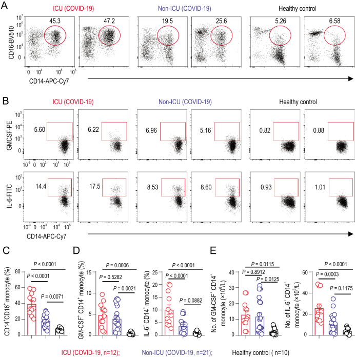
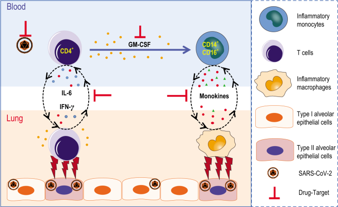
Pathogenic human coronavirus infections, such as severe acute respiratory syndrome CoV (SARS-CoV) and Middle East respiratory syndrome CoV (MERS-CoV), cause high morbidity and mortality [1,2]. Recently, a severe pneumonia-associated respiratory syndrome caused by a new coronavirus (SARS-CoV-2) was reported in December 2019 in the city of Wuhan, Hubei province, China [3–5], which was also named as pneumonia-associated respiratory syndrome (PARS) [6] and can cause coronavirus disease 2019 (COVID-19) to seriously endanger human health. Up to 24 February 2020, at least 77 779 cases had been reported, with 2666 fatal cases according to the report from China CDC. However, the immune mechanism that potentially orchestrates acute mortality from COVID-19 patients is still unknown. Here, we show that, after the SARS-CoV-2 infection, CD4+ T lymphocytes are rapidly activated to become pathogenic T helper (Th) 1 cells and generate GM-CSF, etc. The cytokine environment induces inflammatory CD14+CD16+ monocytes with a high expression of IL-6 and accelerates the inflammation. Given that a large number of inflammatory-cell infiltrations have been observed in lungs from severe COVID-19 patients [7,8], these aberrant pathogenic Th1 cells and inflammatory monocytes may enter the pulmonary circulation in huge numbers and play an immune-damaging role causing lung functional disability and quick mortality. Our results demonstrate that excessive non-effective host-immune responses by pathogenic T-cells and inflammatory monocytes may be associated with severe lung pathology. Thus, we suggest that monoclonal antibodies targeting GM-CSF or interleukin 6 may be effective in blocking inflammatory storms and, therefore, be a promising treatment for severe COVID-19 patients. Coronavirus, including SARS and MERS, has caused two large-scale pandemics in the last two decades [1,2]. Although viral evasion of host-immune responses and virus-induced cytopathic effects are believed to be critical in disease severity, studies from humans who died of SARS and animal models have suggested that an excessive and aberrant host-cytokine storm results in an exuberant immunopathology and lethal disease [9–11]. Inflammatory cytokine storm refers to the immune system gone awry and an excessive inflammatory response flaring out of control. Cytokine storms are associated with a wide variety of infectious and non-infectious diseases including graft-versus-host disease, autoimmune disease, severe virus infection, multiple organ dysfunction syndromes and chimeric antigen receptor (CAR) T-cell therapy [12,13]. It has been reported that, following SARS-CoV infection, dysregulated cytokine/chemokine responses and higher virus titers cause an inflammatory cytokine storm with lung immunopathological injury [12,14]. Such inflammation associated with the cytokine storm may begin at one local site but spread farther throughout the body via the systemic circulation [12,14]. Similarly, patients infected with SARS-CoV-2 who have been reported recently have had increased plasma concentrations of inflammation-related cytokines, including interleukins (IL) 2, 7 and 10, granulocyte-colony-stimulating factor (G-CSF), interferon-γ-inducible protein 10 (IP10), monocyte chemoattractant protein 1 (MCP1), macrophage inflammatory protein 1 alpha (MIP1A) and tumour necrosis factor α (TNF-α), especially in moribund patients [15]. Importantly, COVID-19 patients have developed characteristic pulmonary ground glass changes on imaging and peripheral lymphocytes decreasing [14,16,17]. More importantly, a large number of inflammatory immune-cell infiltrations were also found in a COVID-19 patient with pulmonary pathology [7,8]. These phenomena suggest that severe pulmonary inflammation and cytokine storm also exist in SARS-CoV-2 infection. At present, symptomatic treatments with organ support to moribund patients are the mainstays of clinical management [17]. It is urgent to identify the immunopathology mechanism to delay the pulmonary immune injury. In patients infected with SARS-CoV, it has been reported that the severity of pulmonary immune injury correlated with extensive infiltration of neutrophils and macrophages in the lungs [18,19], accompanied by increased numbers of neutrophils and monocytes and lower CD8+ and CD4+ T-cell counts in the peripheral blood samples [20–22]. To identify the immune characteristics of patients infected with SARS-CoV-2, peripheral blood samples from patients with severe pneumonia were collected for immune analysis. Consistently with previous clinical-characteristics reports [23], these hospitalized patients with confirmed SARS-CoV-2 infection involved in the First Affiliated Hospital of University of Science and Technology of China commonly have fever symptoms. Patients in intensive care units (ICUs) have significantly decreased concentrations of haemoglobin and albumin, but increased concentrations of C-reactive protein, alanine aminotransferase, asparta
Overview
The claims and statistics of this paper haven’t been checked yet.
Run the check — free, no account needed. It reads the open-access text and checks the sources it cites.
Integrity signals
Registry
Cites
Statistics
Figures
Verification
Claims the paper makes
The claims and statistics of this paper haven’t been checked yet.
Run the check — free, no account needed. It reads the open-access text and checks the sources it cites.
Claims other papers make about this one
Each is checked against this paper’s own text: does it say what the citing paper says it does?
The check wasn’t confident either way, so it gives no verdict — never disputed.
In the citing paper
“HLA-DR expression on monocytes from severe COVID-19 patients is also reduced relative to those from mild patients109,110,112,122.”
From SARS-CoV-2, COVID-19 and the aging immune system (2021) · cites this paper as [112]
Experimental — accuracy not yet measuredIn the citing paper
“SARS-CoV-2 infection elicits a Th1 CD4+ T cell response, with IFNγ and IL-2 being the primary cytokines produced by these cells and some TNFα production as well112,126-129.”
From SARS-CoV-2, COVID-19 and the aging immune system (2021) · cites this paper as [112]
Self-check
Percentages, sums and reported differences re-computed from the paper’s own numbers.
The self-check needs the paper’s full text.
Citations
Cites
Clinical features of patients infected with 2019 novel coronavirus in Wuhan, China
Huang, C., Wang, Y., Li, X., Ren, L., Zhao, J., Hu, Y., Zhang, L., Fan, G., Xu, J., Gu, X., et al. (2020) · The Lancet · doi:10.1016/s0140-6736(20)30183-5 · Cited by 48,884
Clinical characteristics of coronavirus disease 2019 in China
Guan, W.-J., Ni, Z.-Y., Hu, Y., Liang, W.-H., Ou, C.-Q., He, J.-X., Liu, L., Shan, H., Lei, C.-L., Hui, D. S. C., et al. (2020) · New England Journal of Medicine · doi:10.1056/nejmoa2002032 · Cited by 30,246
A pneumonia outbreak associated with a new coronavirus of probable bat origin
Zhou, P., Yang, X.-L., Wang, X.-G., Hu, B., Zhang, L., Zhang, W., Si, H.-R., Zhu, Y., Li, B., Huang, C.-L., et al. (2020) · Nature · doi:10.1038/s41586-020-2012-7 · Cited by 21,969
Self-citationWang, D., Hu, B., Hu, C., Zhu, F., Liu, X., Zhang, J., Wang, B., Xiang, H., Cheng, Z., Xiong, Y., et al. (2020) · JAMA · doi:10.1001/jama.2020.1585 · Cited by 20,148
Self-citationLa certeza de lo impredecible: Cultura Educación y Sociedad en tiempos de COVID19
Martínez González, M. (2020) · DSPACE System - Metalibrary (University of the Coast) · Cited by 18,463
Martínez–González, M. B. (2020) · DSPACE System - Metalibrary (University of the Coast) · Cited by 18,292
A new coronavirus associated with human respiratory disease in China
Wu, F., Zhao, S., Yu, B., Chen, Y.-M., Wang, W., Song, Z.-G., Hu, Y., Tao, Z.-W., Tian, J.-H., Pei, Y.-Y., et al. (2020) · Nature · doi:10.1038/s41586-020-2008-3 · Cited by 11,688
Pathological findings of COVID-19 associated with acute respiratory distress syndrome
Xu, Z., Shi, L., Wang, Y., Zhang, J., Huang, L., Zhang, C., Liu, S., Zhao, P., Liu, H., Zhu, L., et al. (2020) · The Lancet Respiratory Medicine · doi:10.1016/s2213-2600(20)30076-x · Cited by 8,545
A novel coronavirus outbreak of global health concern
Wang, C., Horby, P. W., Hayden, F. G., & Gao, G. F. (2020) · The Lancet · doi:10.1016/s0140-6736(20)30185-9 · Cited by 7,700
Identification of a novel coronavirus in patients with severe acute respiratory syndrome
Drosten, C., Günther, S., Preiser, W., van der Werf, S., Brodt, H.-R., Becker, S., Rabenau, H., Panning, M., Kolesnikova, L., Fouchier, R. A. M., et al. (2003) · New England Journal of Medicine · doi:10.1056/nejmoa030747 · Cited by 4,574
Research works printed but not linked: 3
- Tian, 2020, J Thorac Oncol
- Li, 2020, J Med Virol, 92
- Wang, 2020, JAMA, 323
Cited by
Sorted by citations
Items 801–900 of 1,000
How can Biology of Aging Explain the Severity of COVID-19 in Older Adults
Gallo, A., Pero, E., Pellegrino, S., Macerola, N., Murace, C. A., Ibba, F., Agnitelli, M. C., Landi, F., & Montalto, M. (2022) · Clinics in Geriatric Medicine · doi:10.1016/j.cger.2022.04.002 · Cited by 9
Durkee-Shock, J. R., Lazarski, C. A., Jensen-Wachspress, M. A., Zhang, A., Son, A., Kankate, V. V., Field, N. E., Webber, K., Lang, H., Conway, S. R., et al. (2022) · Molecular Therapy — Methods & Clinical Development · doi:10.1016/j.omtm.2022.04.013 · Cited by 9
Choto, T. A., Makupe, I., Cakana, A., Sibanda, E., & Mduluza, T. (2022) · PLoS ONE · doi:10.1371/journal.pone.0273186 · Cited by 7
Chatterjee, A. N., Basir, F. A., Biswas, D., & Abraha, T. (2022) · Vaccines · doi:10.3390/vaccines10111846 · Cited by 9
Some insights of novel COVID 19 virus: structure, pathogenicity and immunity aspects
Al-AAlim, A. M., Hamad, M. A., & AL-ledani, A. A. (2020) · Al-mağallaẗ al-ʻirāqiyyaẗ li-l-ʻulūm al-bayṭariyyaẗ/Iraqi journal of veterinary sciences · doi:10.33899/ijvs.2020.126898.1408 · Cited by 9
Immunotherapies and COVID-19 related Neurological manifestations: A Comprehensive Review Article
Anwar, M. M. (2020) · Journal of Immunoassay and Immunochemistry · doi:10.1080/15321819.2020.1865400 · Cited by 9
Persson, J., Andersson, B., van Veen, S., Haks, M. C., Obudulu, O., Torkzadeh, S., Ottenhoff, T. H. M., Kanberg, N., Gisslén, M., Andersson, L., et al. (2022) · Molecular Immunology · doi:10.1016/j.molimm.2022.03.004 · Cited by 9
Vasculopathy in patients with severe COVID-19 infection
Beketova, T. V., & Nasonov, E. (2020) · Clinical Medicine (Russian Journal) · doi:10.30629/0023-2149-2020-98-5-325-333 · Cited by 8
Road Map to Understanding SARS-CoV-2 Clinico-Immunopathology and COVID-19 Disease Severity
Karmakar, D., Lahiri, B., Ranjan, P., Chatterjee, J., Lahiri, P., & Sengupta, S. (2020) · Pathogens · doi:10.3390/pathogens10010005 · Cited by 8
Relationship between Vitamin D and Immunity in Older People with COVID-19
Lauretani, F., Salvi, M., Zucchini, I., Testa, C., Cattabiani, C., Arisi, A., & Maggio, M. G. (2023) · International Journal of Environmental Research and Public Health · doi:10.3390/ijerph20085432 · Cited by 8
Dewi, I. P., Dachriyanus, D., Aldi, Y., Ismail, N. H., Hefni, D., Susanti, M., Putra, P. P., & Wahyuni, F. S. (2023) · Journal of Ethnopharmacology · doi:10.1016/j.jep.2023.117381 · Cited by 9
Kulkovienė, G., Narauskaitė, D., Tunaitytė, A., Volkevičiūtė, A., Balion, Z., Kutakh, O., Gečys, D., Kairytė, M., Uldukytė, M., Stankevıčıus, E., et al. (2024) · International Journal of Molecular Sciences · doi:10.3390/ijms25063188 · Cited by 8
Monocyte Production of C1q Potentiates CD8+ T-Cell Function Following Respiratory Viral Infection
Eddens, T. J., Parks, O. B., Lou, D., Fan, L., Sojati, J., Ramsey, M. J., Schmitt, L. A., Salgado, C. M., Reyes‐Múgica, M., Evans, A. N., et al. (2024) · American Journal of Respiratory Cell and Molecular Biology · doi:10.1165/rcmb.2024-0004oc · Cited by 8
Chakraborty, C., Bhattacharya, M., Das, A., & Saha, A. (2025) · Reviews in Medical Virology · doi:10.1002/rmv.70026 · Cited by 8
An immunopathogenic perspective of interleukin-1 signaling
Lin, X., Twelkmeyer, T., Wang, S., Xu, R., Wang, F., Zhang, C., & Tang, H. (2020) · Cellular and Molecular Immunology · doi:10.1038/s41423-020-0475-y · Cited by 8
Chaudhari, S., Pereira, S. D., Asare-Werehene, M., Naha, R., Kabekkodu, S. P., Tsang, B. K., & Satyamoorthy, K. (2021) · Journal of Ovarian Research · doi:10.1186/s13048-021-00787-z · Cited by 8
Overview of the Haematological Effects of COVID-19 Infection
Wiggill, T. M., Mayne, E. S., Vaughan, J., & Louw, S. J. (2021) · Advances in experimental medicine and biology · doi:10.1007/978-3-030-59261-5_14 · Cited by 8
EFFECT OF SARS-COV-2 ON PREGNANCY AND FETUS (LITERATURE REVIEW)
Shkolnyk, O. S., Шлемкевич, А., Маланчук, О., Sharhorodska, Y., & Akopyan, H. (2021) · Актуальні проблеми сучасної медицини Вісник Української медичної стоматологічної академії · doi:10.31718/2077-1096.21.2.208 · Cited by 8
Zaffina, S., Lanteri, P., Gilardi, F., Garbarino, S., Santoro, A., Vinci, M. R., Carsetti, R., Scorpecci, A., Raponi, M., Magnavita, N., et al. (2021) · Frontiers in Human Neuroscience · doi:10.3389/fnhum.2021.666468 · Cited by 8
Yi, J., Miao, J., Zuo, Q., Owusu, F., Dong, Q., Lin, P., Wang, Q., Gao, R., Kong, X., & Yang, L. (2022) · Frontiers in Immunology · doi:10.3389/fimmu.2022.978619 · Cited by 9
Is SARS-CoV-2 a Risk Factor of Bipolar Disorder?—A Narrative Review
Lorkiewicz, P., & Waszkiewicz, N. (2022) · Journal of Clinical Medicine · doi:10.3390/jcm11206060 · Cited by 8
Functionality of immune cells in COVID-19 infection: development of cell-based therapeutics
Sayahinouri, M., Firouz, S. M., Sadrabadi, A. E., Masoudnia, M., Abdolahi, M., Jafarzadeh, F., Nouripour, M., Mirzazadeh, S., Zangeneh, N., Jalili, A., et al. (2023) · Bioimpacts · doi:10.34172/bi.2023.23839 · Cited by 8
Kumari, D., Singh, S., Kumari, M., Gupta, H., Chauhan, D., Singh, K., Eslavath, M. R., Bhushan, B., Dogra, V., Bargotya, M., et al. (2023) · Immunobiology · doi:10.1016/j.imbio.2023.152392 · Cited by 8
Fan, J., Lü, C., Zhang, Q., Sun, G., & Sun, W. (2023) · Microchemical Journal · doi:10.1016/j.microc.2023.109372 · Cited by 8
Fisiopatología del daño multiorgánico en la infección por SARS-Cov2
Pérez, G. T. L., Sandoval, M. D. L. P. R., & Altamirano, M. S. T. (2020) · Acta Pediátrica de México · doi:10.18233/apm41no4s1pps27-s412042 · Cited by 8
Characteristics and Developments in Mesenchymal Stem Cell Therapy for COVID-19: An Update
Sang, L., Guo, X., Shi, J., Hou, S., Fan, H., & Lv, Q. (2021) · Stem Cells International · doi:10.1155/2021/5593584 · Cited by 8
Vianello, A., Turco, S. D., Babboni, S., Silvestrini, B., Ragusa, R., Caselli, C., Melani, L., Fanucci, L., & Basta, G. (2021) · Biomedicines · doi:10.3390/biomedicines9070710 · Cited by 8
Two-Step In Vitro Model to Evaluate the Cellular Immune Response to SARS-CoV-2
Melgaço, J. G., Azamor, T., da Silva, A. M. V., Linhares, J., Santos, T. P., Mendes, Y. S., de Lima, S. M. B., Fernandes, C. B., da Silva, J., de Souza, A. P., et al. (2021) · Cells · doi:10.3390/cells10092206 · Cited by 8
Noll, J., Reichert, M., Dietrich, M., Riedel, J. G., Hecker, M., Padberg, W. M., Weigand, M. A., & Hecker, A. (2022) · Langenbeck s Archives of Surgery · doi:10.1007/s00423-022-02495-8 · Cited by 8
Biologics in COVID-19 So Far: Systematic Review
Arias, M., Oliveros, H., Lechtig, S., & Bustos, R. (2022) · Pharmaceuticals · doi:10.3390/ph15070783 · Cited by 9
Kojicone C, new anti-inflammatory polyketide from insect-associated fungus Aspergillus versicolor
Li, T., Zhang, C., Shi, F., Ye, Y., Huang, J.-L., Wu, L., & Xu, C. (2022) · Phytochemistry Letters · doi:10.1016/j.phytol.2022.11.005 · Cited by 7
Zhou, J.-R., Liao, Y., Cao, L., Ma, R., He, Y., Li, N., Zhu, D., Zhao, X., Huang, X., & Sun, Y. (2025) · The Journal of Infectious Diseases · doi:10.1093/infdis/jiaf041 · Cited by 7
COVID-19-Omics Report: From Individual Omics Approaches to Precision Medicine
Louis, I. V.-S., Fang, D., Amer, Y., & Mohei, H. (2023) · Reports — Medical Cases Images and Videos · doi:10.3390/reports6040045 · Cited by 8
Szabo, I., Badii, M. O., Gaal, O. I., Szabó, R., Popp, R. A., Joosten, L. A. B., Crișan, T. O., & Rednic, S. (2023) · International Journal of Molecular Sciences · doi:10.3390/ijms241914438 · Cited by 7
Ng, A. H. C., Hu, H., Wang, K., Scherler, K., Warren, S. E., Zollinger, D. R., McKay-Fleisch, J., Sorg, K., Beechem, J., Ragaglia, E., et al. (2023) · Cell Reports · doi:10.1016/j.celrep.2023.113212 · Cited by 7
An Overview of SARS-CoV-2 Etiopathogenesis and Recent Developments in COVID-19 Vaccines
Mathew, D. S., Pandya, T., Pandya, H., Vaghela, Y., & Subbian, S. (2023) · Biomolecules · doi:10.3390/biom13111565 · Cited by 7
Keturakis, V., Narauskaitė, D., Balion, Z., Gečys, D., Kulkovienė, G., Kairytė, M., Žukauskaitė, I., Benetis, R., Stankevıčıus, E., & Jekabsone, A. (2023) · International Journal of Molecular Sciences · doi:10.3390/ijms242316554 · Cited by 7
He, X., Cui, X., Zhao, Z., Wu, R., Zhang, Q., Xue, L., Zhang, H., Ge, Q., & Leng, Y. (2024) · Frontiers in Immunology · doi:10.3389/fimmu.2024.1372539 · Cited by 7
Agamah, F. E., Ederveen, T. H. A., Skelton, M., Martin, D. P., Chimusa, E. R., & Hoen, P. A. C. ’. (2024) · Frontiers in Molecular Biosciences · doi:10.3389/fmolb.2024.1393240 · Cited by 8
COVID-19-Associated Sepsis: Potential Role of Phytochemicals as Functional Foods and Nutraceuticals
Gonçalves, B. D. S., Sangani, D., Nayyar, A., Puri, R., Irtiza, M., Nayyar, A., Khalyfa, A., Sodhi, K., & Pillai, S. S. (2024) · International Journal of Molecular Sciences · doi:10.3390/ijms25158481 · Cited by 7
Deus, M. D. C., Gadotti, A. C., Dias, E. S., Alegre, J. B. M., Spitzenbergen, B. A. K. V., Bohnen, G., Tozoni, S. S., Stocco, R. B., Olandoski, M., Tuon, F. F., et al. (2024) · International Journal of Molecular Sciences · doi:10.3390/ijms251910578 · Cited by 7
Is susceptibility to severe COVID ‐19 disease an inborn error of metabolism?
Clayton, P. T. (2020) · Journal of Inherited Metabolic Disease · doi:10.1002/jimd.12280 · Cited by 7
Blood purification methods in complex treatment of patients with a new coronavirus infection
Yaroustovsky, M., SHUKEVICH, D. L., Ушакова, Н. Д., Sokolov, A. A., & Rey, S. I. (2020) · Russian Journal of Anesthesiology and Reanimatology · doi:10.17116/anaesthesiology202005147 · Cited by 7
Iraci, N., Corsaro, C., Giofrè, S. V., Neri, G., Mezzasalma, A. M., Vacalebre, M., Speciale, A., Saija, A., Cimino, F., & Fazio, E. (2022) · Biomolecules · doi:10.3390/biom12081060 · Cited by 7
Mucosal Immunity After Novel COVID-19 Infection – Virus-Induced Immunosuppression: Preliminary Study
Agafonova, E. V., Решетникова, И. Д., & Фаритовна, Р. Ф. (2022) · BioNanoScience · doi:10.1007/s12668-022-01020-x · Cited by 7
Mohammad, M. G., Ashmawy, N. S., Al-Rawi, A. M., Abu-Qiyas, A., Hamoda, A. M., Hamdy, R., Dakalbab, S., Arikat, S., Salahat, D., Madkour, M. I., et al. (2023) · PLoS ONE · doi:10.1371/journal.pone.0280592 · Cited by 7
Lesteberg, K. E., Araya, P., Waugh, K. A., Chauhan, L., Espinosa, J. M., & Beckham, J. D. (2023) · Frontiers in Immunology · doi:10.3389/fimmu.2023.1113932 · Cited by 7
Hussien, S. M. (2023) · European journal of medical research · doi:10.1186/s40001-023-00999-7 · Cited by 7
Liontos, A., Asimakopoulos, A., Markopoulos, G. S., Biros, D., Athanasiou, L., Tsourlos, S., Dova, L., Rapti, I.-C., Tsiakas, I., Ntzani, E., et al. (2023) · Pathogens · doi:10.3390/pathogens12030414 · Cited by 7
Thakur, A. (2023) · Life · doi:10.3390/life13030840 · Cited by 7
You, X., Wang, N., Shen, X., Liu, X., Liu, H., & Liu, Y. (2023) · Clinical and Experimental Medicine · doi:10.1007/s10238-023-01114-0 · Cited by 7
Immunoregulatory effects of nanocurcumin in inflammatory milieu: Focus on COVID-19
Arab, F. L., Hoseinzadeh, A., Mohammadi, F. S., Rajabian, A., Faridzadeh, A., & Mahmoudi, M. (2024) · Biomedicine & Pharmacotherapy · doi:10.1016/j.biopha.2024.116131 · Cited by 8
Mascolo, S., Carleo, M. A., Contieri, M., Izzo, S., Perna, A., Luca, A. D., & Esposito, V. (2020) · Journal of Medical Virology · doi:10.1002/jmv.26651 · Cited by 7
Covid-19: fisiopatología, historia natural y diagnóstico
Valverde, A. J. S., Temoche, C. E. M., Caicedo, C. R. C., Hernández, N. B. A., & Padilla, T. M. T. (2021) · REVISTA EUGENIO ESPEJO · doi:10.37135/ee.04.11.13 · Cited by 7
Immunological Approaches to the Treatment of Novel Coronavirus Infection (Review)
Lyubavina, N. A., Сальцев, С. Г., Викторович, М. Н., Валерьевна, Т. Л., Пластинина, С. С., Левановна, Ш. М., TULICHEV, A. A., Юрьевна, М. М., & Макарова, Е. В. (2021) · Современные технологии в медицине · doi:10.17691/stm2021.13.3.09 · Cited by 7
SARS-CoV-2 effects on male reproduction: should men be worried??
Farsimadan, M., & Motamedifar, M. (2021) · Human Fertility · doi:10.1080/14647273.2021.1962986 · Cited by 7
Kilcoyne, A., Jordan, E., Zhou, A., Thomas, K., Pepper, A. N., Chappell, D. B., Amarapala, M., Hughes, A., & Thompson, M. (2022) · Journal of Medical Economics · doi:10.1080/13696998.2022.2030148 · Cited by 7
Fabry disease patients have an increased risk of stroke in the COVID-19 ERA. A hypothesis
Reisin, R. C., Rozenfeld, P. A., & Bonardo, P. (2020) · Medical Hypotheses · doi:10.1016/j.mehy.2020.110282 · Cited by 7
Bhattacharya, R., Mondal, M., Naiya, S. B., Lyngdoh, L., Mukherjee, R., & Singh, P. K. (2020) · International Journal of Research in Medical Sciences · doi:10.18203/2320-6012.ijrms20204010 · Cited by 7
Lores, P., Costa, M., Saravia, A., Landeira, M., da Costa, V., Rodríguez-Zraquia, S. A., Cedrés, M. E., Oliva, J., Rado, G., García, I., et al. (2025) · Immunology Letters · doi:10.1016/j.imlet.2025.107052 · Cited by 6
Betts, B. C., & Young, J. W. (2020) · Critical Care Explorations · doi:10.1097/cce.0000000000000138 · Cited by 6
Post–COVID-19 Afferent Baroreflex Failure
Elkholey, K., Wahba, A., Paranjape, S. Y., Saleem, M., Kirabo, A., Joos, K. M., Diedrich, A., Shibao, C. A., & Biaggioni, I. (2023) · Hypertension · doi:10.1161/hypertensionaha.123.20316 · Cited by 6
COVID-19. Biology, pathophysiology, and immunology: a pathologist view
Chilosi, M., Doglioni, C., Ravaglia, C., Piciucchi, S., Dubini, A., Stefanizzi, L., & Poletti, V. E. (2023) · Pathologica · doi:10.32074/1591-951x-954 · Cited by 7
Lisowska, K. A., Ciesielska-Figlon, K., Komorniczak, M., Bułło‐Piontecka, B., Dębska-Ślizień, A. M., & Wardowska, A. (2024) · International Journal of Molecular Sciences · doi:10.3390/ijms25158278 · Cited by 6
Diagnostic Usefulness of Serum Hyaluronic Acid in Patients with SARS-CoV-2 Infection
Cylwik, B., Gan, K., Kazberuk, M., Gruszewska, E., Panasiuk, A., & Chrostek, L. (2024) · Journal of Clinical Medicine · doi:10.3390/jcm13237471 · Cited by 6
Tizazu, A. M., Gize, A., & Ali, S. (2025) · Scientific Reports · doi:10.1038/s41598-025-95722-3 · Cited by 6
The Intersection of SARS-CoV-2 and Diabetes
Nichols, J. H., Smith, A. M., & Jonsson, C. B. (2025) · Microorganisms · doi:10.3390/microorganisms13061390 · Cited by 6
COVID-19 and the Rationale for Pharmacotherapy: A South African Perspective
Richards, G. A., Mer, M., Schleicher, G., & Stacey, S. L. (2020) · Wits Journal of Clinical Medicine · doi:10.18772/26180197.2020.v2nsia2 · Cited by 6
Potential treatment of COVID-19
Şen, N., Ayten, Ö., Özdemir, C., & Aktürk, Ü. A. (2020) · Eurasian Journal of Pulmonology · doi:10.4103/ejop.ejop_61_20 · Cited by 6
Perspectives About Modulating Host Immune System in Targeting SARS-CoV-2 in India
Majumdar, S., Verma, R., Saha, A., Bhattacharyya, P., Maji, P., Surjit, M., Kundu, M., Basu, J., & Saha, S. (2021) · Frontiers in Genetics · doi:10.3389/fgene.2021.637362 · Cited by 6
Peripheral and central smell regions in COVID-19 positive patients: an MRI evaluation
Burulday, V., Sayar, M. S., & Muluk, N. B. (2021) · Acta Radiologica · doi:10.1177/02841851211034043 · Cited by 6
Fitriah, M., Tambunan, B. A., Kahar, H., Nugraha, J., Aulia, F. A., Aryati, A., Yudhawati, R., Sudarsono, S., Tinduh, D., Prakoeswa, C. R. S., et al. (2022) · Research Journal of Pharmacy and Technology · doi:10.52711/0974-360x.2022.00365 · Cited by 6
Galindo, L. F., Acosta, A., Sarmiento, M. E., Norazmi, M. N., Ginori, M., de Armas, Y., & Calderón, E. J. (2022) · Tropical Medicine and Infectious Disease · doi:10.3390/tropicalmed7080157 · Cited by 6
Prospects of animal models and their application in studies on adaptive immunity to SARS-CoV-2
Wei, X., Rong, N., & Liu, J. (2022) · Frontiers in Immunology · doi:10.3389/fimmu.2022.993754 · Cited by 6
Serum biomarker panel for disease severity and prognosis in patients with COVID ‐19
Li, J., Tang, M., Liu, D., Xie, Z., Wang, F., & Yang, Y. (2023) · Journal of Clinical Laboratory Analysis · doi:10.1002/jcla.24831 · Cited by 6
Manaças, L. R. A., de Amorim, R. L. O., Aguila, A., Novo, P., & Badin, R. C. (2024) · PLoS ONE · doi:10.1371/journal.pone.0297490 · Cited by 6
scParser: sparse representation learning for scalable single-cell RNA sequencing data analysis
Zhao, K., So, H., & Lin, Z. (2024) · Genome biology · doi:10.1186/s13059-024-03345-0 · Cited by 6
COVID‑19 in China: From epidemiology to treatment (Review)
Chen, H., Wu, S., & Zhang, X. (2020) · Experimental and Therapeutic Medicine · doi:10.3892/etm.2020.9353 · Cited by 6
Therapeutic and Vaccine Options for COVID-19: Status after Six Months of the Disease Outbreak
Maciorowski, D., Ogaugwu, C., Durvasula, S. R., Durvasula, R. V., & Kunamneni, A. (2020) · SLAS DISCOVERY · doi:10.1177/2472555220979579 · Cited by 6
The immune dysregulations in COVID-19: Implications for the management of rheumatic diseases
Xiao, F., Han, M., Zhu, X., Tang, Y., Huang, E., Zou, H., Jiang, Q., & Lu, L. (2021) · Modern Rheumatology · doi:10.1080/14397595.2020.1868673 · Cited by 6
Pérez‐Pulido, A. J., Cortés, G. A., Brokate-Llanos, A. M., Brea‐Calvo, G., Rodríguez-Griñolo, R., Garzón, A., & Muñoz, M. J. (2020) · Briefings in Bioinformatics · doi:10.1093/bib/bbaa419 · Cited by 6
Guillain Barrè syndrome and myelitis associated with SARS-CoV-2 infection
Canavero, I., Ravaglia, S., Valentino, F., & Micieli, G. (2021) · Neuroscience Letters · doi:10.1016/j.neulet.2021.136040 · Cited by 6
Shareef, R. H., Zwain, Z. D., & Mahbuba, W. A. (2021) · Heliyon · doi:10.1016/j.heliyon.2021.e08412 · Cited by 6
Miripour, Z. S., Aminifar, M., Akbari, M. E., Abbasvandi, F., Miraghaie, S. H., Hoseinpour, P., Javadi, M. R., Dabbagh, N., Mohajerzadeh, L., Aghdam, M. K., et al. (2021) · Journal of Pharmaceutical and Biomedical Analysis · doi:10.1016/j.jpba.2021.114488 · Cited by 6
Decrypting the cellular and molecular intricacies associated with COVID-19-induced chronic pain
Rani, M., Uniyal, A., Akhilesh, A., & Tiwari, V. (2022) · Metabolic Brain Disease · doi:10.1007/s11011-022-01048-8 · Cited by 6
Simas, M. C. D. C., Costa, S. M., Gomes, P. D. S. F. C., da Cruz, N. V. G., Corrêa, I. A., de Souza, M. R. M., Dornelas-Ribeiro, M., Nogueira, T. L. S., dos Santos, C. G. M., Hoffmann, L., et al. (2023) · Viruses · doi:10.3390/v15030801 · Cited by 6
It’s in the blood: a review of the hematological system in SARS-CoV-2-associated COVID-19
Harte, J. V., Coleman-Vaughan, C., Crowley, M. P., & Mykytiv, V. (2023) · Critical Reviews in Clinical Laboratory Sciences · doi:10.1080/10408363.2023.2232010 · Cited by 6
Excess Ascorbate is a Chemical Stress Agent against Proteins and Cells
Lehene, M., Fischer‐Fodor, E., Scurtu, F., Hădade, N. D., Gál, E., Mot, A. C., Matei, A. O., & Silaghi-Dumitrescu, R. L. (2020) · Pharmaceuticals · doi:10.3390/ph13060107 · Cited by 6
Coagulopathies in novel coronavirus (SARS-CoV-2) pandemic: Emerging evidence for hematologists
Al‐Subaie, A. M. (2020) · Saudi Journal of Biological Sciences · doi:10.1016/j.sjbs.2020.11.006 · Cited by 6
Meng, L., Xu, J., Zhang, J., Zhang, K., Shang, P., Li, Q., Li, Y., Wang, L., & Cai, B. (2021) · Aging · doi:10.18632/aging.202701 · Cited by 6
Dysregulated liver function in SARS-CoV-2 infection: Current understanding and perspectives
Huang, Y., Li, Y., Li, B., Wang, P., & Wang, Q. (2021) · World Journal of Gastroenterology · doi:10.3748/wjg.v27.i27.4358 · Cited by 6
The Impact of COVID-19 in Gastroenterology and Hepatology
Ramkissoon, R. A., & Wang, X. (2021) · Journal of Clinical Gastroenterology · doi:10.1097/mcg.0000000000001600 · Cited by 6
Cytokine storm syndrome in SARS-CoV-2: a review
Wahid, B., Rani, N., & Idrees, M. (2021) · Zeitschrift für Naturforschung C · doi:10.1515/znc-2021-0062 · Cited by 6
Lv, B., Zhong, R., Wang, Y., Li, G., Zhang, S., Yang, L., Cai, M., Zhang, J., Lu, Z., Li, H., et al. (2022) · ACS Agricultural Science & Technology · doi:10.1021/acsagscitech.2c00208 · Cited by 6
Temporal Orchestration of Immunity: How Circadian Clocks Coordinate Immune Responses
Jiang, Y., Liu, C., & Zhong, Y. (2025) · Frontiers in Bioscience-Landmark · doi:10.31083/fbl39948 · Cited by 5
Xi, A., Luo, Y., Guan, J., Wang, W., & Xu, Z. (2022) · Inflammopharmacology · doi:10.1007/s10787-022-01105-9 · Cited by 5
Unterberger, S., Terrazzini, N., & Sacre, S. M. (2025) · Human Immunology · doi:10.1016/j.humimm.2025.111249 · Cited by 5
Zhou, Y., Wang, P., Zhang, H., Wang, T., Han, S., Ma, X., Liang, S., Bai, M.-H., Fan, P., Wang, L., et al. (2025) · Journal of Advanced Research · doi:10.1016/j.jare.2025.03.051 · Cited by 5
Self-citationThe effect of drugs used in rheumatology for treating SARS-CoV2 infection
Atzeni, F., Gerratana, E., Giallanza, M., Corte, L. L., Nucera, V., Miceli, G., Sangari, D., & Masala, I. F. (2020) · Expert Opinion on Biological Therapy · doi:10.1080/14712598.2020.1817372 · Cited by 5
GeneTrail: A Framework for the Analysis of High-Throughput Profiles
Gerstner, N., Kehl, T., Lenhof, K., Eckhart, L., Schneider, L., Stöckel, D., Backes, C., Meese, E. U., Keller, A., & Lenhof, H. (2021) · Frontiers in Molecular Biosciences · doi:10.3389/fmolb.2021.716544 · Cited by 5
Metadata
- Authors
- Zhou, Y., Fu, B., Zheng, X., Wang, D., Zhao, C., Qi, Y., Sun, R., Tian, Z., Xu, X., & Wei, H.
- Affiliations
- University of Science and Technology of China (CN)
- Corresponding author
- Xu, Xiaoling
- Wei, Haiming
- ORCID
- Venue
- National Science Review
- Publisher
- Oxford University Press (OUP)
- Published
- 2020-03-13
- Volume
- 7
- Issue
- 6
- Pages
- 998-1002
- ISSN
- 2053-714X (ISSN-L)
- 2095-5138
- PMC
- PMC7108005
- OpenAlex
- W3011230498
- Cited by
- 1,295
- References
- 36
- Citations by year
- 2026: 14 · 2025: 48 · 2024: 78 · 2023: 131 · 2022: 201 · 2021: 392 · 2020: 431
- FWCI
- 37.89
- Type
- journal article
- Language
- en
- Open access
- gold
- doi.org
- Licence
- Locations
- National Science Review (publishedVersion, cc-by)
- bioRxiv (Cold Spring Harbor Laboratory) (acceptedVersion)
- Europe PMC (PubMed Central) (publishedVersion, cc-by)
- PubMed Central (publishedVersion, cc-by)
- PubMed (publishedVersion)
- Funders
- China National Center for Biotechnology Development (2020YFC0843800)
- Natural Science Foundation of China (81788101, 81922028)
- Youth Innovation Promotion Association of Chinese Academy of Sciences (2019442)
- Topics
- Health Sciences › Medicine › Infectious Diseases › COVID-19 Clinical Research Studies
- Health Sciences › Medicine › Infectious Diseases › SARS-CoV-2 and COVID-19 Research
- Life Sciences › Immunology and Microbiology › Immunology › Inflammation biomarkers and pathways
- Keywords
- COVID-19 pandemic, SARS-CoV-2, CD4+ T cells, inflammatory monocytes, CD14+CD16+ monocytes, interleukin-6, GM-CSF, cytokine storm, lung inflammation, immunopathology, monoclonal antibodies, IL-6 inhibition, severe pneumonia
About these checks
Registry, retraction, Cites and statistics results are computed, not judged, with one exception: a printed reference that no index lists is read once by a model before it counts as a citation error. Claims are extracted by a model and kept only when their quote appears verbatim in the text; each cited source is then checked, with a model’s help, for whether it says what the claim relies on. Those verdicts can be wrong — read the claim’s quote and the cited source before relying on one.
Statistics and arithmetic are recomputed, not judged.
Confidence bands are shown only for fields calibrated on our annotated test set. None is calibrated yet, so no band is shown.
What counts as a claim ›How we measure confidence ›How we check this ›
Are you an author, or do you see a problem with this page? Request a review
