

The DOI matches its Crossref record; 3 of the 3 references it cites were found in a registry or an index.
Hepatocyte nuclear factor 4α negatively regulates connective tissue growth factor during liver regeneration
Open accessAbstract
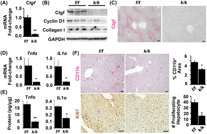
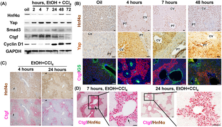
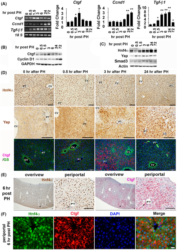
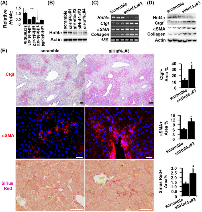
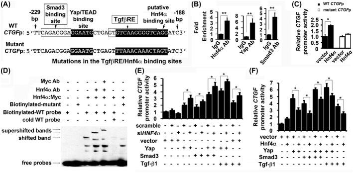
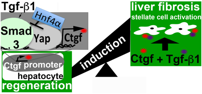
Liver regeneration after injury requires fine‐tune regulation of connective tissue growth factor (Ctgf). It also involves dynamic expression of hepatocyte nuclear factor (Hnf)4α, Yes‐associated protein (Yap), and transforming growth factor (Tgf)‐β. The upstream inducers of Ctgf, such as Yap, etc, are well‐known. However, the negative regulator of Ctgf remains unclear. Here, we investigated the Hnf4α regulation of Ctgf post‐various types of liver injury. Both wild‐type animals and animals contained siRNA‐mediated Hnf4α knockdown and Cre‐mediated Ctgf conditional deletion were used. We observed that Ctgf induction was associated with Hnf4α decline, nuclear Yap accumulation, and Tgf‐β upregulation during early stage of liver regeneration. The Ctgf promoter contained an Hnf4α binding sequence that overlapped with the cis ‐regulatory element for Yap and Tgf‐β. Ctgf loss attenuated inflammation, hepatocyte proliferation, and collagen synthesis, whereas Hnf4α knockdown enhanced Ctgf induction and liver fibrogenesis. These findings provided a new mechanism about fine‐tuned regulation of Ctgf through Hnf4α antagonism of Yap and Tgf‐β activities to balance regenerative and fibrotic signals.
Overview
The claims and statistics of this paper haven’t been checked yet.
Run the check — free, no account needed. It reads the open-access text and checks the sources it cites.
Integrity signals
Registry
Cites
Statistics
Figures
Verification
Claims the paper makes
The claims and statistics of this paper haven’t been checked yet.
Run the check — free, no account needed. It reads the open-access text and checks the sources it cites.
Claims other papers make about this one
Each is checked against this paper’s own text: does it say what the citing paper says it does?
The source backs part of the claim, not all of it — or backs it for a different population, condition or comparison.
In the citing paper
“In particular, inflammation is promoted by hepatocytes through the activation of mechanotransduction proteins known as yes‐associated protein 1 (YAP1) and WW‐domain‐containing transcription regulator 1 (WWTR1), or TAZ, in liver fibrosis [6, 7].”
From Mechanical stiffness orchestrates distinct regulation of MRTFs and YAP / TAZ transcriptional cofactors in hepatocytes (2026) · cites this paper as [7]
Experimental — accuracy not yet measuredThe cited source says what the claim attributes to it.
In the citing paper
“The regulator gene of inflammation, the Ctgf gene, showed a tendency mainly in RT‐PCR; despite being described to be strongly regulated during liver fibrosis (Fig. S5D) [6, 7].”
From Mechanical stiffness orchestrates distinct regulation of MRTFs and YAP / TAZ transcriptional cofactors in hepatocytes (2026) · cites this paper as [7]
The cited source says what the claim attributes to it.
In the citing paper
“The proinflammatory Ctgf gene was partially upregulated in the analysis, as previously described in liver fibrosis [6, 7].”
From Mechanical stiffness orchestrates distinct regulation of MRTFs and YAP / TAZ transcriptional cofactors in hepatocytes (2026) · cites this paper as [7]
Self-check
Percentages, sums and reported differences re-computed from the paper’s own numbers.
The self-check needs the paper’s full text.
Citations
Cites
Varghese, F., Bukhari, A. B., Malhotra, R., & De, A. (2014) · PLoS ONE · doi:10.1371/journal.pone.0096801 · Cited by 1,318
Schober, J. M., Chen, N., Grzeszkiewicz, T. M., Jovanovic, I., Emeson, E. E., Ugarova, T. P., Ye, R. D., Lau, L. F., & Lam, S. C.-T. (2002) · Blood · doi:10.1182/blood.v99.12.4457 · Cited by 242
Nishiyama, K., Nakashima, H., Ikarashi, M., Kinoshita, M., Nakashima, M., Aosasa, S., Seki, S., & Yamamoto, J. (2015) · PLOS ONE · doi:10.1371/journal.pone.0136774 · Cited by 82
Research works printed but not linked: 42
- 10.1016/j.joim.2018.11.008 · DOI
- 10.1016/j.jhep.2012.04.016 · DOI · Paper page
- 10.1016/j.joim.2018.05.002 · DOI
- 10.1016/j.jhep.2018.05.008 · DOI · Paper page
- 10.1002/hep.30405 · DOI
- 10.1053/j.gastro.2016.10.047 · DOI
- 10.1002/hep.24587 · DOI
- 10.1101/gad.1664408 · DOI · Paper page
- 10.1111/j.1478-3231.2008.01826.x · DOI
- 10.1038/ncb826 · DOI
- 10.1002/hep.23102 · DOI
- 10.1186/s12864-016-2492-x · DOI
- 10.1073/pnas.122359899 · DOI
- 10.1002/hep.27425 · DOI
- 10.3389/fphys.2018.00138 · DOI
- 10.1002/art.30074 · DOI
- 10.1053/j.gastro.2005.03.081 · DOI
- 10.1111/j.1530-0277.2011.01720.x · DOI
- 10.1096/fj.11-200154 · DOI
- Lin S, 2018, Mettl1/Wdr4‐mediated m(7)G tRNA methylome is required for normal mRNA translation and embryonic stem cell self‐renewal and differentiation, Mol Cell, 71, e245
- 10.1093/bioinformatics/bts635 · DOI · Paper page
- 10.1093/bioinformatics/btu638 · DOI · Paper page
- 10.1093/bioinformatics/btp616 · DOI · Paper page
- 10.1038/nprot.2008.211 · DOI · Paper page
- 10.1038/s41467-018-06898-4 · DOI
- 10.1136/mp.54.3.180 · DOI
- 10.3390/biom6010005 · DOI
- 10.1152/ajpgi.00012.2008 · DOI
- 10.1093/toxsci/kfq280 · DOI
- 10.1016/S0022-2275(20)32068-X · DOI
- Grotendorst GR, 1996, A novel transforming growth factor beta response element controls the expression of the connective tissue growth factor gene, Cell Growth Differ, 7, 469
- 10.1084/jem.20111653 · DOI · Paper page
- 10.1002/hep.28911 · DOI
- 10.1038/srep27398 · DOI
- 10.1016/j.joim.2018.05.001 · DOI
- 10.1002/hep.24815 · DOI
- 10.1371/journal.pone.0092515 · DOI
- 10.1016/S1097-2765(00)80253-1 · DOI
- 10.1210/me.2006-0300 · DOI
- 10.1159/000430195 · DOI
- 10.1096/fj.15-280511 · DOI
- 10.1016/j.biomaterials.2017.12.004 · DOI
Cited by
Sorted by citations
Torres‐Ortiz, B. S., Pérez‐Calixto, D., Carretero‐Ortega, J., Guzman‐López, A. G., Pinto‐Dueñas, D. C., Hernández‐Juárez, M., Jiménez‐Escobar, A., Rodríguez‐Hernández, A., Díaz‐Bello, B., Serna‐Márquez, N., et al. (2026) · The FEBS Journal · doi:10.1111/febs.70570
Metadata
- Authors
- Zhou, J., Sun, X., Yang, L., Wang, L., Ran, G., Wang, J., Cao, Q., Wu, L., Bryant, A., Ling, C., & Pi, L.
- Affiliations
- University of Florida Health (US)
- Beckman Research Institute (US)
- Fudan University (CN)
- University of Maryland, Baltimore (US)
- Corresponding author
- Ling, Chen
- Pi, Liya
- ORCID
- Venue
- The FASEB Journal
- Publisher
- Wiley
- Published
- 2020-02-14
- Volume
- 34
- Issue
- 4
- Pages
- 4970-4983
- ISSN
- 0892-6638 (ISSN-L)
- 1530-6860
- PMC
- PMC7722640
- OpenAlex
- W3005993270
- Cited by
- 16
- References
- 3
- Citations by year
- 2026: 3 · 2025: 2 · 2024: 3 · 2023: 1 · 2022: 4 · 2021: 2 · 2020: 1
- FWCI
- 1.18
- Type
- journal article
- Language
- en
- Open access
- hybrid
- doi.org
- Licence
- Locations
- The FASEB Journal (publishedVersion, cc-by-nc)
- PubMed Central (publishedVersion, cc-by-nc)
- PubMed (publishedVersion)
- Lanzhou University Institutional Repository (submittedVersion, cc-by-nc-sa)
- Lanzhou University Institutional Repository (submittedVersion)
- OSTI OAI (U.S. Department of Energy Office of Scientific and Technical Information) (submittedVersion)
- Funders
- National Institute on Alcohol Abuse and Alcoholism (KO1AA024174, R01AA028035)
- Research and Development (2018YFA0109400)
- Topics
- Health Sciences › Medicine › Hepatology › Liver physiology and pathology
- Life Sciences › Biochemistry, Genetics and Molecular Biology › Molecular Biology › Connective Tissue Growth Factor Research
- Life Sciences › Biochemistry, Genetics and Molecular Biology › Cell Biology › Hippo pathway signaling and YAP/TAZ
- Keywords
- HNF4α, connective tissue growth factor, liver regeneration, liver fibrosis, YAP, TGF-beta, hepatocyte proliferation, collagen synthesis, liver injury, inflammation
- MeSH terms
- YAP-Signaling Proteins, Animals, Collagen, Humans, Liver Regeneration, Protein Binding, Up-Regulation, Transforming Growth Factor beta, Cell Cycle Proteins, Hepatocytes, Adaptor Proteins, Signal Transducing, Cell Proliferation, Mice, Hepatocyte Nuclear Factor 4, Connective Tissue Growth Factor, Hep G2 Cells, HEK293 Cells
About these checks
Registry, retraction, Cites and statistics results are computed, not judged, with one exception: a printed reference that no index lists is read once by a model before it counts as a citation error. Claims are extracted by a model and kept only when their quote appears verbatim in the text; each cited source is then checked, with a model’s help, for whether it says what the claim relies on. Those verdicts can be wrong — read the claim’s quote and the cited source before relying on one.
Statistics and arithmetic are recomputed, not judged.
Confidence bands are shown only for fields calibrated on our annotated test set. None is calibrated yet, so no band is shown.
What counts as a claim ›How we measure confidence ›How we check this ›
Are you an author, or do you see a problem with this page? Request a review
How to cite
Cite CoChat for the checks on this page, and the paper for its own findings.
- This page
- CoChat. (2026). Citation check: Hepatocyte nuclear factor 4α negatively regulates connective tissue growth factor during liver regeneration. https://app.cochat.ai/papers/10.1096/fj.201902382r
- The paper
- Zhou, J., Sun, X., Yang, L., Wang, L., Ran, G., Wang, J., Cao, Q., Wu, L., Bryant, A., Ling, C., & Pi, L. (2020). Hepatocyte nuclear factor 4α negatively regulates connective tissue growth factor during liver regeneration. The FASEB Journal. https://doi.org/10.1096/fj.201902382r
