

The DOI matches its Crossref record; 84 of the 85 references it cites were found in a registry or an index.
Wide-field optical mapping of neural activity and brain haemodynamics: considerations and novel approaches
Open accessAbstract
Although modern techniques such as two-photon microscopy can now provide cellular-level three-dimensional imaging of the intact living brain, the speed and fields of view of these techniques remain limited. Conversely, two-dimensional wide-field optical mapping (WFOM), a simpler technique that uses a camera to observe large areas of the exposed cortex under visible light, can detect changes in both neural activity and haemodynamics at very high speeds. Although WFOM may not provide single-neuron or capillary-level resolution, it is an attractive and accessible approach to imaging large areas of the brain in awake, behaving mammals at speeds fast enough to observe widespread neural firing events, as well as their dynamic coupling to haemodynamics. Although such wide-field optical imaging techniques have a long history, the advent of genetically encoded fluorophores that can report neural activity with high sensitivity, as well as modern technologies such as light emitting diodes and sensitive and high-speed digital cameras have driven renewed interest in WFOM. To facilitate the wider adoption and standardization of WFOM approaches for neuroscience and neurovascular coupling research, we provide here an overview of the basic principles of WFOM, considerations for implementation of wide-field fluorescence imaging of neural activity, spectroscopic analysis and interpretation of results. This article is part of the themed issue ‘Interpreting BOLD: a dialogue between cognitive and cellular neuroscience’.
Overview
The claims and statistics of this paper haven’t been checked yet.
Run the check — free, no account needed. It reads the open-access text and checks the sources it cites.
Integrity signals
Registry
Cites
Statistics
Figures
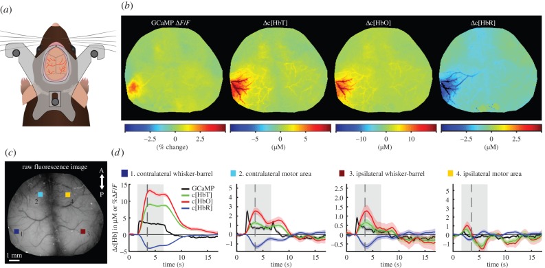
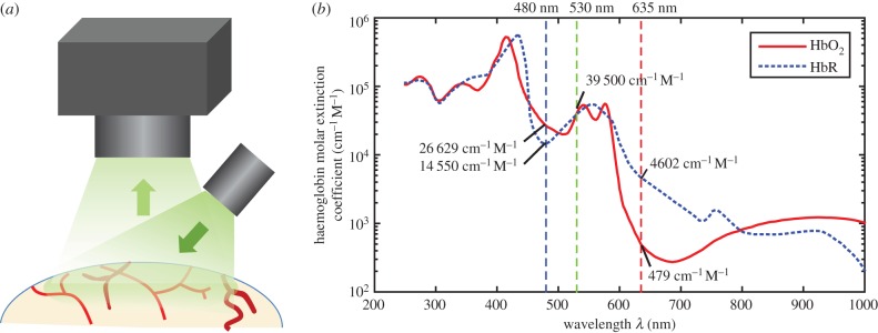
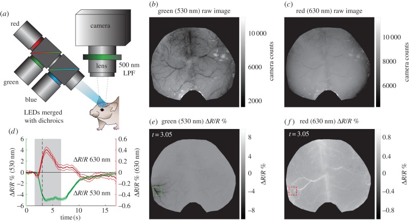
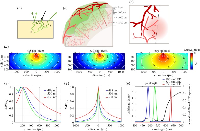
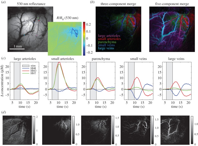
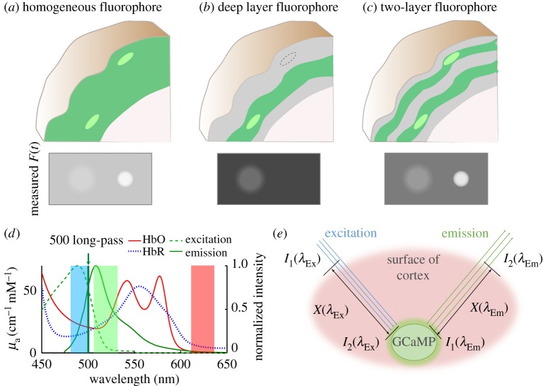
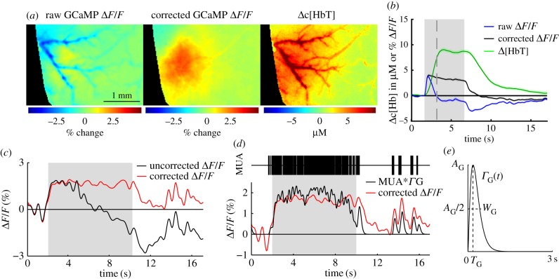
Verification
Claims the paper makes
The claims and statistics of this paper haven’t been checked yet.
Run the check — free, no account needed. It reads the open-access text and checks the sources it cites.
Claims other papers make about this one
Each is checked against this paper’s own text: does it say what the citing paper says it does?
The source backs part of the claim, not all of it — or backs it for a different population, condition or comparison.
In the citing paper
“Violet to yellow excitation and emission light are well absorbed by the blood within the vasculature. Therefore, when fluorescent proteins are diffusely expressed in brain cells, blood vessels that lack these proteins appear as dark shadows36,37.”
From Energy paradox in REM sleep: balancing supply and consumption in brain metabolism (2026) · cites this paper as [37]
Experimental — accuracy not yet measuredThe cited source says what the claim attributes to it.
In the citing paper
“Vessel dilation consequently reduces the detectable parenchymal fluorescence.”
From Energy paradox in REM sleep: balancing supply and consumption in brain metabolism (2026) · cites this paper as [37]
Self-check
Percentages, sums and reported differences re-computed from the paper’s own numbers.
The self-check needs the paper’s full text.
Citations
Cites
Ultrasensitive fluorescent proteins for imaging neuronal activity
Chen, T.-W., Wardill, T. J., Sun, Y., Pulver, S. R., Renninger, S. L., Baohan, A., Schreiter, E. R., Kerr, R. A., Orger, M. B., Jayaraman, V., et al. (2013) · Nature · doi:10.1038/nature12354 · Cited by 6,474
Millisecond-timescale, genetically targeted optical control of neural activity
Boyden, E. S., Zhang, F., Bamberg, E., Nagel, G., & Deisseroth, K. A. (2005) · Nature Neuroscience · doi:10.1038/nn1525 · Cited by 5,015
MCML—Monte Carlo modeling of light transport in multi-layered tissues
Wang, L. V., Jacques, S. L., & Zheng, L. (1995) · Computer Methods and Programs in Biomedicine · doi:10.1016/0169-2607(95)01640-f · Cited by 3,330
Capillary pericytes regulate cerebral blood flow in health and disease
Hall, C. N., Reynell, C., Gesslein, B., Hamilton, N. B., Mishra, A., Sutherland, B. A., O’Farrell, F. M., Buchan, A. M., Lauritzen, M., & Attwell, D. (2014) · Nature · doi:10.1038/nature13165 · Cited by 1,870
Functional architecture of cortex revealed by optical imaging of intrinsic signals
Grinvald, A., Lieke, E. E., Frostig, R. D., Gilbert, C. D., & Wiesel, T. N. (1986) · Nature · doi:10.1038/324361a0 · Cited by 1,243
Diversity of astrocyte functions and phenotypes in neural circuits
Khakh, B. S., & Sofroniew, M. V. (2015) · Nature Neuroscience · doi:10.1038/nn.4043 · Cited by 1,239
Functional imaging with cellular resolution reveals precise micro-architecture in visual cortex
Ohki, K., Chung, S., Ch'ng, Y. H., Kara, P., & Reid, R. C. (2005) · Nature · doi:10.1038/nature03274 · Cited by 1,191
Miniaturized integration of a fluorescence microscope
Ghosh, K. K., Burns, L. D., Cocker, E. D., Nimmerjahn, A., Ziv, Y., Gamal, A. E., & Schnitzer, M. J. (2011) · Nature Methods · doi:10.1038/nmeth.1694 · Cited by 1,130
Malonek, D., & Grinvald, A. (1996) · Science · doi:10.1126/science.272.5261.551 · Cited by 1,109
Akerboom, J., Calderón, N. C., Tian, L., Wabnig, S., Prigge, M., Tolö, J., Gordus, A. G., Orger, M. B., Severi, K. E., Macklin, J. J., et al. (2013) · Frontiers in Molecular Neuroscience · doi:10.3389/fnmol.2013.00002 · Cited by 736
Cited by
Sorted by citations
Ma, Y., Shaik, M. A., Kozberg, M. G., Kim, S. H., Portes, J., Timerman, D., & Hillman, E. M. C. (2016) · Proceedings of the National Academy of Sciences · doi:10.1073/pnas.1525369113 · Cited by 325
Self-citationGlymphatic influx and clearance are accelerated by neurovascular coupling
Holstein-Rønsbo, S., Gan, Y., Giannetto, M. J., Rasmussen, M. K., Sigurdsson, B., Beinlich, F. R. M., Rose, L., Untiet, V., Hablitz, L. M., Kelley, D. H., et al. (2023) · Nature Neuroscience · doi:10.1038/s41593-023-01327-2 · Cited by 248
Task-Dependent Changes in the Large-Scale Dynamics and Necessity of Cortical Regions
Pinto, L., Rajan, K., DePasquale, B., Thiberge, S. Y., Tank, D. W., & Brody, C. D. (2019) · Neuron · doi:10.1016/j.neuron.2019.08.025 · Cited by 244
Renaudin, N., Demené, C., Dizeux, A., Ialy-Radio, N., Pezet, S., & Tanter, M. (2022) · Nature Methods · doi:10.1038/s41592-022-01549-5 · Cited by 204
Linking brain vascular physiology to hemodynamic response in ultra-high field MRI
Uludaǧ, K., & Blinder, P. (2017) · NeuroImage · doi:10.1016/j.neuroimage.2017.02.063 · Cited by 175
Simultaneous cortex-wide fluorescence Ca2+ imaging and whole-brain fMRI
Lake, E. M. R., Ge, X., Shen, X., Hermán, P., Hyder, F., Cardin, J. A., Higley, M. J., Scheinost, D., Papademetris, X., Crair, M. C., et al. (2020) · Nature Methods · doi:10.1038/s41592-020-00984-6 · Cited by 168
Imaging Cortical Dynamics in GCaMP Transgenic Rats with a Head-Mounted Widefield Macroscope
Scott, B. B., Thiberge, S. Y., Guo, C., Tervo, D. G. R., Brody, C. D., Karpova, A. Y., & Tank, D. W. (2018) · Neuron · doi:10.1016/j.neuron.2018.09.050 · Cited by 166
Movement and Performance Explain Widespread Cortical Activity in a Visual Detection Task
Salkoff, D. B., Zagha, E., McCarthy, E., & McCormick, D. A. (2019) · Cerebral Cortex · doi:10.1093/cercor/bhz206 · Cited by 158
Clancy, N. T., Jones, G., Maier‐Hein, L., Elson, D. S., & Stoyanov, D. V. (2020) · Medical Image Analysis · doi:10.1016/j.media.2020.101699 · Cited by 158
Inhibition stabilization is a widespread property of cortical networks
Sanzeni, A., Akitake, B., Goldbach, H. C., Leedy, C. E., Brunel, N., & Histed, M. H. (2020) · eLife · doi:10.7554/elife.54875 · Cited by 159
Lights, fiber, action! A primer on in vivo fiber photometry
Simpson, E. H., Akam, T., Patriarchi, T., Blanco-Pozo, M., Burgeno, L. M., Mohebi, A., Cragg, S. J., & Walton, M. E. (2023) · Neuron · doi:10.1016/j.neuron.2023.11.016 · Cited by 160
Mesoscopic Imaging: Shining a Wide Light on Large-Scale Neural Dynamics
Cardin, J. A., Crair, M. C., & Higley, M. J. (2020) · Neuron · doi:10.1016/j.neuron.2020.09.031 · Cited by 135
Vanni, M. P., Chan, A. W., Balbi, M., Silasi, G., & Murphy, T. H. (2017) · Journal of Neuroscience · doi:10.1523/jneurosci.3560-16.2017 · Cited by 152
Spatiotemporally heterogeneous coordination of cholinergic and neocortical activity
Lohani, S., Moberly, A. H., Benisty, H., Landa, B., Jing, M., Li, Y., Higley, M. J., & Cardin, J. A. (2022) · Nature Neuroscience · doi:10.1038/s41593-022-01202-6 · Cited by 146
Simultaneous mesoscopic and two-photon imaging of neuronal activity in cortical circuits
Barson, D. G., Hamodi, A. S., Shen, X., Lür, G., Constable, R. T., Cardin, J. A., Crair, M. C., & Higley, M. J. (2019) · Nature Methods · doi:10.1038/s41592-019-0625-2 · Cited by 135
Behavioral Strategy Determines Frontal or Posterior Location of Short-Term Memory in Neocortex
Gilad, A., Gallero-Salas, Y., Groos, D., & Helmchen, F. (2018) · Neuron · doi:10.1016/j.neuron.2018.07.029 · Cited by 140
Mapping cortical mesoscopic networks of single spiking cortical or sub-cortical neurons
Xiao, D., Vanni, M. P., Mitelut, C., Chan, A. W., LeDue, J. M., Xie, Y., Chen, A. C. N., Swindale, N. V., & Murphy, T. H. (2017) · eLife · doi:10.7554/elife.19976 · Cited by 134
Norepinephrine links astrocytic activity to regulation of cortical state
Reitman, M. E., Tse, V., Mi, X., Willoughby, D. D., Peinado, A., Aivazidis, A., Myagmar, B., Simpson, P. C., Bayraktar, O. A., Yu, G., et al. (2023) · Nature Neuroscience · doi:10.1038/s41593-023-01284-w · Cited by 133
Sensory coding and the causal impact of mouse cortex in a visual decision
Zatka-Haas, P., Steinmetz, N. A., Carandini, M., & Harris, K. D. (2021) · eLife · doi:10.7554/elife.63163 · Cited by 122
Functional connectivity structure of cortical calcium dynamics in anesthetized and awake mice
Wright, P. W., Brier, L. M., Bauer, A. Q., Baxter, G. A., Kraft, A. W., Reisman, M. D., Bice, A. R., Snyder, A. Z., Lee, J., & Culver, J. P. (2017) · PLoS ONE · doi:10.1371/journal.pone.0185759 · Cited by 121
Monitoring norepinephrine release in vivo using next-generation GRABNE sensors
Feng, J., Dong, H., Lischinsky, J. E., Zhou, J., Deng, F., Zhuang, C., Miao, X., Wang, H., Li, G., Cai, R., et al. (2024) · Neuron · doi:10.1016/j.neuron.2024.03.001 · Cited by 122
Cerebral oxygenation during locomotion is modulated by respiration
Zhang, Q., Roche, M., Gheres, K. W., Chaigneau, E. M., Kedarasetti, R. T., Haselden, W. D., Charpak, S., & Drew, P. J. (2019) · Nature Communications · doi:10.1038/s41467-019-13523-5 · Cited by 125
Aging drives cerebrovascular network remodeling and functional changes in the mouse brain
Bennett, H. C., Zhang, Q., Wu, Y.-T., Manjila, S. B., Chon, U., Shin, D., Vanselow, D. J., Pi, H.-J., Drew, P. J., & Kim, Y. (2024) · Nature Communications · doi:10.1038/s41467-024-50559-8 · Cited by 115
Montgomery, M. K., Kim, S. H., Dovas, A., Zhao, H., Goldberg, A., Xu, W., Yagielski, A. J., Cambareri, M. K., Patel, K., Mela, A., et al. (2020) · Cell Reports · doi:10.1016/j.celrep.2020.03.064 · Cited by 104
Self-citationChronic, cortex-wide imaging of specific cell populations during behavior
Couto, J., Musall, S., Sun, X. R., Khanal, A., Gluf, S., Saxena, S., Kinsella, I., Abe, T., Cunningham, J. P., Paninski, L., et al. (2021) · Nature Protocols · doi:10.1038/s41596-021-00527-z · Cited by 111
Advances in Monte Carlo Simulation for Light Propagation in Tissue
Periyasamy, V., & Pramanik, M. (2017) · IEEE Reviews in Biomedical Engineering · doi:10.1109/rbme.2017.2739801 · Cited by 99
Separation of hemodynamic signals from GCaMP fluorescence measured with wide-field imaging
Valley, M. T., Moore, M. G., Zhuang, J., Mesa, N., Castelli, D., Sullivan, D., Reimers, M., & Waters, J. (2019) · Journal of Neurophysiology · doi:10.1152/jn.00304.2019 · Cited by 99
Low-Dimensional Spatiotemporal Dynamics Underlie Cortex-wide Neural Activity
MacDowell, C. J., & Buschman, T. J. (2020) · Current Biology · doi:10.1016/j.cub.2020.04.090 · Cited by 92
Localized semi-nonnegative matrix factorization (LocaNMF) of widefield calcium imaging data
Saxena, S., Kinsella, I., Musall, S., Kim, S. H., Mészáros, J., Thibodeaux, D. N., Kim, C. Y., Cunningham, J. P., Hillman, E. M. C., Churchland, A. K., et al. (2020) · PLoS Computational Biology · doi:10.1371/journal.pcbi.1007791 · Cited by 86
Self-citationCharacterizing Cortex-Wide Dynamics with Wide-Field Calcium Imaging
Ren, C., & Komiyama, T. (2021) · Journal of Neuroscience · doi:10.1523/jneurosci.3003-20.2021 · Cited by 90
Spreading depression as an innate antiseizure mechanism
Tamim, I., Chung, D. Y., Morais, A., Loonen, I. C. M., Qin, T., Misra, A., Schlunk, F., Endres, M., Schiff, S. J., & Ayata, C. (2021) · Nature Communications · doi:10.1038/s41467-021-22464-x · Cited by 83
Improved green and red GRAB sensors for monitoring spatiotemporal serotonin release in vivo
Deng, F., Wan, J., Li, G., Dong, H., Xia, X., Wang, Y., Li, X., Zhuang, C., Zheng, Y. R., Liu, L. D., et al. (2024) · Nature Methods · doi:10.1038/s41592-024-02188-8 · Cited by 95
A human-specific modifier of cortical connectivity and circuit function
Schmidt, E. R. E., Zhao, H. T., Park, J. M., Dipoppa, M., Monsalve-Mercado, M. M., Dahan, J. B., Rodgers, C. C., Lejeune, A., Hillman, E. M. C., Miller, K. D., et al. (2021) · Nature · doi:10.1038/s41586-021-04039-4 · Cited by 82
Self-citationDesigning a large field-of-view two-photon microscope using optical invariant analysis
Bumstead, J., Park, J. J., Rosen, I. A., Kraft, A. W., Wright, P. W., Reisman, M. D., Côté, D. C., & Culver, J. P. (2018) · Neurophotonics · doi:10.1117/1.nph.5.2.025001 · Cited by 75
Cramer, J. V., Gesierich, B. G., Roth, S., Dichgans, M., Duering, M., & Liesz, A. (2019) · NeuroImage · doi:10.1016/j.neuroimage.2019.06.014 · Cited by 75
Mascaro, A. L. A., Conti, E., Lai, S., Giovanna, A. P. D., Spalletti, C., Alia, C., Panarese, A., Scaglione, A., Sacconi, L., Micera, S., et al. (2019) · Cell Reports · doi:10.1016/j.celrep.2019.08.062 · Cited by 75
Neuronal Origin of the Temporal Dynamics of Spontaneous BOLD Activity Correlation
Matsui, T., Murakami, T., & Ohki, K. (2018) · Cerebral Cortex · doi:10.1093/cercor/bhy045 · Cited by 79
Cortical State Fluctuations during Sensory Decision Making
Jacobs, E. A. K., Steinmetz, N. A., Peters, A. J., Carandini, M., & Harris, K. D. (2020) · Current Biology · doi:10.1016/j.cub.2020.09.067 · Cited by 70
Interpreting BOLD: towards a dialogue between cognitive and cellular neuroscience
Hall, C. N., Howarth, C., Kurth-Nelson, Z. L., & Mishra, A. (2016) · Philosophical Transactions of the Royal Society B Biological Sciences · doi:10.1098/rstb.2015.0348 · Cited by 80
Rapid fluctuations in functional connectivity of cortical networks encode spontaneous behavior
Benisty, H., Barson, D., Moberly, A. H., Lohani, S., Tang, L., Coifman, R. R., Crair, M. C., Mishne, G., Cardin, J. A., & Higley, M. J. (2024) · Nature Neuroscience · doi:10.1038/s41593-023-01498-y · Cited by 65
Arousal as a universal embedding for spatiotemporal brain dynamics
Raut, R. V., Rosenthal, Z. P., Wang, X., Miao, H., Zhang, Z., Lee, J., Raichle, M. E., Bauer, A. Q., Brunton, S. L., Brunton, B. W., et al. (2025) · Nature · doi:10.1038/s41586-025-09544-4 · Cited by 87
Wide-Field Calcium Imaging of Dynamic Cortical Networks during Locomotion
West, S. L., Aronson, J. D., Popa, L. S., Feller, K. D., Carter, R. E., Chiesl, W. M., Gerhart, M. L., Shekhar, A. C., Ghanbari, L., Kodandaramaiah, S. B., et al. (2021) · Cerebral Cortex · doi:10.1093/cercor/bhab373 · Cited by 65
Oršolić, I., Rio, M., Mrsic‐Flogel, T. D., & Znamenskiy, P. (2021) · Neuron · doi:10.1016/j.neuron.2021.03.031 · Cited by 63
McGirr, A., LeDue, J. M., Chan, A. W., Xie, Y., & Murphy, T. H. (2017) · Brain · doi:10.1093/brain/awx142 · Cited by 60
Borden, P. Y., Wright, N. C., Morrissette, A. E., Jaeger, D., Haider, B., & Stanley, G. B. (2022) · Neuron · doi:10.1016/j.neuron.2022.06.008 · Cited by 54
Zhang, W., Chao, T.-H. H., Yang, Y., Wang, T., Lee, S.-H., Oyarzabal, E. A., Zhou, J., Nonneman, R., Pégard, N. C., Hongtu Zhu, B., et al. (2022) · Cell Reports Methods · doi:10.1016/j.crmeth.2022.100243 · Cited by 58
The impact of bilateral ongoing activity on evoked responses in mouse cortex
Shimaoka, D., Steinmetz, N. A., Harris, K. D., & Carandini, M. (2019) · eLife · doi:10.7554/elife.43533 · Cited by 56
Differential effects of anesthetics on resting state functional connectivity in the mouse
Xie, H., Chung, D. Y., Kura, S., Sugimoto, K., Aykan, S. A., Wu, Y., Sakadžić, S., Yaseen, M. A., Boas, D. A., & Ayata, C. (2019) · Journal of Cerebral Blood Flow & Metabolism · doi:10.1177/0271678x19847123 · Cited by 56
Emerging imaging methods to study whole-brain function in rodent models
Markicevic, M., Savvateev, I., Grimm, C., & Zerbi, V. (2021) · Translational Psychiatry · doi:10.1038/s41398-021-01575-5 · Cited by 55
Kim, E., Anguluan, E., & Kim, J. G. (2017) · Scientific Reports · doi:10.1038/s41598-017-13572-0 · Cited by 52
Kisler, K., Lazić, D., Sweeney, M. D., Plunkett, S., Khatib, M. E., Vinogradov, S. A., Boas, D. A., Sakadžić, S., & Zloković, B. V. (2018) · Nature Protocols · doi:10.1038/nprot.2018.034 · Cited by 52
Müllenbroich, M. C., Turrini, L., Silvestri, L., Alterini, T., Gheisari, A., Tiso, N., Vanzi, F., Sacconi, L., & Pavone, F. S. (2018) · Frontiers in Cellular Neuroscience · doi:10.3389/fncel.2018.00315 · Cited by 50
Wide-Field Calcium Imaging of Neuronal Network Dynamics In Vivo
Nietz, A. K., Popa, L. S., Streng, M. L., Carter, R. E., Kodandaramaiah, S. B., & Ebner, T. J. (2022) · Biology · doi:10.3390/biology11111601 · Cited by 53
Chao, T.-H. H., Lee, B., Hsu, L.-M., Cerri, D. H., Zhang, W.-T., Wang, T.-W. W., Ryali, S., Menon, V., & Shih, Y.-Y. I. (2023) · Science Advances · doi:10.1126/sciadv.ade5732 · Cited by 49
Shahsavarani, S., Thibodeaux, D. N., Xu, W., Kim, S. H., Lodgher, F., Nwokeabia, C., Cambareri, M. K., Yagielski, A. J., Zhao, H., Handwerker, D. A., et al. (2023) · Cell Reports · doi:10.1016/j.celrep.2023.112527 · Cited by 49
Self-citationCortical signatures of wakeful somatosensory processing
Song, C., Piscopo, D. M., Niell, C. M., & Knöpfel, T. (2018) · Scientific Reports · doi:10.1038/s41598-018-30422-9 · Cited by 49
Neurovascular dynamics of repeated cortical spreading depolarizations after acute brain injury
Zhao, H., Tuohy, M. C., Chow, D. S., Kozberg, M. G., Kim, S. H., Shaik, M. A., & Hillman, E. M. C. (2021) · Cell Reports · doi:10.1016/j.celrep.2021.109794 · Cited by 46
Self-citationRelating Pupil Diameter and Blinking to Cortical Activity and Hemodynamics across Arousal States
Turner, K. L., Gheres, K. W., & Drew, P. J. (2022) · Journal of Neuroscience · doi:10.1523/jneurosci.1244-22.2022 · Cited by 42
Zhong, X., Liang, Y., Wang, X., Lan, H., Bai, X., Jin, L., & Guan, B. (2024) · Light Science & Applications · doi:10.1038/s41377-023-01348-3 · Cited by 47
Urban, A., Golgher, L., Brunner, C., Gdalyahu, A., Har-Gil, H., Kain, D., Montaldo, G., Sironi, L., & Blinder, P. (2017) · Advanced Drug Delivery Reviews · doi:10.1016/j.addr.2017.07.018 · Cited by 46
Buchanan, E. K., Kinsella, I., Zhou, D. X., Zhu, R., Zhou, P., Gerhard, F., Ferrante, J., Ma, Y., Kim, S. H., Shaik, M., et al. (2018) · bioRxiv (Cold Spring Harbor Laboratory) · doi:10.1101/334706 · Cited by 47
Self-citationLong-wavelength traveling waves of vasomotion modulate the perfusion of cortex
Broggini, T., Duckworth, J. C., Ji, X., Liu, R., Xia, X., Mächler, P., Shaked, I., Munting, L. P., Iyengar, S., Kotlikoff, M. I., et al. (2024) · Neuron · doi:10.1016/j.neuron.2024.04.034 · Cited by 43
Real-time non-invasive in vivo visible light detection of cortical spreading depolarizations in mice
Chung, D. Y., Sugimoto, K., Fischer, P., Böhm, M., Takizawa, T., Sadeghian, H., Morais, A., Harriott, A. M., Oka, F., Qin, T., et al. (2018) · Journal of Neuroscience Methods · doi:10.1016/j.jneumeth.2018.09.001 · Cited by 44
Separability of calcium slow waves and functional connectivity during wake, sleep, and anesthesia
Brier, L. M., Landsness, E. C., Snyder, A. Z., Wright, P. W., Baxter, G. A., Bauer, A. Q., Lee, J., & Culver, J. P. (2019) · Neurophotonics · doi:10.1117/1.nph.6.3.035002 · Cited by 48
Bethge, P., Carta, S., Lorenzo, D. A., Egolf, L., Goniotaki, D., Madisen, L., Voigt, F. F., Chen, J. L., Schneider, B. L., Ohkura, M., et al. (2017) · PLoS ONE · doi:10.1371/journal.pone.0179460 · Cited by 43
Lu, X., Wang, Y., Liu, Z., Gou, Y., Jaeger, D., & St-Pierre, F. (2023) · Nature Communications · doi:10.1038/s41467-023-41975-3 · Cited by 43
Krolak, T., Kaplan, L., Navas, K., Chen, L., Birmingham, A., Ryvkin, D., Izsa, V., Powell, M., Wu, Z., Deverman, B. E., et al. (2025) · Cell · doi:10.1016/j.cell.2025.06.030 · Cited by 44
Kura, S., Xie, H., Fu, B., Ayata, C., Boas, D. A., & Sakadžić, S. (2018) · Journal of Neural Engineering · doi:10.1088/1741-2552/aaafe4 · Cited by 38
Multimodal Noninvasive Functional Neurophotonic Imaging of Murine Brain‐Wide Sensory Responses
Chen, Z., Zhou, Q., Deán‐Ben, X. L., Gezginer, I., Ni, R., Reiss, M., Shoham, S., & Razansky, D. (2022) · Advanced Science · doi:10.1002/advs.202105588 · Cited by 37
A human-specific enhancer fine-tunes radial glia potency and corticogenesis
Liu, J., Mosti, F., Zhao, H., Lollis, D., Sotelo-Fonseca, J. E., Escobar-Tomlienovich, C. F., Musso, C. M., Mao, Y., Massri, A. J., Doll, H. M., et al. (2025) · Nature · doi:10.1038/s41586-025-09002-1 · Cited by 39
Self-citationAntipsychotic drugs selectively decorrelate long-range interactions in deep cortical layers
Heindorf, M., & Keller, G. B. (2023) · eLife · doi:10.1101/2022.01.31.478462 · Cited by 64
He, Y., Shi, J., Maslov, K., Cao, R., & Wang, L. V. (2020) · Journal of Biomedical Optics · doi:10.1117/1.jbo.25.6.066501 · Cited by 36
Chung, D. Y., Oka, F., Jin, G., Harriott, A. M., Kura, S., Aykan, S. A., Qin, T., Edmiston, W. J., Lee, H., Yaseen, M. A., et al. (2020) · Journal of Cerebral Blood Flow & Metabolism · doi:10.1177/0271678x20940152 · Cited by 36
Babij, R., Ferrer, C., Donatelle, A., Wacks, S., Buch, A. M., Niemeyer, J. E., Ma, H., Duan, Z. R. S., Fetcho, R. N., Che, A., et al. (2023) · Neuron · doi:10.1016/j.neuron.2022.10.037 · Cited by 35
Zhang, X., Landsness, E. C., Chen, W., Miao, H., Tang, M., Brier, L. M., Culver, J. P., Lee, J., & Anastasio, M. A. (2021) · Journal of Neuroscience Methods · doi:10.1016/j.jneumeth.2021.109421 · Cited by 36
Cortical glutamatergic projection neuron types contribute to distinct functional subnetworks
Mohan, H., An, X., Xu, X. H., Kondo, H., Zhao, S., Matho, K. S., Wang, B., Musall, S., Mitra, P. P., & Huang, Z. J. (2023) · Nature Neuroscience · doi:10.1038/s41593-022-01244-w · Cited by 40
Spatiotemporal patterns of spontaneous brain activity: a mini-review
Meyer-Baese, L., Watters, H. N., & Keilholz, S. D. (2022) · Neurophotonics · doi:10.1117/1.nph.9.3.032209 · Cited by 34
Wide-field calcium imaging of cortex-wide activity in awake, head-fixed mice
Ren, C., & Komiyama, T. (2021) · STAR Protocols · doi:10.1016/j.xpro.2021.100973 · Cited by 33
Любашина, О. А., Mamontov, O. V., Volynsky, M. A., Zaytsev, V. V., & Kamshilin, A. A. (2019) · Frontiers in Neuroscience · doi:10.3389/fnins.2019.01235 · Cited by 31
Decoding cortical brain states from widefield calcium imaging data using visibility graph
Zhu, L., Lee, C. R., Margolis, D. J., & Najafizadeh, L. (2018) · Biomedical Optics Express · doi:10.1364/boe.9.003017 · Cited by 32
Jones-Tabah, J., Mohammad, H., Hadj-Youssef, S., Kim, L., Martin, R., Benaliouad, F., Tanny, J. C., Clarke, P. B. S., & Hébert, T. E. (2020) · Scientific Reports · doi:10.1038/s41598-020-71121-8 · Cited by 31
Шумкова, В. В., Sitdikova, V., Rechapov, I., Leukhin, A., & Minlebaev, M. (2021) · Scientific Reports · doi:10.1038/s41598-021-88461-8 · Cited by 30
Nakajima, R., & Baker, B. J. (2018) · Journal of Physics D Applied Physics · doi:10.1088/1361-6463/aae2e3 · Cited by 30
Matsui, T., & Yamashita, K.-I. (2022) · Brain Connectivity · doi:10.1089/brain.2022.0044 · Cited by 27
Swanson, R. A., Chinigò, E., Levenstein, D., Vöröslakos, M., Mousavi, N., Wang, X., Basu, J., & Buzsáki, G. (2025) · Neuron · doi:10.1016/j.neuron.2024.12.019 · Cited by 30
Distributed and Localized Dynamics Emerge in the Mouse Neocortex during Reach-to-Grasp Behavior
Quarta, E., Scaglione, A., Lucchesi, J., Sacconi, L., Mascaro, A. L. A., & Pavone, F. S. (2021) · Journal of Neuroscience · doi:10.1523/jneurosci.0762-20.2021 · Cited by 27
Song, X., Guo, Y., Li, H., Chen, C., Lee, J. H., Zhang, Y., Schmidt, Z., & Wang, X. (2022) · Nature Communications · doi:10.1038/s41467-022-29864-7 · Cited by 26
Padawer-Curry, J. A., Krentzman, O. J., Kuo, C., Wang, X., Bice, A. R., Nicol, G. E., Snyder, A. Z., Siegel, J. S., McCall, J. G., & Bauer, A. Q. (2025) · Nature Neuroscience · doi:10.1038/s41593-025-02069-z · Cited by 29
Munting, L. P., Bonnar, O., Kozberg, M. G., Auger, C. A., Hirschler, L., Hou, S. S., Greenberg, S. M., Bacskai, B. J., & van Veluw, S. J. (2023) · Journal of Cerebral Blood Flow & Metabolism · doi:10.1177/0271678x231152550 · Cited by 28
Self-citationDCC Is Required for the Development of Nociceptive Topognosis in Mice and Humans
da Silva, R. V., Johannssen, H. C., Wyss, M. T., Roome, R. B., Bourojeni, F. B., Stifani, N., Marsh, A. P. L., Ryan, M. M., Lockhart, P. J., Leventer, R. J., et al. (2018) · Cell Reports · doi:10.1016/j.celrep.2018.01.004 · Cited by 26
Rehani, R., Atamna, Y., Tiroshi, L., Chiu, W., Buendia, J. D. J. A., Martins, G. J., Jacobson, G. A., & Goldberg, J. A. (2019) · eNeuro · doi:10.1523/eneuro.0351-18.2018 · Cited by 25
Computing hemodynamic response functions from concurrent spectral fiber-photometry and fMRI data
Chao, T.-H. H., Zhang, W., Hsu, L.-M., Cerri, D. H., Wang, T.-W. W., & Shih, Y.-Y. I. (2022) · Neurophotonics · doi:10.1117/1.nph.9.3.032205 · Cited by 21
Chen, W., Park, K., Pan, Y., Koretsky, A. P., & Du, C. (2020) · NeuroImage · doi:10.1016/j.neuroimage.2020.116554 · Cited by 24
De-scattering with Excitation Patterning enables rapid wide-field imaging through scattering media
Zheng, C., Park, J. K., Yıldırım, M., Boivin, J. R., Xue, Y., Sur, M., So, P. T. C., & Wadduwage, D. N. (2021) · Science Advances · doi:10.1126/sciadv.aay5496 · Cited by 23
Cortex-wide transcranial localization microscopy with fluorescently labeled red blood cells
Zhou, Q., Glück, C., Tang, L., Glandorf, L., Droux, J., Amki, M. E., Wegener, S., Weber, B., Razansky, D., & Chen, Z. (2024) · Nature Communications · doi:10.1038/s41467-024-47892-3 · Cited by 22
Giannoni, L., Lange, F., Sajic, M., Smith, K. J., & Tachtsidis, I. (2021) · IEEE Journal of Selected Topics in Quantum Electronics · doi:10.1109/jstqe.2021.3053634 · Cited by 22
Mesoscopic calcium imaging in a head-unrestrained male non-human primate using a lensless microscope
Wu, J., Chen, Y., Veeraraghavan, A., Seidemann, E., & Robinson, J. T. (2024) · Nature Communications · doi:10.1038/s41467-024-45417-6 · Cited by 22
Optogenetic Spreading Depolarizations Do Not Worsen Acute Ischemic Stroke Outcome
Sugimoto, K., Yang, J. C., Fischer, P., Takizawa, T., Mulder, I. A., Qin, T., Erdogan, T. D., Yaseen, M. A., Sakadžić, S., Chung, D. Y., et al. (2023) · Stroke · doi:10.1161/strokeaha.122.041351 · Cited by 21
Chronic brain functional ultrasound imaging in freely moving rodents performing cognitive tasks
Hady, A. E., Takahashi, D. Y., Sun, R., Akinwale, O., Boyd-Meredith, T., Zhang, Y. S., Charles, A. S., & Brody, C. D. (2023) · Journal of Neuroscience Methods · doi:10.1016/j.jneumeth.2023.110033 · Cited by 20
Mesoscale brain explorer, a flexible python-based image analysis and visualization tool
Haupt, D., Vanni, M. P., Bolaños, F., Mitelut, C., LeDue, J. M., & Murphy, T. H. (2017) · Neurophotonics · doi:10.1117/1.nph.4.3.031210 · Cited by 21
Metadata
- Authors
- Ma, Y., Shaik, M. A., Kim, S. H., Kozberg, M. G., Thibodeaux, D. N., Zhao, H. T., Yu, H., & Hillman, E. M. C.
- Affiliations
- Mortimer B. Zuckerman Mind Brain Behavior Institute (US)
- Corresponding author
- Hillman, Elizabeth M. C.
- ORCID
- Venue
- Philosophical Transactions of the Royal Society B: Biological Sciences
- Publisher
- The Royal Society
- Published
- 2016-10-05
- Volume
- 371
- Issue
- 1705
- Pages
- 20150360
- ISSN
- 0962-8436 (ISSN-L)
- 1471-2970
- PMC
- PMC5003860
- OpenAlex
- W2513047215
- Cited by
- 335
- References
- 85
- Citations by year
- 2026: 24 · 2025: 54 · 2024: 52 · 2023: 47 · 2022: 40 · 2021: 31 · 2020: 24 · 2019: 21 · 2018: 24 · 2017: 15 · 2016: 3
- FWCI
- 3.25
- Type
- journal article
- Language
- en
- Update notices
- correction (2017-02-19)
- Open access
- hybrid
- doi.org
- Locations
- Philosophical Transactions of the Royal Society B Biological Sciences (publishedVersion, cc-by)
- PubMed Central (publishedVersion, cc-by)
- PubMed (publishedVersion)
- Funders
- National Science Foundation (0954796)
- U.S. Department of Defense (MURI W911NF-12-1-0594)
- National Institute of Neurological Disorders and Stroke (1R01NS063226, 5U01NS094296, 1R21NS053684, 1R01NS076628)
- National Eye Institute (1R01EY019500)
- Topics
- Health Sciences › Medicine › Radiology, Nuclear Medicine and Imaging › Optical Imaging and Spectroscopy Techniques
- Life Sciences › Biochemistry, Genetics and Molecular Biology › Biophysics › Advanced Fluorescence Microscopy Techniques
- Life Sciences › Neuroscience › Cognitive Neuroscience › Neural dynamics and brain function
- Keywords
- neuronal activity imaging, neurovascular coupling, genetically encoded fluorescent indicators, wide-field fluorescence imaging, spectroscopic analysis, cortical imaging, BOLD signal
- MeSH terms
- Animals, Brain, Brain Mapping, Hemodynamics, Humans, Neurons, Mice, Rats, Optical Imaging
About these checks
Registry, retraction, Cites and statistics results are computed, not judged, with one exception: a printed reference that no index lists is read once by a model before it counts as a citation error. Claims are extracted by a model and kept only when their quote appears verbatim in the text; each cited source is then checked, with a model’s help, for whether it says what the claim relies on. Those verdicts can be wrong — read the claim’s quote and the cited source before relying on one.
Statistics and arithmetic are recomputed, not judged.
Confidence bands are shown only for fields calibrated on our annotated test set. None is calibrated yet, so no band is shown.
What counts as a claim ›How we measure confidence ›How we check this ›
Are you an author, or do you see a problem with this page? Request a review
