

The DOI matches its Crossref record; 1 of the 1 references it cites were found in a registry or an index.
Nucleus size and its effect on the chromatin structure in living cells
Open accessPreprint — published as 10.1016/j.bpj.2022.09.019 · Paper page
Abstract
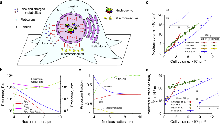
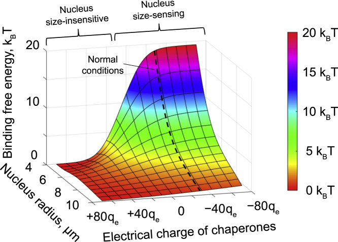
DNA-architectural proteins play a major role in organization of chromosomal DNA in living cells by packaging it into chromatin, whose spatial conformation is determined by an intricate interplay between the DNA-binding properties of architectural proteins and physical constraints applied to the DNA by a tight nuclear space. Yet, the exact effects of the cell nucleus size on DNA-protein interactions and chromatin structure currently remain obscure. Furthermore, there is even no clear understanding of molecular mechanisms responsible for the nucleus size regulation in living cells. To find answers to these questions, we developed a general theoretical framework based on a combination of polymer field theory and transfer-matrix calculations, which showed that the nucleus size is mainly determined by the difference between the surface tensions of the nuclear envelope and the endoplasmic reticulum membrane as well as the osmotic pressure exerted by cytosolic macromolecules on the nucleus. In addition, the model demonstrated that the cell nucleus functions as a piezoelectric element, changing its electrostatic potential in a size-dependent manner. This effect has been found to have a profound impact on stability of nucleosomes, revealing a previously unknown link between the nucleus size and chromatin structure. Overall, our study provides new insights into the molecular mechanisms responsible for regulation of the cell nucleus size, as well as the potential role of nuclear organization in shaping the cell response to environmental cues. SIGNIFICANCE STATEMENT: The cell nucleus plays a central role in the life of eukaryotic cells, providing the highest level of control of intracellular processes. Depending on the stage of the cell cycle and / or surrounding environment, the size of the cell nucleus may undergo changes that are believed to cause chromatin reorganization, affecting gene transcription. At present, however, there is no clear understanding of the molecular mechanisms that may be responsible for such regulation, whose exact effect on chromatin structure remains unclear. In this study, by developing an advanced computational approach, we explore these issues from a physical perspective, revealing previously unknown mechanisms contributing to organization of the cell nucleus and chromatin.
Overview
The claims and statistics of this paper haven’t been checked yet.
Run the check — free, no account needed. It reads the open-access text and checks the sources it cites.
Integrity signals
Registry
Cites
Statistics
Figures
Verification
Claims the paper makes
The claims and statistics of this paper haven’t been checked yet.
Run the check — free, no account needed. It reads the open-access text and checks the sources it cites.
Self-check
Percentages, sums and reported differences re-computed from the paper’s own numbers.
The self-check needs the paper’s full text.
Citations
Cites
Milo, R. (2013) · BioEssays · doi:10.1002/bies.201300066 · Cited by 614
Research works printed but not linked: 132
- 10.1242/jcs.037333 · DOI
- 10.1016/j.ceb.2014.01.003 · DOI
- 10.1080/19491034.2016.1162933 · DOI
- 2019, Curr. Genet, 65, 1281, 10.1007/s00294-019-00999-3 · DOI
- 1999, Biophys. Chem, 80, 199, 10.1016/S0301-4622(99)00079-4 · DOI
- J. Keener and J. Sneyd ,Mathematical physiology I: cellular physiology., 2nd ed. (Springer, New York, United States of America, 2009). · DOI
- 2017, Front. Cell Dev. Biol, 5, 41, 10.3389/fcell.2017.00041 · DOI
- 10.1007/BF01930444 · DOI
- 10.1083/jcb.115.4.941 · DOI
- 10.1083/jcb.200708054 · DOI
- 10.1073/pnas.1705179114 · DOI
- 10.1128/MCB.00981-07 · DOI
- 10.1038/nature08448 · DOI
- 10.1126/science.1191241 · DOI
- 10.1016/j.bpj.2013.01.006 · DOI
- 10.1038/ncomms5619 · DOI
- 2017, Proc. Natl. Acad. Sci. U.S.A, 115, E4741
- 2018, Mol. Biol. Cell, 29, 3039, 10.1091/mbc.E18-04-0256 · DOI
- 10.1038/nrm2594 · DOI
- 10.1146/annurev-biophys-042910-155319 · DOI
- 2012, Bipohys. J, 103, 1416, 10.1016/j.bpj.2012.08.041 · DOI
- 10.1038/nmat4729 · DOI
- 10.1038/nrm.2017.101 · DOI
- 10.1126/sciadv.aay9095 · DOI
- 2021, Nat. Commun, 12, 1756, 10.1038/s41467-021-22010-9 · DOI
- 10.1016/0014-4827(85)90240-X · DOI
- 10.1006/jmbi.1995.0498 · DOI
- 10.1016/j.bbrc.2004.09.175 · DOI
- 10.1016/j.jsb.2003.12.008 · DOI
- 10.1529/biophysj.105.062554 · DOI
- 10.1038/emboj.2009.340 · DOI
- 10.1038/srep08512 · DOI
- 10.1073/pnas.92.7.2710 · DOI
- 10.1371/journal.pcbi.1000153 · DOI
- 10.1126/science.1181369 · DOI · Paper page
- 10.1103/PhysRevLett.106.168102 · DOI
- 10.1007/s10577-010-9177-0 · DOI
- 10.1073/pnas.1204799109 · DOI · Paper page
- 10.1093/nar/gkt1353 · DOI · Paper page
- 10.1038/srep29775 · DOI
- 10.1073/pnas.1613607113 · DOI
- 10.1073/pnas.1714980114 · DOI
- 10.1073/pnas.1717730115 · DOI
- 10.1073/pnas.1812268115 · DOI
- 2019, Semin. Cell Dev. Biol, 90, 19, 10.1016/j.semcdb.2018.07.012 · DOI
- 2020, Biophys. J, 118, 1479, 10.1016/j.bpj.2020.01.034 · DOI
- 10.1073/pnas.0702576104 · DOI
- 10.1038/sj.emboj.7601728 · DOI
- 2016, Phys. Rev.E, 94, 032404, 10.1103/PhysRevE.94.032404 · DOI
- 2017, Polymers, 9, 74, 10.3390/polym9020074 · DOI
- 2018, Nucleic Acids Res, 46, 6504, 10.1093/nar/gky478 · DOI
- 10.1038/nsmb1318 · DOI
- 10.1038/nrm.2016.159 · DOI
- 10.1016/j.bbagrm.2011.09.007 · DOI
- 1970, J. Cell Sci, 7, 5, 10.1242/jcs.7.1.5 · DOI
- 10.1073/pnas.89.1.241 · DOI
- 2018, Sci. Rep, 8, 17818, 10.1038/s41598-018-36127-3 · DOI
- 10.1038/38444 · DOI · Paper page
- 2000, Acta Crystallogr. Sect. D - Biol. Crystallogr, D56, 1513
- 10.1074/jbc.M511619200 · DOI
- K. Rippe , J. Mazurkiewicz , and N. Kepper , in DNA interactions with polymers and surfactants., edited by R. Dias and B. Lindman (John Wiley & Sons, Hoboken, United States of America, 2008) Chap. 6.
- 10.1074/jbc.M805918200 · DOI
- 10.1016/j.molcel.2010.01.037 · DOI
- 2021, Biophys.J, 120, 2040, 10.1016/j.bpj.2021.03.016 · DOI
- 10.1038/nature04979 · DOI
- 10.1371/journal.pcbi.1000216 · DOI
- 10.1038/nature07667 · DOI
- 10.1371/journal.pcbi.1000891 · DOI
- 10.1371/journal.pcbi.1003698 · DOI
- G. H. Fredrickson , The equilibrium theory of inhomogeneous polymers. (Oxford University Press, Oxford, United Kingdom, 2006). · DOI
- 10.1137/S1052623496303470 · DOI
- 10.1126/science.8079175 · DOI
- 10.1016/S0006-3495(97)78780-0 · DOI
- Y. Saad , Numerical methods for large eigenvalue problems. (SIAM, Philadelphia, United States of America, 2011). · DOI · Paper page
- 10.1103/PhysRevLett.106.028102 · DOI
- 10.1073/pnas.1233721100 · DOI
- 10.1016/j.virol.2006.01.003 · DOI
- 10.1126/science.1127981 · DOI
- 10.1073/pnas.1321637111 · DOI
- 10.1073/pnas.241486298 · DOI
- 10.1529/biophysj.104.052738 · DOI
- 2009, Phys. Rev. E, 79, 011924, 10.1103/PhysRevE.79.011924 · DOI
- 2019, Phys. Rev. E, 100, 012503
- 10.1242/jcs.01357 · DOI
- 2009, Ann.Biomed. Eng, 37, 477, 10.1007/s10439-008-9618-5 · DOI
- 10.1038/323560a0 · DOI
- 10.1242/jcs.022020 · DOI
- 10.1083/jcb.111.6.2247 · DOI
- 10.1083/jcb.139.5.1077 · DOI
- 10.1083/jcb.200805140 · DOI
- 10.1073/pnas.1902035116 · DOI
- 10.1002/cm.10037 · DOI
- 10.1073/pnas.1300801110 · DOI
- 10.1091/mbc.E17-06-0393 · DOI
- 10.1007/s00018-012-1034-1 · DOI
- 1994, J. Neu-rochem, 63, 1609
- 10.1073/pnas.0901462106 · DOI
- 10.1002/jcb.22183 · DOI
- 10.1016/S0006-3495(76)85713-X · DOI
- 10.1016/S0006-3495(03)74697-9 · DOI
- R. Phillips , J. Kondev , J. Theriot , and H. G. Garcia , Physical biology of the cell., 2nd ed. (Garland Science, New York, United States of America, 2013). · DOI
- 10.1016/0014-4827(63)90164-2 · DOI
- 10.1038/343764a0 · DOI
- 10.1091/mbc.E06-09-0800 · DOI
- 10.1093/nar/gkv215 · DOI
- 10.1016/j.cell.2010.09.012 · DOI
- 10.1083/jcb.201406004 · DOI
- 10.1016/j.cell.2018.05.047 · DOI
- 2020, Mol. Biol. Cell, 31, 1315, 10.1091/mbc.E18-10-0636 · DOI
- D. Deviri and S. A. Safran , bioRxiv (2021).
- 10.1073/pnas.1513189113 · DOI
- 10.1073/pnas.1702226114 · DOI
- 2004, Nat. Rev.Cancer, 4, 677, 10.1038/nrc1430 · DOI
- 2012, Nat. Rev.Cancer, 12, 196, 10.1038/nrc3219 · DOI
- 10.1007/978-1-4899-8032-8_25 · DOI
- 10.1083/jcb.128.4.467 · DOI
- 10.1038/ng.2305 · DOI
- 2020, BMC Biol, 18, 42, 10.1186/s12915-020-00770-y · DOI
- 10.1126/science.451539 · DOI
- 10.1007/BF01324188 · DOI
- 10.1007/s004180050138 · DOI
- 10.1634/stemcells.2004-0159 · DOI
- 10.1126/science.1240104 · DOI
- 10.1038/cr.2011.22 · DOI · Paper page
- 10.1146/annurev.biochem.67.1.265 · DOI · Paper page
- 10.1093/emboj/cdf647 · DOI
- 10.1074/jbc.M106845200 · DOI
- 10.1016/S1097-2765(01)00218-0 · DOI
- 10.1016/j.molcel.2017.12.011 · DOI
- 10.1083/jcb.131.1.33 · DOI
- 10.1007/PL00000813 · DOI
- 10.1016/j.jmb.2003.08.063 · DOI
Metadata
- Authors
- Efremov, A. K., Hovan, L., & Yan, J.
- Affiliations
- National University of Singapore (SG)
- University of Geneva (CH)
- Corresponding author
- Efremov, Artem K.
- Venue
- Biophysical Journal
- Publisher
- openRxiv
- Published
- 2021-07-27
- Volume
- 121
- Issue
- 21
- Pages
- 4189-4204
- ISSN
- 0006-3495 (ISSN-L)
- PMC
- PMC9675033
- OpenAlex
- W4296799747
- Cited by
- 21
- References
- 1
- Citations by year
- 2026: 4 · 2025: 6 · 2024: 8 · 2023: 1 · 2022: 2
- FWCI
- 1.39
- Type
- journal article
- Language
- en
- Open access
- hybrid
- doi.org
- Licence
- cc-by
- Locations
- Biophysical Journal (publishedVersion, cc-by)
- bioRxiv (Cold Spring Harbor Laboratory) (acceptedVersion, cc-by)
- PubMed Central (publishedVersion, cc-by)
- PubMed (publishedVersion)
- Funders
- National Research Foundation
- National Research Foundation Singapore
- Ministry of Education - Singapore
- Mechanobiology Institute, Singapore
- Shenzhen Bay Laboratory
- Topics
- Life Sciences › Biochemistry, Genetics and Molecular Biology › Molecular Biology › Nuclear Structure and Function
- Life Sciences › Biochemistry, Genetics and Molecular Biology › Molecular Biology › Genomics and Chromatin Dynamics
- Life Sciences › Biochemistry, Genetics and Molecular Biology › Molecular Biology › Protein Structure and Dynamics
- Keywords
- nuclear size, nucleosome stability, chromatin structure, endoplasmic reticulum membrane, osmotic pressure, piezoelectric properties, polymer field theory, nuclear organization
- MeSH terms
- Cell Nucleus, Chromatin, DNA, Nucleosomes, Cell Nucleus Size
About these checks
Registry, retraction, Cites and statistics results are computed, not judged, with one exception: a printed reference that no index lists is read once by a model before it counts as a citation error. Claims are extracted by a model and kept only when their quote appears verbatim in the text; each cited source is then checked, with a model’s help, for whether it says what the claim relies on. Those verdicts can be wrong — read the claim’s quote and the cited source before relying on one.
Statistics and arithmetic are recomputed, not judged.
Confidence bands are shown only for fields calibrated on our annotated test set. None is calibrated yet, so no band is shown.
What counts as a claim ›How we measure confidence ›How we check this ›
Are you an author, or do you see a problem with this page? Request a review
