PreprintCited by 10Antifungal resistance and susceptibilityFungal Plant Pathogen ControlMicrobial Natural Products and Biosynthesis

The DOI matches its Crossref record; 1 of the 1 references it cites were found in a registry or an index.

Characterization of a selective, iron-chelating antifungal compound that disrupts fungal metabolism and synergizes with fluconazole

Open access
Corrales, Jeanne, Ramos-Alonso, Lucía, González-Sabín, Javier, Ríos-Lombardía, Nicolás, Trevijano-Contador, Nuria, Berg, Henriette Engen, Skottvoll, Frøydis Sved, Moris, Francisco, Zaragoza, Óscar, Chymkowitch, Pierre, Garcia, Ignacio, Enserink, Jorrit M.·2023·Microbiology Spectrum

Preprint — published as 10.1128/spectrum.02594-23

Abstract

Fungal infections are a growing global health concern due to the limited number of available antifungal therapies as well as the emergence of fungi that are resistant to first-line antimicrobials, particularly azoles and echinocandins. Development of novel, selective antifungal therapies is challenging due to similarities between fungal and mammalian cells. An attractive source of potential antifungal treatments is provided by ecological niches co-inhabited by bacteria, fungi and multicellular organisms, where complex relationships between multiple organisms has resulted in evolvement of a wide variety of selective antimicrobials. Here, we characterized several analogs of the one such natural compound, Collismycin A. We show that NR-6226C has antifungal activity against several pathogenic Candida species, including C. albicans and C. glabrata , whereas it only has little toxicity against mammalian cells. Mechanistically, NR-6226C selectively chelates iron, which is a limiting factor for pathogenic fungi during infection. As a result, NR-6226C treatment causes severe mitochondrial dysfunction, leading to formation of reactive oxygen species, metabolic reprogramming and a severe reduction in ATP levels. Using an in vivo model for fungal infections, we show that NR-6226C significantly increases survival of Candida -infected Galleria mellonella larvae. Finally, our data indicate that NR-6226C synergizes strongly with fluconazole in inhibition of C. albicans . Taken together, NR-6226C is a promising antifungal compound that acts by chelating iron and disrupting mitochondrial functions. Importance statement: Drug-resistant fungal infections are an emerging global threat, and pan-resistance to current antifungal therapies is an increasing problem. Clearly, there is a need for new antifungal drugs. In this study, we characterized a novel antifungal agent, the Collismycin analog NR-6226C. NR-6226C has a favorable toxicity profile for human cells, which is essential for further clinical development. We unraveled the mechanism of action of NR-6226C and found that it disrupts iron homeostasis and thereby depletes fungal cells of energy. Importantly, NR-6226C strongly potentiates the antifungal activity of fluconazole, thereby providing inroads for combination therapy that may reduce or prevent azole resistance. Thus, NR-6226C is a promising compound for further development into antifungal treatment.

Overview

The claims and statistics of this paper haven’t been checked yet.

Run the check — free, no account needed. It reads the open-access text and checks the sources it cites.

A few free checks a day · papers already verified open instantly · results are public

Integrity signals

Registry

Crossref · 10.1101/2023.08.09.552590

Cites

1 of 1 references verified0 citation errors · 0 not checkable

Statistics

Figures

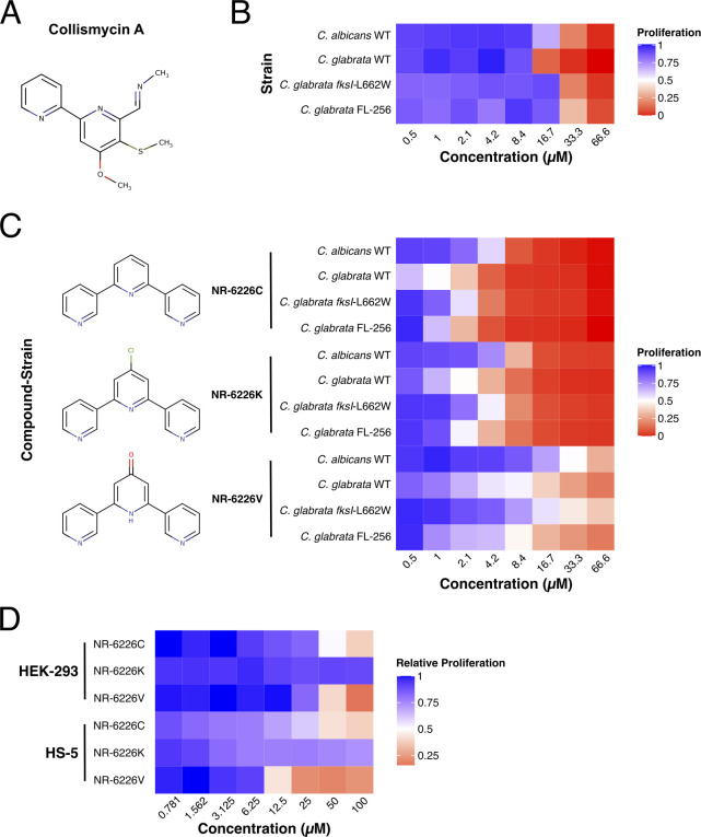
Fig 1 Identification of the ColA analog NR-6226C as an antifungal agent with a potential therapeutic window. (A) ColA compound structure. (B) Heat map of relative proliferation of several wild-type (WT) and echinocandin- and azole-resistant Candida strains. The indicated strains were incubated in synthetic defined medium (SD) with increasing concentrations of ColA for 24 h, after which proliferation was analyzed by measuring the optical density (OD600). Data were normalized to dimethyl sulfoxide (DMSO)-treated control samples. (C) Heat map of Candida proliferation in the presence of the ColA analogs NR-6226C, NR-6226K, and NR-6226V. The indicated fungal strains were incubated in the presence of increasing compound concentrations in yeast nitrogen base (YNB) −iron (0.69% yeast nitrogen base without amino acids or iron, with 2% dextrose) media for 24 h, after which proliferation was measured using OD600. (D) Human HEK-293 and HS-5 cells are considerably less sensitive to ColA analogs than Candida spp. Cells were incubated with increasing concentrations of the indicated compounds for 24 h, after which relative cell viability was assayed using CellTiter-Glo. Data were normalized to DMSO controls.
Fig 1 Identification of the ColA analog NR-6226C as an antifungal agent with a potential therapeutic window. (A) ColA compound structure. (B) Heat map of relative proliferation of several wild-type (WT) and echinocandin- and azole-resistant Candida strains. The indicated strains were incubated in synthetic defined medium (SD) with increasing concentrations of ColA for 24 h, after which proliferation was analyzed by measuring the optical density (OD600). Data were normalized to dimethyl sulfoxide (DMSO)-treated control samples. (C) Heat map of Candida proliferation in the presence of the ColA analogs NR-6226C, NR-6226K, and NR-6226V. The indicated fungal strains were incubated in the presence of increasing compound concentrations in yeast nitrogen base (YNB) −iron (0.69% yeast nitrogen base without amino acids or iron, with 2% dextrose) media for 24 h, after which proliferation was measured using OD600. (D) Human HEK-293 and HS-5 cells are considerably less sensitive to ColA analogs than Candida spp. Cells were incubated with increasing concentrations of the indicated compounds for 24 h, after which relative cell viability was assayed using CellTiter-Glo. Data were normalized to DMSO controls.CC BY · PubMed Central
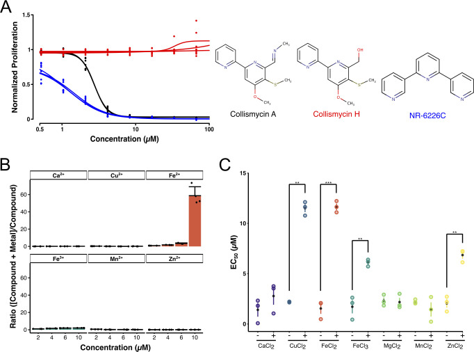
Fig 2 NR-6226C is an iron chelator that inhibits Candida proliferation by sequestering iron. (A) ColH, which does not bind iron, does not inhibit growth of wild-type C. glabrata (JEY10028). Fungal cells were treated with ColA, ColH, and NR-6226C in SD media for 24 h, after which proliferation was measured using OD600. Data were normalized to DMSO control samples. (B) NR-6226C selectively binds Fe2+ in vitro. The interaction of NR-6226C with increasing concentrations of the indicated metal ions; n = 3. (C) Exogenous Fe2+ rescues the antiproliferative effect of NR-6226C. Wild-type C. glabrata was incubated for 24 h with NR-6226C in presence of 5 µM CaCl2, CuCl2, FeCl2, FeCl3, MgCl2, MnCl2, or ZnCl2, respectively. Proliferation was measured using OD600, and values were normalized to DMSO controls. Resulting EC50 values were calculated, and statistical significance was analyzed using unpaired two-sample t-tests in R; ***P ≤ 0.001; **P ≤ 0.01; n = 3.
Fig 2 NR-6226C is an iron chelator that inhibits Candida proliferation by sequestering iron. (A) ColH, which does not bind iron, does not inhibit growth of wild-type C. glabrata (JEY10028). Fungal cells were treated with ColA, ColH, and NR-6226C in SD media for 24 h, after which proliferation was measured using OD600. Data were normalized to DMSO control samples. (B) NR-6226C selectively binds Fe2+ in vitro. The interaction of NR-6226C with increasing concentrations of the indicated metal ions; n = 3. (C) Exogenous Fe2+ rescues the antiproliferative effect of NR-6226C. Wild-type C. glabrata was incubated for 24 h with NR-6226C in presence of 5 µM CaCl2, CuCl2, FeCl2, FeCl3, MgCl2, MnCl2, or ZnCl2, respectively. Proliferation was measured using OD600, and values were normalized to DMSO controls. Resulting EC50 values were calculated, and statistical significance was analyzed using unpaired two-sample t-tests in R; ***P ≤ 0.001; **P ≤ 0.01; n = 3.CC BY · PubMed Central
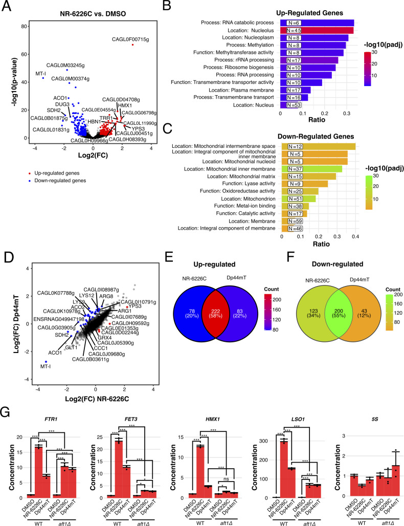
Fig 3 NR-6226C treatment induces an iron starvation response. (A) Wild-type C. glabrata cells were incubated with either DMSO or 10 µM NR-6226C for 1 h, after which RNA levels were analyzed by RNA sequencing. The volcano plot shows transcripts below (blue) or above (red) a 1.4-fold change (log2 fold change <−0.5 or >0.5) and an adjusted P-value <0.01 threshold in NR-6226C-treated cells relative to the DMSO control. (B and C) GO analysis of genes that are either upregulated (B) or downregulated (C) by NR-6226C treatment. (D) Volcano plot showing commonly regulated genes based on the threshold for the calculated mean difference and standard deviation in log fold change between C. glabrata cells treated with either NR-6226C or Dp44mT (log2 fold change <−0.683 or >0.635). (E and F) Venn diagram showing the count and respective percentages of upregulated (E) or downregulated (F) genes between C. glabrata cells treated with either NR-6226C or Dp44mT. (G) Validation of a panel of selected genes by quantitative reverse transcription PCR (RT-qPCR) shows that NR-6226C treatment activates the transcription factor Aft1. Wild-type or aft1Δ S. cerevisiae strains were treated with DMSO, 10 µM NR-6226C, or 10 µM Dp44mT for 24 h, after which RNA levels were analyzed by RT-qPCR. cDNA concentrations were normalized to DMSO controls, followed by calculation of the means, sample standard deviations, and statistical significance using one-way analysis of variance (ANOVA) and Tukey’s HSD for post hoc analyses in R. ***P ≤ 0.001; **P ≤ 0.01; ns, not significant; n = 3.
Fig 3 NR-6226C treatment induces an iron starvation response. (A) Wild-type C. glabrata cells were incubated with either DMSO or 10 µM NR-6226C for 1 h, after which RNA levels were analyzed by RNA sequencing. The volcano plot shows transcripts below (blue) or above (red) a 1.4-fold change (log2 fold change <−0.5 or >0.5) and an adjusted P-value <0.01 threshold in NR-6226C-treated cells relative to the DMSO control. (B and C) GO analysis of genes that are either upregulated (B) or downregulated (C) by NR-6226C treatment. (D) Volcano plot showing commonly regulated genes based on the threshold for the calculated mean difference and standard deviation in log fold change between C. glabrata cells treated with either NR-6226C or Dp44mT (log2 fold change <−0.683 or >0.635). (E and F) Venn diagram showing the count and respective percentages of upregulated (E) or downregulated (F) genes between C. glabrata cells treated with either NR-6226C or Dp44mT. (G) Validation of a panel of selected genes by quantitative reverse transcription PCR (RT-qPCR) shows that NR-6226C treatment activates the transcription factor Aft1. Wild-type or aft1Δ S. cerevisiae strains were treated with DMSO, 10 µM NR-6226C, or 10 µM Dp44mT for 24 h, after which RNA levels were analyzed by RT-qPCR. cDNA concentrations were normalized to DMSO controls, followed by calculation of the means, sample standard deviations, and statistical significance using one-way analysis of variance (ANOVA) and Tukey’s HSD for post hoc analyses in R. ***P ≤ 0.001; **P ≤ 0.01; ns, not significant; n = 3.CC BY · PubMed Central
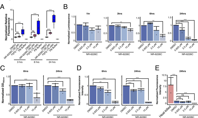
Fig 4 NR-6226C treatment impairs mitochondrial functions and induces ROS formation. (A) C. glabrata cells were preloaded with the fluorescent ROS sensor H2DCF-DA, after which changes in fluorescence were measured after 3, 6, and 24 h after exposure to DMSO, 30 µM NR-6226C, or 75 µM H2O2. (B) C. glabrata cells were treated for 1, 3, 6, and 24 h with DMSO, 0.625 µM, 2.5 µM, or 10 µM NR-6226C, after which relative ATP levels were measured by luminescence using CellTiter-Glo. An equal number of cells was harvested for each experiment, and luminescence was normalized to DMSO. (C) C. glabrata cells treated with DMSO, 0.625 µM, 2.5 µM, or 10 µM NR-6226C for 6 and 24 h and then treated with XTT-phenazine methosulfate. Colorimetric changes were measured using OD492nm. An equal number of cells were harvested for each experiment, after which the OD492nm was normalized to DMSO. (D) C. glabrata cells were treated with DMSO, 0.625 µM, 2.5 µM, or 10 µM NR-6226C for 6 and 24 h, after which they were loaded with MitoTracker and measured for fluorescence using a plate reader. The autofluorescence from unstained treated samples was subtracted from the raw data and then normalized to DMSO. (E) C. glabrata cells were treated with DMSO, 0.625 µM, 2.5 µM, or 10 µM NR-6226C for 24 h and then stained with propidium iodide and measured for fluorescence using a plate reader. The autofluorescence from unstained samples was subtracted from the raw data and then normalized to DMSO. Statistical analyses for all panels were performed using one-way ANOVA and Tukey’s HSD for post hoc analyses; ***P ≤ 0.001; **P ≤ 0.01; ns, not significant; n = 3.
Fig 4 NR-6226C treatment impairs mitochondrial functions and induces ROS formation. (A) C. glabrata cells were preloaded with the fluorescent ROS sensor H2DCF-DA, after which changes in fluorescence were measured after 3, 6, and 24 h after exposure to DMSO, 30 µM NR-6226C, or 75 µM H2O2. (B) C. glabrata cells were treated for 1, 3, 6, and 24 h with DMSO, 0.625 µM, 2.5 µM, or 10 µM NR-6226C, after which relative ATP levels were measured by luminescence using CellTiter-Glo. An equal number of cells was harvested for each experiment, and luminescence was normalized to DMSO. (C) C. glabrata cells treated with DMSO, 0.625 µM, 2.5 µM, or 10 µM NR-6226C for 6 and 24 h and then treated with XTT-phenazine methosulfate. Colorimetric changes were measured using OD492nm. An equal number of cells were harvested for each experiment, after which the OD492nm was normalized to DMSO. (D) C. glabrata cells were treated with DMSO, 0.625 µM, 2.5 µM, or 10 µM NR-6226C for 6 and 24 h, after which they were loaded with MitoTracker and measured for fluorescence using a plate reader. The autofluorescence from unstained treated samples was subtracted from the raw data and then normalized to DMSO. (E) C. glabrata cells were treated with DMSO, 0.625 µM, 2.5 µM, or 10 µM NR-6226C for 24 h and then stained with propidium iodide and measured for fluorescence using a plate reader. The autofluorescence from unstained samples was subtracted from the raw data and then normalized to DMSO. Statistical analyses for all panels were performed using one-way ANOVA and Tukey’s HSD for post hoc analyses; ***P ≤ 0.001; **P ≤ 0.01; ns, not significant; n = 3.CC BY · PubMed Central
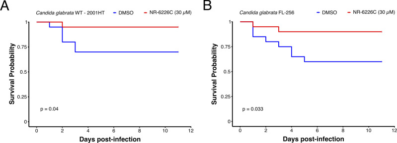
Fig 5 NR-6226C treatment improves the survival of C. glabrata-infected G. mellonella larvae. (A and B) G. mellonella larvae were infected with either C. glabrata 2001HT (A) or C. glabrata FL-256 (B) and treated with either 0.15% DMSO or 30 µM NR-6226C, after which overall survival was monitored over time. Survival curves were generated using the Kaplan-Meier formula. Statistical significance was calculated using log-rank tests in R.
Fig 5 NR-6226C treatment improves the survival of C. glabrata-infected G. mellonella larvae. (A and B) G. mellonella larvae were infected with either C. glabrata 2001HT (A) or C. glabrata FL-256 (B) and treated with either 0.15% DMSO or 30 µM NR-6226C, after which overall survival was monitored over time. Survival curves were generated using the Kaplan-Meier formula. Statistical significance was calculated using log-rank tests in R.CC BY · PubMed Central
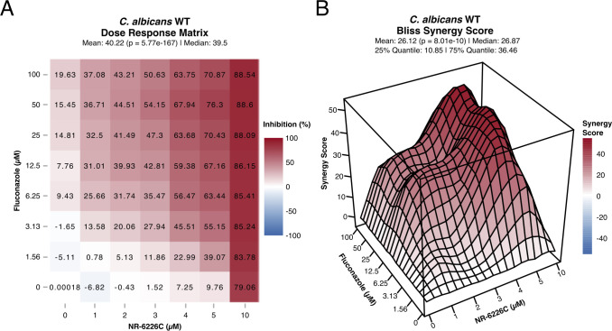
Fig 6 Fluconazole and NR-6226C have a synergistic effect. (A) Dose-response matrix of C. albicans WT cells treated in combination with fluconazole and NR-6226C. Relative cell numbers were quantified as described in Fig. 1B. (B) 3D visualization of the synergy matrix shown in panel A using the Bliss synergy model. Panels A and B were generated using SynergyFinder in R.
Fig 6 Fluconazole and NR-6226C have a synergistic effect. (A) Dose-response matrix of C. albicans WT cells treated in combination with fluconazole and NR-6226C. Relative cell numbers were quantified as described in Fig. 1B. (B) 3D visualization of the synergy matrix shown in panel A using the Bliss synergy model. Panels A and B were generated using SynergyFinder in R.CC BY · PubMed Central

Verification

Claims the paper makes

The claims and statistics of this paper haven’t been checked yet.

Run the check — free, no account needed. It reads the open-access text and checks the sources it cites.

A few free checks a day · papers already verified open instantly · results are public

Self-check

Percentages, sums and reported differences re-computed from the paper’s own numbers.

The self-check needs the paper’s full text.

Citations

Cites

Research works printed but not linked: 64

  1. 2017, Global and Multi-National Prevalence of Fungal Diseases-Estimate Precision, J Fungi (Basel, 3, 57, 10.3390/jof3040057 · DOI · Paper page
  2. 10.1146/annurev-micro-030117-020345 · DOI · Paper page
  3. 10.1038/s41579-022-00720-1 · DOI · Paper page
  4. 2021, Uncharted territories in the discovery of antifungal and antivirulence natural products from bacteria, Comput Struct Biotechnol J, 19, 1244, 10.1016/j.csbj.2021.02.003 · DOI
  5. 10.1038/ncomms6675 · DOI
  6. 10.1073/pnas.0812082106 · DOI
  7. Seipke RF , Grüschow S , Goss RJM , Hutchings MI . 2012. Isolating Antifungals from Fungus-Growing Ant Symbionts Using a Genome-Guided Chemistry Approach, p. 47–70. In Methods in Enzymology. Elsevier. · DOI
  8. 10.7164/antibiotics.47.1385 · DOI
  9. 10.1016/j.chembiol.2013.06.014 · DOI
  10. 10.1021/jm00142a012 · DOI
  11. 2016, Proteomic profiling reveals that collismycin A is an iron chelator, Sci Rep, 6, 38385, 10.1038/srep38385 · DOI
  12. 2010, Design, synthesis, and biological properties of triazole derived compounds and their transition metal complexes, J Enzyme Inhib Med Chem, 25, 737, 10.3109/14756360903389906 · DOI
  13. 10.1038/nrmicro.2017.15 · DOI · Paper page
  14. 10.1074/jbc.R114.588814 · DOI
  15. 2023, Mixed heavy metal stress induces global iron starvation response, ISME J, 17, 382, 10.1038/s41396-022-01351-3 · DOI
  16. 10.1182/blood-2004-03-0868 · DOI
  17. 10.1002/mbo3.303 · DOI
  18. 10.1242/jcs.03229 · DOI
  19. 10.1016/0092-8674(94)90345-X · DOI · Paper page
  20. 10.1111/j.1365-2958.2004.04214.x · DOI · Paper page
  21. 10.1534/genetics.106.066944 · DOI
  22. 1989, Candida glabrata metallothioneins. Cloning and sequence of the genes and characterization of proteins, J Biol Chem, 264, 19747, 10.1016/S0021-9258(19)47175-5 · DOI
  23. 10.1158/0008-5472.CAN-11-1218 · DOI
  24. 10.1128/EC.00354-07 · DOI · Paper page
  25. 10.3389/fmicb.2020.582830 · DOI
  26. 10.1002/j.1460-2075.1995.tb07106.x · DOI · Paper page
  27. 10.1371/journal.pone.0004357 · DOI · Paper page
  28. 10.1016/0022-1759(93)90011-U · DOI · Paper page
  29. 10.4161/viru.2.6.18520 · DOI
  30. 2022, Antifungal Combinations against Candida Species: From Bench to Bedside, J Fungi (Basel, 8, 1077, 10.3390/jof8101077 · DOI
  31. 2022, SynergyFinder Plus: Toward Better Interpretation and Annotation of Drug Combination Screening Datasets, Genomics, Proteomics & Bioinformatics, 20, 587, 10.1016/j.gpb.2022.01.004 · DOI · Paper page
  32. 10.1093/nar/gkac382 · DOI
  33. 10.1093/jac/dkq136 · DOI
  34. 10.1177/15648265070284S405 · DOI
  35. 10.1021/bi061429r · DOI
  36. 2019, The regulation of iron homeostasis in the fungal human pathogen Candida glabrata, Microbiology, 165, 1041, 10.1099/mic.0.000807 · DOI
  37. 10.1016/j.freeradbiomed.2010.08.020 · DOI
  38. 2004, Transcriptional Remodeling in Response to Iron Deprivation in Saccharomyces cerevisiae, MBoC, 15, 1233, 10.1091/mbc.e03-09-0642 · DOI · Paper page
  39. 2002, The Saccharomyces cerevisiae mitochondrial succinate:ubiquinone oxidoreductase, Biochimica et Biophysica Acta (BBA) - Bioenergetics, 1553, 102, 10.1016/S0005-2728(01)00229-8 · DOI
  40. 10.1126/science.1106391 · DOI
  41. 10.1021/cr9001676 · DOI
  42. 2012, The role of mitochondria in cellular iron–sulfur protein biogenesis and iron metabolism, Biochimica et Biophysica Acta (BBA) - Molecular Cell Research, 1823, 1491, 10.1016/j.bbamcr.2012.05.009 · DOI
  43. 10.1371/journal.pgen.1007476 · DOI
  44. 10.1016/j.cell.2004.11.032 · DOI · Paper page
  45. 10.1093/femsre/fux050 · DOI
  46. 10.1371/journal.pone.0111585 · DOI
  47. 2018, A mitochondrial proteomics view of complex I deficiency in Candida albicans, Mitochondrion, 38, 48, 10.1016/j.mito.2017.08.003 · DOI · Paper page
  48. 10.1073/pnas.1911560116 · DOI
  49. 2022, Disruption of Iron Homeostasis and Mitochondrial Metabolism Are Promising Targets to Inhibit Candida auris, Microbiol Spectr, 10, e00100
  50. 2023, Combined Transcriptome and Metabolome Analysis Reveals That the Potent Antifungal Pyrylium Salt Inhibits Mitochondrial Complex I in Candida albicans, Microbiol Spectr, 11, e03209
  51. 2022, Transcriptional regulation of ergosterol biosynthesis genes in response to iron deficiency, Environ Microbiol, 24, 5248, 10.1111/1462-2920.16157 · DOI
  52. 10.1016/0378-1119(96)00132-1 · DOI
  53. 10.1002/(SICI)1097-0061(19980130)14:2<115::AID-YEA204>3.0.CO;2-2 · DOI
  54. 10.1038/nature00935 · DOI · Paper page
  55. 2017, Self-packed core shell nano liquid chromatography columns and silica-based monolithic trap columns for targeted proteomics, Journal of Chromatography A, 1498, 111, 10.1016/j.chroma.2017.03.043 · DOI
  56. 2023, Mitotic chromosome condensation resets chromatin to safeguard transcriptional homeostasis during interphase, Proc Natl Acad Sci USA, 120, e2210593120, 10.1073/pnas.2210593120 · DOI
  57. 10.14806/ej.17.1.200 · DOI · Paper page
  58. 10.1093/bioinformatics/bts635 · DOI · Paper page
  59. 10.1093/bioinformatics/btu638 · DOI · Paper page
  60. 10.1186/gb-2010-11-10-r106 · DOI · Paper page
  61. 10.1186/s13059-014-0550-8 · DOI · Paper page
  62. 10.2307/2346101 · DOI
  63. 10.1016/j.celrep.2021.110186 · DOI
  64. 2015, A rapid method to assess reactive oxygen species in yeast using H 2 DCF-DA, Anal Methods, 7, 8572, 10.1039/C5AY02278A · DOI

Cited by

Sorted by citations

See all 10

Metadata

Authors
Corrales, J., Ramos-Alonso, L., González-Sabín, J., Ríos-Lombardía, N., Trevijano-Contador, N., Berg, H. E., Skottvoll, F. S., Moris, F., Zaragoza, Ó., Chymkowitch, P., Garcia, I., & Enserink, J. M.
Affiliations
  • University of Oslo (NO)
  • Instituto de Salud Carlos III (ES)
Corresponding author
Enserink, Jorrit M.
Venue
Microbiology Spectrum
Publisher
openRxiv
Published
2023-08-09
Volume
12
Issue
2
Pages
e0259423-e0259423
ISSN
2165-0497 (ISSN-L)
OpenAlex
W4390954798
Cited by
10
References
1
Citations by year
2026: 2 · 2025: 4 · 2024: 3 · 2023: 1
FWCI
3.10
Type
journal article
Language
en
Open access
Licence
cc-by
Locations
  • Microbiology Spectrum (publishedVersion, cc-by)
  • bioRxiv (Cold Spring Harbor Laboratory) (acceptedVersion, cc-by-nd)
  • PubMed Central (publishedVersion, cc-by)
  • PubMed (publishedVersion)
  • DOAJ (DOAJ: Directory of Open Access Journals) (submittedVersion, cc-by-sa)
  • NORA - Norwegian Open Research Archives (submittedVersion, other-oa)
Funders
  • Kreftforeningen (182524, 208012, 182524, 208012)
  • Agence Nationale de la Recherche (ANR-10-INBS-0009, ANR-10)
  • Helse- og Omsorgsdepartementet (2018012, 2019096, 2017072, 2017064)
  • Ministerio de Ciencia e Innovación (MCIN/AEI/10, .13039/501100011033, / AEI/10, PID2020-114546RB, 501100011033, PID2020)
  • Norges Forskningsråd (301268, #262652, 261936, MCIN/AEI/10.13039/501100011033, 261936, 301268, 262652)
  • Agencia Estatal de Investigación (AEI//10.13039/501100011033/, 501100011033, by MCIN/AEI/10.13039/501100011033, 10.13039/501100011033, MCIN/AEI/10.13039/501 100 011 033, 13039, .13039/501100011033, 10.13039, PID2020-114546RB, AEI/10)
Topics
  • Health Sciences › Medicine › Infectious Diseases › Antifungal resistance and susceptibility
  • Life Sciences › Agricultural and Biological Sciences › Plant Science › Fungal Plant Pathogen Control
  • Life Sciences › Immunology and Microbiology › Applied Microbiology and Biotechnology › Microbial Natural Products and Biosynthesis
Keywords
iron chelation, antifungal agents, Candida albicans, Candida glabrata, fluconazole synergy, azole resistance, mitochondrial dysfunction, reactive oxygen species, metabolic reprogramming, ATP depletion, iron homeostasis, Galleria mellonella infection model, fungal drug resistance, natural product derivatives
MeSH terms
Animals, Anti-Infective Agents, Antifungal Agents, Azoles, Candida, Candida albicans, Humans, Iron, Iron Chelating Agents, Mammals, Microbial Sensitivity Tests, Mycoses, Fluconazole, Drug Resistance, Fungal, Candida glabrata

About these checks

Registry, retraction, Cites and statistics results are computed, not judged, with one exception: a printed reference that no index lists is read once by a model before it counts as a citation error. Claims are extracted by a model and kept only when their quote appears verbatim in the text; each cited source is then checked, with a model’s help, for whether it says what the claim relies on. Those verdicts can be wrong — read the claim’s quote and the cited source before relying on one.

Statistics and arithmetic are recomputed, not judged.

Confidence bands are shown only for fields calibrated on our annotated test set. None is calibrated yet, so no band is shown.

What counts as a claim ›How we measure confidence ›How we check this ›

Are you an author, or do you see a problem with this page? Request a review