

The DOI matches its Crossref record; 1 of the 1 references it cites were found in a registry or an index.
Cryo-EM structures of human magnesium channel MRS2 reveal gating and regulatory mechanisms
Open accessPreprint — published as 10.1038/s41467-023-42599-3 · Paper page
Abstract
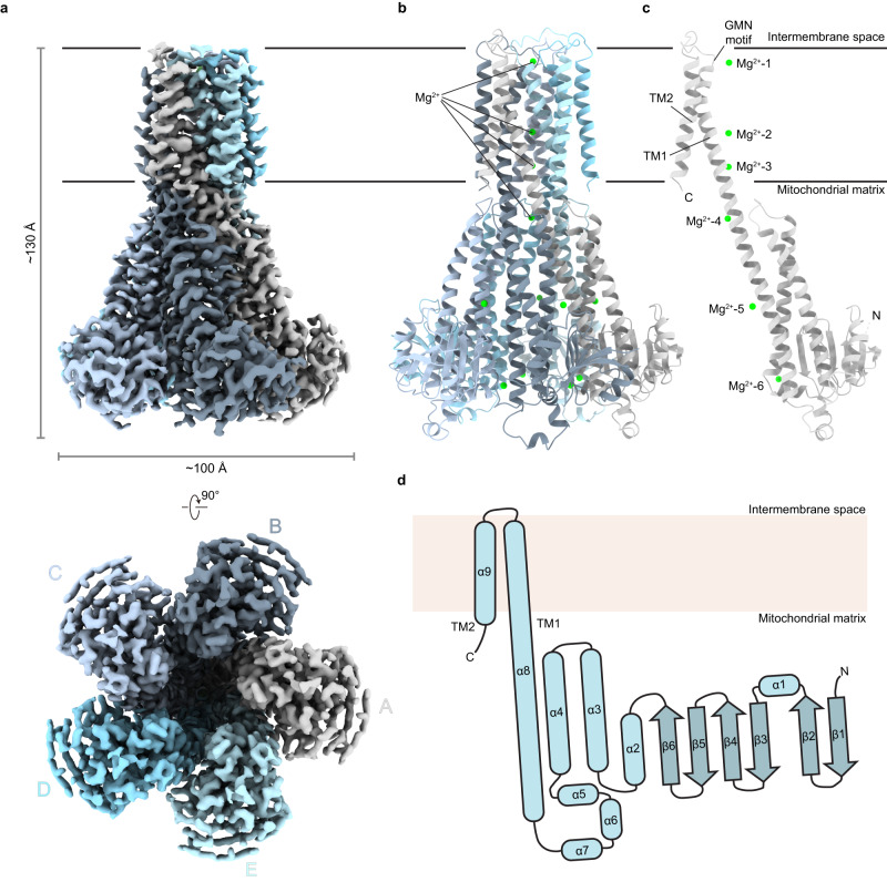
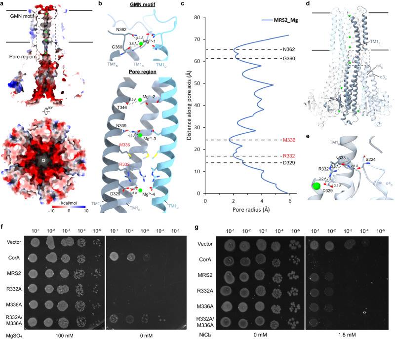
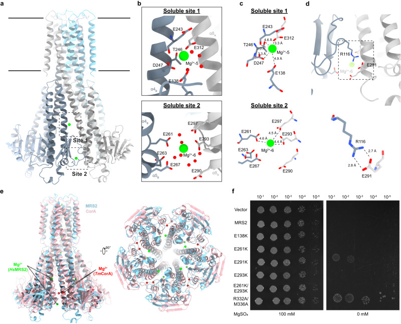
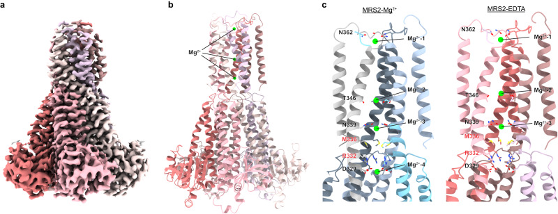
Magnesium ions (Mg 2+ ) play an essential role in cellular physiology. In mitochondria, protein and ATP synthesis and various metabolic pathways are directly regulated by Mg 2+ . MRS2, a magnesium channel located in the inner mitochondrial membrane, mediates the influx of Mg 2+ into the mitochondrial matrix and regulates Mg 2+ homeostasis. Knockdown of MRS2 in human cells leads to reduced uptake of Mg 2+ into mitochondria and disruption of the mitochondrial metabolism. Despite the importance of MRS2, the Mg 2+ translocation and regulation mechanisms of MRS2 are still unclear. Here, using cryo-EM we determined the structure of human MRS2 in the presence and absence of Mg 2+ at 2.8 Å and 3.3 Å, respectively. From the homo-pentameric structures, we identified R332 and M336 as major gating residues, which were then tested using mutagenesis and two cellular divalent ion uptake assays. A network of hydrogen bonds was found connecting the gating residue R332 to the soluble domain, potentially regulating the gate. Two Mg 2+ -binding sites were identified in the MRS2 soluble domain, distinct from the two sites previously reported in CorA, a homolog of MRS2 in prokaryotes. Altogether, this study provides the molecular basis for understanding the Mg 2+ translocation and regulatory mechanisms of MRS2.
Overview
The claims and statistics of this paper haven’t been checked yet.
Run the check — free, no account needed. It reads the open-access text and checks the sources it cites.
Integrity signals
Registry
Cites
Statistics
Figures
Verification
Claims the paper makes
The claims and statistics of this paper haven’t been checked yet.
Run the check — free, no account needed. It reads the open-access text and checks the sources it cites.
Self-check
Percentages, sums and reported differences re-computed from the paper’s own numbers.
The self-check needs the paper’s full text.
Citations
Cites
Highly accurate protein structure prediction with AlphaFold
Jumper, J., Evans, R., Pritzel, A., Green, T., Figurnov, M., Ronneberger, O., Tunyasuvunakool, K., Bates, R., Žídek, A., Potapenko, A., et al. (2021) · Nature · doi:10.1038/s41586-021-03819-2 · Cited by 43,764
Research works printed but not linked: 55
- 10.1093/ndtplus/sfr163 · DOI · Paper page
- 10.1152/physrev.00012.2014 · DOI · Paper page
- 10.1007/s10534-021-00328-7 · DOI
- 10.1136/openhrt-2018- · DOI
- 10.1016/S0009-8981(99)00258-2 · DOI
- 10.3390/ijms20143439 · DOI
- 10.1016/j.abb.2011.05.010 · DOI
- 10.1074/jbc.273.14.7850 · DOI
- 10.1155/2017/6797460 · DOI · Paper page
- 10.1113/jphysiol.2013.268235 · DOI
- 10.1074/jbc.274.29.20438 · DOI
- 10.1006/geno.2000.6407 · DOI
- 10.1093/emboj/cdg122 · DOI
- 10.1529/biophysj.107.112318 · DOI
- 10.1111/j.1582-4934.2008.00328.x · DOI
- 10.1038/srep30027 · DOI · Paper page
- 10.1371/journal.pgen.1001262 · DOI
- 10.1016/j.cell.2020.08.049 · DOI
- 10.1016/j.celrep.2023.112155 · DOI
- 10.1038/ncomms4590 · DOI
- 10.1073/pnas.1209018109 · DOI
- 10.1107/s0907444913011712 · DOI
- 10.26508/lsa.202201742 · DOI · Paper page
- 10.26508/lsa.201900429 · DOI
- 10.1038/s41587-021-01156-3 · DOI
- 10.1074/mcp.M114.043083 · DOI
- 10.1016/j.cell.2015.12.055 · DOI
- 10.1038/sj.emboj.7601269 · DOI
- 10.1038/nature04642 · DOI
- 10.1126/science.1127121 · DOI
- 10.1016/j.str.2017.05.024 · DOI
- 10.1073/pnas.1210076109 · DOI
- 10.1038/s41467-017-01483-7 · DOI
- 10.1016/j.ijbiomac.2021.06.130 · DOI
- 10.1021/ja4087769 · DOI
- 10.1073/pnas.1319054111 · DOI
- 10.1016/S0263-7855(97)00009-X · DOI
- 10.1074/jbc.M707889200 · DOI
- 10.1016/j.bpj.2009.11.009 · DOI
- 10.1371/journal.pcbi.1004303 · DOI
- 10.1371/journal.pbio.3001231 · DOI
- 10.1038/emboj.2009.288 · DOI · Paper page
- 10.1038/s41467-017-00082-w · DOI
- 10.1021/acs.jcim.1c00261 · DOI
- 10.1038/s41467-023-40516-2 · DOI
- 10.1042/BJ20121745 · DOI
- 10.1016/B978-0-12-418687-3.00018-5 · DOI
- 10.1016/j.jsb.2005.07.007 · DOI
- 10.7554/eLife.35383 · DOI
- 10.1038/nmeth.4169 · DOI · Paper page
- 10.1002/jcc.20084 · DOI
- 10.1107/S0907444910007493 · DOI · Paper page
- 10.1107/S2059798318006551 · DOI
- 10.1002/pro.3330 · DOI · Paper page
- 10.1002/pro.3235 · DOI · Paper page
Metadata
- Authors
- Lai, L. T. F., Balaraman, J., Zhou, F., & Matthies, D.
- Affiliations
- National Institutes of Health (US)
- Corresponding author
- Matthies, Doreen
- Venue
- Nature Communications
- Publisher
- openRxiv
- Published
- 2023-08-23
- Volume
- 14
- Issue
- 1
- Pages
- 7207-7207
- ISSN
- 2041-1723 (ISSN-L)
- PMC
- PMC10632456
- OpenAlex
- W4388500134
- Cited by
- 30
- References
- 1
- Citations by year
- 2026: 9 · 2025: 8 · 2024: 13
- FWCI
- 2.59
- Type
- journal article
- Language
- en
- Open access
- gold
- doi.org
- Licence
- cc-by
- Locations
- Nature Communications (publishedVersion, cc-by)
- bioRxiv (Cold Spring Harbor Laboratory) (acceptedVersion)
- PubMed Central (publishedVersion, cc-by)
- PubMed (publishedVersion)
- PubMed Central (submittedVersion)
- RePEc: Research Papers in Economics (submittedVersion)
- DOAJ (DOAJ: Directory of Open Access Journals) (submittedVersion, cc-by-sa)
- Funders
- U.S. Department of Health and Human Services (ZIA HD008998)
- National Institutes of Health
- National Institute of Child Health and Human Development
- Eunice Kennedy Shriver National Institute of Child Health and Human Development (ZIAHD008998)
- NHLBI Division of Intramural Research
- Topics
- Life Sciences › Biochemistry, Genetics and Molecular Biology › Molecular Biology › Ion channel regulation and function
- Life Sciences › Biochemistry, Genetics and Molecular Biology › Molecular Biology › ATP Synthase and ATPases Research
- Life Sciences › Biochemistry, Genetics and Molecular Biology › Structural Biology › Advanced Electron Microscopy Techniques and Applications
- Keywords
- MRS2, magnesium channel, magnesium homeostasis, channel gating, magnesium binding sites, inner mitochondrial membrane, hydrogen bond network
About these checks
Registry, retraction, Cites and statistics results are computed, not judged, with one exception: a printed reference that no index lists is read once by a model before it counts as a citation error. Claims are extracted by a model and kept only when their quote appears verbatim in the text; each cited source is then checked, with a model’s help, for whether it says what the claim relies on. Those verdicts can be wrong — read the claim’s quote and the cited source before relying on one.
Statistics and arithmetic are recomputed, not judged.
Confidence bands are shown only for fields calibrated on our annotated test set. None is calibrated yet, so no band is shown.
What counts as a claim ›How we measure confidence ›How we check this ›
Are you an author, or do you see a problem with this page? Request a review
