PreprintCited by 1Lysosomal Storage Disorders ResearchGlycogen Storage Diseases and MyoclonusProteoglycans and glycosaminoglycans research

The DOI matches its Crossref record; 1 of the 1 references it cites were found in a registry or an index.

Structure of the human heparan-α-glucosaminide N -acetyltransferase (HGSNAT)

Open access
Navratna, Vikas, Kumar, Arvind, Rana, Jaimin K., Mosalaganti, Shyamal·2023·bioRxiv (Cold Spring Harbor Laboratory)

Preprint — published as 10.7554/elife.93510 · Paper page

Abstract

Degradation of heparan sulfate (HS), a glycosaminoglycan (GAG) comprised of repeating units of N -acetylglucosamine and glucuronic acid, begins in the cytosol and is completed in the lysosomes. Acetylation of the terminal non-reducing amino group of α-D-glucosamine of HS is essential for its complete breakdown into monosaccharides and free sulfate. Heparan-α-glucosaminide N -acetyltransferase (HGSNAT), a resident of the lysosomal membrane, catalyzes this essential acetylation reaction by accepting and transferring the acetyl group from cytosolic acetyl-CoA to terminal α-D-glucosamine of HS in the lysosomal lumen. Mutation-induced dysfunction in HGSNAT causes abnormal accumulation of HS within the lysosomes and leads to an autosomal recessive neurodegenerative lysosomal storage disorder called mucopolysaccharidosis IIIC (MPS IIIC). There are no approved drugs or treatment strategies to cure or manage the symptoms of, MPS IIIC. Here, we use cryo-electron microscopy (cryo-EM) to determine a high-resolution structure of the HGSNAT-acetyl-CoA complex, the first step in HGSNAT catalyzed acetyltransferase reaction. In addition, we map the known MPS IIIC mutations onto the structure and elucidate the molecular basis for mutation-induced HGSNAT dysfunction.

Overview

The claims and statistics of this paper haven’t been checked yet.

Run the check — free, no account needed. It reads the open-access text and checks the sources it cites.

A few free checks a day · papers already verified open instantly · results are public

Integrity signals

Registry

Crossref · 10.1101/2023.10.23.563672

Cites

1 of 1 references verified0 citation errors · 0 not checkable

Statistics

Figures

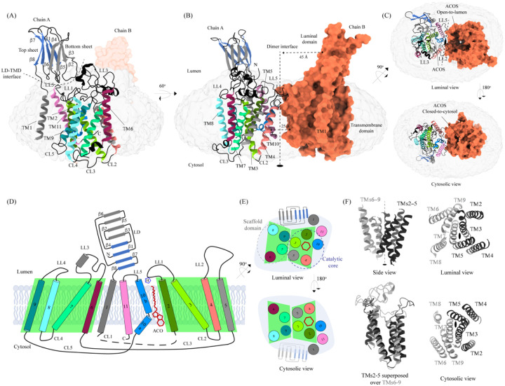
Figure 1: Structure of HGSNAT Panels (A) and (B) show two different orientations of HGSNAT dimer that highlight (dashed lines) the LD-TMD interface and dimer interface respectively. Micelle is displayed in gray. Chain A is displayed as a cartoon and chain B as orange surface. All the luminal loops (LLs), cytosolic loops (CLs), and the loops that connect β-sheets are shown in black. The top and bottom sheets in the luminal domain (LD) are colored blue and gray, respectively. The two-fold rotation axis is displayed as a dashed line with an ellipsoid. (C) Luminal (top) and cytosolic (bottom) views of the protein. The surface representation of chain B suggests that the acetyl-CoA binding site (ACOS) is more accessible from the luminal side (top) than the cytosolic side (bottom). (D) 2D topology of HGSNAT and YeiB family. The helices and strands in the topology are colored similarly to the 3D structure. TMs 2–5 and 6–9 form two bundles (4+4), highlighted by green parallelograms, that are related to each other by a 2-fold rotation parallel to the plane of the membrane. TMs 1, 10, and 11 do not seem involved in this internal symmetry, with TM10 being bent in the plane of the membrane into two halves TM10a and TM10b. The relative position of bound ACO and active site H269 of LL1 are indicated. (E) Luminal (top) and cytosolic (bottom) views of the protein topology. TMs 2–5 and TM10 enclose ACOS (red hexagon) and are referred to as catalytic core (blue dashed oval). TMs 6–9 will be referred to as scaffold domain (gray dashed oval). (F) 4+4 bundle formed by TMs 2–5 (black) and TMs 6–9 (gray) are related by a 2-fold rotation. The last sub-panel (bottom left) shows a superposition of TMs 2–5 on TMs 6–9.
Figure 1: Structure of HGSNAT Panels (A) and (B) show two different orientations of HGSNAT dimer that highlight (dashed lines) the LD-TMD interface and dimer interface respectively. Micelle is displayed in gray. Chain A is displayed as a cartoon and chain B as orange surface. All the luminal loops (LLs), cytosolic loops (CLs), and the loops that connect β-sheets are shown in black. The top and bottom sheets in the luminal domain (LD) are colored blue and gray, respectively. The two-fold rotation axis is displayed as a dashed line with an ellipsoid. (C) Luminal (top) and cytosolic (bottom) views of the protein. The surface representation of chain B suggests that the acetyl-CoA binding site (ACOS) is more accessible from the luminal side (top) than the cytosolic side (bottom). (D) 2D topology of HGSNAT and YeiB family. The helices and strands in the topology are colored similarly to the 3D structure. TMs 2–5 and 6–9 form two bundles (4+4), highlighted by green parallelograms, that are related to each other by a 2-fold rotation parallel to the plane of the membrane. TMs 1, 10, and 11 do not seem involved in this internal symmetry, with TM10 being bent in the plane of the membrane into two halves TM10a and TM10b. The relative position of bound ACO and active site H269 of LL1 are indicated. (E) Luminal (top) and cytosolic (bottom) views of the protein topology. TMs 2–5 and TM10 enclose ACOS (red hexagon) and are referred to as catalytic core (blue dashed oval). TMs 6–9 will be referred to as scaffold domain (gray dashed oval). (F) 4+4 bundle formed by TMs 2–5 (black) and TMs 6–9 (gray) are related by a 2-fold rotation. The last sub-panel (bottom left) shows a superposition of TMs 2–5 on TMs 6–9.CC BY · PubMed Central
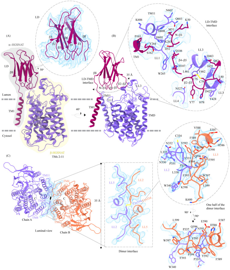
Figure 2: Domain organization, and LD-TMD and dimer interfaces of HGSNAT (A) HGSNAT is predicted to be proteolyzed into two chains of unequal size – α-HGSNAT (dark magenta cartoon, gray shaded area) and β-HGSNAT (purple cartoon, yellow shaded area). The site for proteolysis remains debated. Based on our structure and prediction of HGSNAT structures from other kingdoms (Fig S4), we have represented α- and β-HGSNAT fragments as shown in panel A. The inset (dashed oval) shows the luminal domain (dark magenta) fit to cryo-EM density (blue; display level 0.21 of the composite map in ChimeraX) (Fig S3). The lysosomal membrane is shown as a dashed gray line. (B) LD-TMD interface is highlighted (dashed line). Inset highlights the residues that interact at the LD-TMD interface, and cryo-EM density for the same (blue; display level 0.25 of the 3.26 Å C2 refined map in ChimeraX). C76-C79 disulfide of β2-β3 turn is shown as yellow sticks, while the residue sidechains are colored the same as their secondary structure elements, with heteroatoms highlighted. (C) Luminal-view of the protein with dimer interface highlighted (dashed line). Inset (dashed rectangle) highlights LL2 and LL5 that line the dimer interface, and the C334-C334 inter-chain disulfide (yellow) between the chains A (purple) and B (orange). The dashed oval inset shows one-half of the dimer interface with LL2 and LL5 of chains A and B, respectively, contributing other hydrophobic interactions that stabilize the dimer interface. The cryo-EM density in panel C is displayed as blue mesh (display level 0.22 of the C2 refine map in ChimeraX).
Figure 2: Domain organization, and LD-TMD and dimer interfaces of HGSNAT (A) HGSNAT is predicted to be proteolyzed into two chains of unequal size – α-HGSNAT (dark magenta cartoon, gray shaded area) and β-HGSNAT (purple cartoon, yellow shaded area). The site for proteolysis remains debated. Based on our structure and prediction of HGSNAT structures from other kingdoms (Fig S4), we have represented α- and β-HGSNAT fragments as shown in panel A. The inset (dashed oval) shows the luminal domain (dark magenta) fit to cryo-EM density (blue; display level 0.21 of the composite map in ChimeraX) (Fig S3). The lysosomal membrane is shown as a dashed gray line. (B) LD-TMD interface is highlighted (dashed line). Inset highlights the residues that interact at the LD-TMD interface, and cryo-EM density for the same (blue; display level 0.25 of the 3.26 Å C2 refined map in ChimeraX). C76-C79 disulfide of β2-β3 turn is shown as yellow sticks, while the residue sidechains are colored the same as their secondary structure elements, with heteroatoms highlighted. (C) Luminal-view of the protein with dimer interface highlighted (dashed line). Inset (dashed rectangle) highlights LL2 and LL5 that line the dimer interface, and the C334-C334 inter-chain disulfide (yellow) between the chains A (purple) and B (orange). The dashed oval inset shows one-half of the dimer interface with LL2 and LL5 of chains A and B, respectively, contributing other hydrophobic interactions that stabilize the dimer interface. The cryo-EM density in panel C is displayed as blue mesh (display level 0.22 of the C2 refine map in ChimeraX).CC BY · PubMed Central
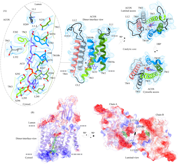
Figure 3: Acetyl-CoA binding site (ACOS) (A) Catalytic core (chain A) of HGSNAT comprised of TMs 2–5 and TM 10. LLs and CLs are shown in black, and the helices are colored as in Fig 1. Acetyl-CoA (ACO) is colored (purple), the same as chain A in Fig 2 with heteroatoms highlighted. The inset (dashed oval) shows ACOS and highlights the amino acids of HGSNAT that interact with ACO. The amino acids are colored same as the corresponding TMs, with heteroatoms highlighted. Cryo-EM density for ACOS is displayed as blue mesh (display level 0.3 of the 3.26 Å C2 refine map in ChimeraX). ACO could be modeled into the densities at chain A and B ACOSs with a mean correlation coefficient (CC) of 0.77. The nucleoside headgroup of ACO plugs in the cytosolic access of ACOS, and the luminal access seems relatively more accessible. (B) Electrostatic potential and surface charge distribution of HGSNAT, with the surface display colored based on the potential contoured from −10 kT (red) to +10 kT (blue). ACO bound at the ACOS is highlighted in golden yellow. Luminal and cytosolic sides of the protein show a conspicuous polarity. The lysosomal membrane is shown as a dashed gray line in both sub-panels.
Figure 3: Acetyl-CoA binding site (ACOS) (A) Catalytic core (chain A) of HGSNAT comprised of TMs 2–5 and TM 10. LLs and CLs are shown in black, and the helices are colored as in Fig 1. Acetyl-CoA (ACO) is colored (purple), the same as chain A in Fig 2 with heteroatoms highlighted. The inset (dashed oval) shows ACOS and highlights the amino acids of HGSNAT that interact with ACO. The amino acids are colored same as the corresponding TMs, with heteroatoms highlighted. Cryo-EM density for ACOS is displayed as blue mesh (display level 0.3 of the 3.26 Å C2 refine map in ChimeraX). ACO could be modeled into the densities at chain A and B ACOSs with a mean correlation coefficient (CC) of 0.77. The nucleoside headgroup of ACO plugs in the cytosolic access of ACOS, and the luminal access seems relatively more accessible. (B) Electrostatic potential and surface charge distribution of HGSNAT, with the surface display colored based on the potential contoured from −10 kT (red) to +10 kT (blue). ACO bound at the ACOS is highlighted in golden yellow. Luminal and cytosolic sides of the protein show a conspicuous polarity. The lysosomal membrane is shown as a dashed gray line in both sub-panels.CC BY · PubMed Central
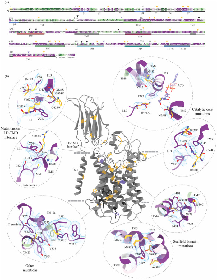
Figure 4: Molecular basis for MPS IIIC mutation-induced dysfunction (A) Evolutionary sequence conservation of HGSNAT. Amino acids are color coded according to the conservation scores generated by ConSurf webserver using a Clustal multiple sequence alignment of homologs identified by PSI-BLAST (Ashkenazy et al, 2016). The positions of the mutations - missense (orange), nonsense (black), and polymorphisms (purple) – are indicated on the sequence by triangles. (B) MPS IIIC-causing mutations mapped on the HGSNAT structure. The color coding of the positions is the same as in panel A. Some of the missense mutants are highlighted in the insets (dashed ovals). We grouped them based on their position within the protein – LD-TMD interface, catalytic core, scaffold domain, and other C-terminal mutations. The insets show the 3D environment of the mutant sites on the wild-type HGSNAT color coded as per their evolutionary sequence conservation scores, and the potential disturbance to it caused by the mutation (orange side chains). The coordinates for mutant side chains were generated based on wild-type HGSNAT structure as input in FoldX webserver (Schymkowitz et al, 2005).
Figure 4: Molecular basis for MPS IIIC mutation-induced dysfunction (A) Evolutionary sequence conservation of HGSNAT. Amino acids are color coded according to the conservation scores generated by ConSurf webserver using a Clustal multiple sequence alignment of homologs identified by PSI-BLAST (Ashkenazy et al, 2016). The positions of the mutations - missense (orange), nonsense (black), and polymorphisms (purple) – are indicated on the sequence by triangles. (B) MPS IIIC-causing mutations mapped on the HGSNAT structure. The color coding of the positions is the same as in panel A. Some of the missense mutants are highlighted in the insets (dashed ovals). We grouped them based on their position within the protein – LD-TMD interface, catalytic core, scaffold domain, and other C-terminal mutations. The insets show the 3D environment of the mutant sites on the wild-type HGSNAT color coded as per their evolutionary sequence conservation scores, and the potential disturbance to it caused by the mutation (orange side chains). The coordinates for mutant side chains were generated based on wild-type HGSNAT structure as input in FoldX webserver (Schymkowitz et al, 2005).CC BY · PubMed Central
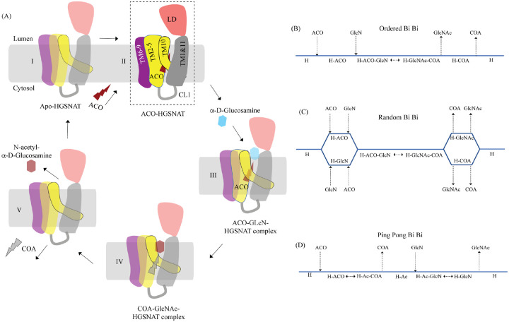
Figure 5: Proposed mechanism of acetyl transfer by HGSNAT (A) HGSNAT (I) catalyzes a bisubstrate reaction of transferring acetyl group from cytosolic acetyl-CoA (ACO, red lightning) to terminal non-reducing α-D-Glucosamine (GlcN, blue hexagon) of luminal heparan sulfate (III and IV). After the acetyl group transfer, COA (gray lightning) and acetylated glucosamine (GlcNAc, red hexagon) are believed to be released to cytosol and lumen respectively (V). Depending on the order of binding and release of substrates and products, enzyme-catalyzed bisubstrate reactions could either be sequential reactions (B and C) or ping pong reactions (D). The mechanism of reaction catalyzed by HGSNAT has been a longstanding debate. We believe that the acetyl-CoA bound HGSNAT structure presented in this work (II, dashed box) is in a cofactor primed conformation which could proceed by any of the bisubstrate reaction mechanisms shown in B-D. The function of LD is unclear, and we believe it plays essential role in recognition of substrate and its positioning at the active site.
Figure 5: Proposed mechanism of acetyl transfer by HGSNAT (A) HGSNAT (I) catalyzes a bisubstrate reaction of transferring acetyl group from cytosolic acetyl-CoA (ACO, red lightning) to terminal non-reducing α-D-Glucosamine (GlcN, blue hexagon) of luminal heparan sulfate (III and IV). After the acetyl group transfer, COA (gray lightning) and acetylated glucosamine (GlcNAc, red hexagon) are believed to be released to cytosol and lumen respectively (V). Depending on the order of binding and release of substrates and products, enzyme-catalyzed bisubstrate reactions could either be sequential reactions (B and C) or ping pong reactions (D). The mechanism of reaction catalyzed by HGSNAT has been a longstanding debate. We believe that the acetyl-CoA bound HGSNAT structure presented in this work (II, dashed box) is in a cofactor primed conformation which could proceed by any of the bisubstrate reaction mechanisms shown in B-D. The function of LD is unclear, and we believe it plays essential role in recognition of substrate and its positioning at the active site.CC BY · PubMed Central

Verification

Claims the paper makes

The claims and statistics of this paper haven’t been checked yet.

Run the check — free, no account needed. It reads the open-access text and checks the sources it cites.

A few free checks a day · papers already verified open instantly · results are public

Self-check

Percentages, sums and reported differences re-computed from the paper’s own numbers.

The self-check needs the paper’s full text.

Citations

Cites

Show all 1 references

Research works printed but not linked: 67

  1. 10.1093/nar/gkw408 · DOI · Paper page
  2. 10.1016/j.ymgme.2005.09.021 · DOI
  3. 1985, Acetyl coenzyme A: alpha-glucosaminide N-acetyltransferase. Evidence for a transmembrane acetylation mechanism, J Biol Chem, 260, 11293, 10.1016/S0021-9258(17)39179-2 · DOI
  4. 1986, Acetyl-coenzyme A:alpha-glucosaminide N-acetyltransferase. Evidence for an active site histidine residue, J Biol Chem, 261, 10127, 10.1016/S0021-9258(18)67500-3 · DOI
  5. 10.1126/science.3090688 · DOI
  6. 2012, Differential expression of matrix metalloproteinases in the serum of patients with mucopolysaccharidoses, JIMD Rep, 3, 59
  7. 10.1016/j.str.2015.07.016 · DOI
  8. 10.1146/annurev.biochem.72.121801.161800 · DOI · Paper page
  9. 10.1016/S0022-2836(84)71582-8 · DOI
  10. 10.1111/j.1399-0004.2010.01525.x · DOI
  11. 2022, De novo design of immunoglobulin-like domains, Nat Commun, 13, 5661, 10.1038/s41467-022-33004-6 · DOI
  12. 10.1155/2012/471325 · DOI
  13. 2010, Analysis of the biogenesis of heparan sulfate acetyl-CoA:alpha-glucosaminide N-acetyltransferase provides insights into the mechanism underlying its complete deficiency in mucopolysaccharidosis IIIC, J Biol Chem, 285, 31233-31242
  14. 10.1107/S0907444910007493 · DOI · Paper page
  15. 10.1371/journal.pone.0024951 · DOI
  16. 10.1086/508068 · DOI
  17. 10.1002/humu.21286 · DOI
  18. 10.1002/humu.20986 · DOI
  19. 10.1371/journal.pone.0007434 · DOI
  20. 10.1371/journal.ppat.1002386 · DOI
  21. 10.1038/nprot.2014.173 · DOI
  22. Hirano Y , Gao YG , Stephenson DJ , Vu NT , Malinina L , Simanshu DK , Chalfant CE , Patel DJ , Brown RE (2019) Structural basis of phosphatidylcholine recognition by the C2-domain of cytosolic phospholipase A(2)alpha. Elife 8 · DOI
  23. 10.1002/pro.4519 · DOI
  24. 10.1006/jmbi.2000.4078 · DOI
  25. 10.1086/508294 · DOI
  26. 2020, Inherited disorders of lysosomal membrane transporters, Biochim Biophys Acta Biomembr, 1862, 183336, 10.1016/j.bbamem.2020.183336 · DOI
  27. Jamali K , Kall L , Zhang R , Brown A , Kimanius D , Scheres SHW (2023) Automated model building and protein identification in cryo-EM maps. bioRxiv · DOI · Paper page
  28. 10.1016/S0006-3495(99)77009-8 · DOI
  29. 10.1093/bioinformatics/btx350 · DOI
  30. 10.1016/j.bbamcr.2017.05.013 · DOI
  31. 10.1016/j.str.2006.01.013 · DOI
  32. 10.1073/pnas.75.10.5185 · DOI
  33. 10.1002/prot.340120403 · DOI
  34. Kretsinger RH , Kretsinger RH , Uversky VN , Permiakov EA , SpringerLink (2013) Encyclopedia of metalloproteins. Springer, New York · DOI
  35. 2014, Quantifying the local resolution of cryo-EM density maps, Nat Methods, 11, 63, 10.1038/nmeth.2727 · DOI
  36. 10.1016/J.GPB.2019.08.002 · DOI
  37. 10.1107/s2059798319011471 · DOI · Paper page
  38. 2021, Update in the Mucopolysaccharidoses, Semin Pediatr Neurol, 37, 100874, 10.1016/j.spen.2021.100874 · DOI
  39. 10.1042/bj3080327 · DOI
  40. Meng EC , Goddard TD , Pettersen EF , Couch GS , Pearson ZJ , Morris JH , Ferrin TE (2023) UCSF ChimeraX: Tools for Structure Building and Analysis. Protein Sci: e4792 · DOI
  41. 10.1038/S41592-022-01488-1 · DOI · Paper page
  42. 10.1016/j.bbapap.2011.07.003 · DOI
  43. 2019, HGSNAT enzyme deficiency results in accumulation of heparan sulfate in podocytes and basement membranes, Histol Histopathol, 34, 1377
  44. Newman KE , Tindall SN , Mader SL , Khalid S , Thomas GH , Van Der Woude MW (2023) A novel fold for acyltransferase-3 (AT3) proteins provides a framework for transmembrane acyl-group transfer. Elife 12 · DOI · Paper page
  45. 2016, The Use of Detergents to Purify Membrane Proteins, Curr Protoc Protein Sci, 84, 8
  46. Pan X , Taherzadeh M , Bose P , Heon-Roberts R , Nguyen ALA , Xu T , Para C , Yamanaka Y , Priestman DA , Platt FM et al (2022) Glucosamine amends CNS pathology in mucopolysaccharidosis IIIC mouse expressing misfolded HGSNAT. J Exp Med 219 · DOI
  47. Parker JL , Deme JC , Wu Z , Kuteyi G , Huo J , Owens RJ , Biggin PC , Lea SM , Newstead S (2021) Cryo-EM structure of PepT2 reveals structural basis for proton-coupled peptide and prodrug transport in mammals. Sci Adv 7 · DOI
  48. 2018, Lysosomal storage diseases. Nat Rev Dis Primers, 4, 27, 10.1038/s41572-018-0025-4 · DOI · Paper page
  49. 10.1093/nar/gky309 · DOI
  50. 2018, Sanfilippo type C disease: pathogenic mechanism and potential therapeutic applications, Expert Opin Orphan D, 6, 635, 10.1080/21678707.2018.1534585 · DOI
  51. 10.1038/nmeth.4169 · DOI · Paper page
  52. 10.1038/s41592-020-00990-8 · DOI
  53. 10.1042/BCJ20180828 · DOI
  54. 1981, Degradation of mucopolysaccharide in intact isolated lysosomes, J Biol Chem, 256, 10763, 10.1016/S0021-9258(19)68506-6 · DOI
  55. 1983, Utilization of exogenously added acetyl coenzyme A by intact isolated lysosomes, J Biol Chem, 258, 3006, 10.1016/S0021-9258(18)32821-7 · DOI
  56. 10.1111/febs.15602 · DOI
  57. 10.1016/j.bbamcr.2008.12.008 · DOI
  58. 10.1093/nar/gkaa1004 · DOI · Paper page
  59. 10.1016/j.arr.2016.04.012 · DOI
  60. 10.1111/tra.12056 · DOI
  61. 10.1093/nar/gki387 · DOI · Paper page
  62. 10.1371/journal.pone.0050300 · DOI
  63. 2012, Proteolytic cleavage of the disease-related lysosomal membrane glycoprotein CLN7, Biochim Biophys Acta, 1822, 1617, 10.1016/j.bbadis.2012.05.015 · DOI
  64. 10.1016/S0167-4838(99)00263-0 · DOI
  65. 10.1016/S0005-2736(01)00299-1 · DOI
  66. 10.1093/NAR/GKAB1061 · DOI · Paper page
  67. 10.1038/s41594-024-01315-5 · DOI

Metadata

Authors
Navratna, V., Kumar, A., Rana, J. K., & Mosalaganti, S.
Affiliations
  • University of Michigan (US)
  • Thermo Fisher Scientific (United States) (US)
Corresponding author
Mosalaganti, Shyamal
Venue
bioRxiv (Cold Spring Harbor Laboratory)
Publisher
openRxiv
Published
2023-10-25
OpenAlex
W4388273628
Cited by
1
References
1
Citations by year
2025: 1
Type
posted content
Language
en
Open access
Licence
cc-by
Locations
  • bioRxiv (Cold Spring Harbor Laboratory) (acceptedVersion, cc-by)
  • PubMed (publishedVersion)
  • PubMed Central (submittedVersion, cc-by)
Funders
National Institutes of Health (1dp2gm150019-01)
Topics
  • Health Sciences › Medicine › Genetics (clinical) › Lysosomal Storage Disorders Research
  • Life Sciences › Biochemistry, Genetics and Molecular Biology › Biochemistry › Glycogen Storage Diseases and Myoclonus
  • Life Sciences › Biochemistry, Genetics and Molecular Biology › Cell Biology › Proteoglycans and glycosaminoglycans research
Keywords
HGSNAT, heparan sulfate degradation, lysosomal storage disease, acetyl-CoA, enzyme structure, lysosomal membrane, glycosaminoglycan catabolism

About these checks

Registry, retraction, Cites and statistics results are computed, not judged, with one exception: a printed reference that no index lists is read once by a model before it counts as a citation error. Claims are extracted by a model and kept only when their quote appears verbatim in the text; each cited source is then checked, with a model’s help, for whether it says what the claim relies on. Those verdicts can be wrong — read the claim’s quote and the cited source before relying on one.

Statistics and arithmetic are recomputed, not judged.

Confidence bands are shown only for fields calibrated on our annotated test set. None is calibrated yet, so no band is shown.

What counts as a claim ›How we measure confidence ›How we check this ›

Are you an author, or do you see a problem with this page? Request a review

How to cite

Cite CoChat for the checks on this page, and the paper for its own findings.

This page
CoChat. (2026). Citation check: Structure of the human heparan-α-glucosaminide N -acetyltransferase (HGSNAT). https://app.cochat.ai/papers/10.1101/2023.10.23.563672
The paper
Navratna, V., Kumar, A., Rana, J. K., & Mosalaganti, S. (2023). Structure of the human heparan-α-glucosaminide N -acetyltransferase (HGSNAT). bioRxiv (Cold Spring Harbor Laboratory). https://doi.org/10.1101/2023.10.23.563672