PreprintCited by 15Genomics and Chromatin DynamicsRNA Research and SplicingNuclear Structure and Function

The DOI matches its Crossref record; 1 of the 1 references it cites were found in a registry or an index.

Structural basis of eukaryotic transcription termination by the Rat1 exonuclease complex

Open access

Preprint — published as 10.1038/s41467-024-52157-0 · Paper page

Abstract

The 5’-3’ exoribonuclease Rat1 (or Xrn2) plays a pivotal role in termination of mRNA transcription by eukaryotic RNA polymerase II (RNAPII). Rat1 forms a complex with its partner proteins, Rai1 and Rtt103, and acts as a “torpedo” to bind transcribing RNAPII and dissociate DNA/RNA from it. Here we report the cryo-electron microscopy structures of a Rat1-Rai1-Rtt103 complex and three Rat1-Rai1-associated RNAPII complexes (type-1, type-1b, and type-2) from the yeast Komagataella phaffii . The Rat1-Rai1-Rtt103 complex structure revealed that Rat1 and Rai1 form a heterotetramer, with a single Rtt103 bound between two Rai1 molecules. In the type-1 complex, Rat1-Rai1 forms a heterodimer and directly binds to the RNA exit site of transcribing RNAPII to extract RNA into the Rat1 nuclease active site. This interaction changes the RNA path in favor of termination (the “pre-termination” state). The type-1b and type-2 complexes have no bound DNA/RNA, likely representing the “post-termination” states. These structures shed light on the mechanism of eukaryotic mRNA transcription termination.

Overview

The claims and statistics of this paper haven’t been checked yet.

Run the check — free, no account needed. It reads the open-access text and checks the sources it cites.

A few free checks a day · papers already verified open instantly · results are public

Integrity signals

Registry

Crossref · 10.1101/2024.03.28.587100

Cites

1 of 1 references verified0 citation errors · 0 not checkable

Statistics

Figures

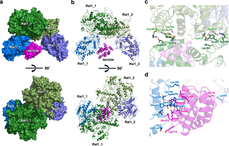
Fig. 1 Structure of the Rat1-Rai1-Rtt103 complex. a, b The K. phaffii Rat1-Rai1 heterotetramer complexed with Rtt103 is represented as a surface and ribbon model. Rat1 is colored green and grass green, Rai1 is colored marine and purple blue and the N-terminal domain of Rtt103 (Rtt103N) is colored magenta. Bottom, view after a 90° rotation about the horizontal axis from the upper panel. c A close-up view of the Rat1 dimer interface. The Rat1 residues involved in dimerization are shown by green stick models. Hydrogen bonding interactions are represented by dotted lines. d A close-up view of the interface between Rai1 and Rtt103N. The Rai1 and Rtt103 residues are shown by blue and magenta stick models, respectively. A transparent ribbon model of the Rat1-Rai1 (blue and green) complexed with Rtt103 (magenta) is shown in the background. Hydrogen bonds and ionic interactions between Rai1 and Rtt103 are represented by dotted lines.
Fig. 1 Structure of the Rat1-Rai1-Rtt103 complex. a, b The K. phaffii Rat1-Rai1 heterotetramer complexed with Rtt103 is represented as a surface and ribbon model. Rat1 is colored green and grass green, Rai1 is colored marine and purple blue and the N-terminal domain of Rtt103 (Rtt103N) is colored magenta. Bottom, view after a 90° rotation about the horizontal axis from the upper panel. c A close-up view of the Rat1 dimer interface. The Rat1 residues involved in dimerization are shown by green stick models. Hydrogen bonding interactions are represented by dotted lines. d A close-up view of the interface between Rai1 and Rtt103N. The Rai1 and Rtt103 residues are shown by blue and magenta stick models, respectively. A transparent ribbon model of the Rat1-Rai1 (blue and green) complexed with Rtt103 (magenta) is shown in the background. Hydrogen bonds and ionic interactions between Rai1 and Rtt103 are represented by dotted lines.CC BY-NC-ND · PubMed Central
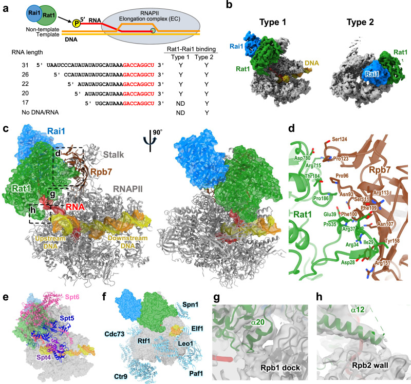
Fig. 2 Structure of the RNAPII pre-termination complex (type 1). a Schematic representation of the EC-Rat1-Rai1 complex and the RNA sequences used in this study. Y, the indicated complex was observed in cryo-EM analysis; ND, not detected (Supplementary Fig. 17). b Structures of the type-1 and type-2 RNAPII-Rat1-Rai1 complexes. The composite maps were generated from the overall-masked maps and the Rat1-Rai-masked focused maps by Phenix64. The maps are colored to match the structural models (gray, RNAP; green, Rat1, blue, Rai1; yellow, DNA; red, RNA). c Structure of the elongation complex (EC) engaged with Rat1-Rai1 at the RNA exit (the type-1 complex). The structural model is displayed in two orientations. RNAPII, template DNA, non-template DNA, RNA, Rat1, and Rai1 are colored gray (Rpb7 in brown), yellow, orange, green, and blue, respectively. Transparent surfaces of DNA/RNA, Rat1, and Rai1 are overlaid. d Close-up view of the Rpb7-Rat1 interface. e The Rat1-binding site overlaps with the binding sites of the elongation factors Spt5 and Spt6. The structural model of the type-1 complex is shown in transparent surface, colored as in (c). The structural model of RNAPII EC (PDB 7XN7) is superimposed with the type-1 complex by the Rpb1 subunit. Elongation factors Spt4, Spt5 and Spt6 are shown in cartoon representation and colored purple, blue, and pink, respectively. f The Rat1-binding site does not overlap with the binding sites of the Paf1 complex (Paf1, Leo1, Ctr9, Cdc73, and Rtf1), Elf1, and Spn1. The structural model of the type-1 complex is shown in transparent surface, colored as in (c). The structural model of RNAPII EC (PDB 7XN7) is superimposed with the type-1 complex by the Rpb1 subunit, and the elongation factors are shown in cartoon representation colored in light blue. g A close-up view of the interaction between the Rpb1 dock domain and the Rat1 α20 helix. Cryo-EM map (transparent surfaces) is overlaid on the structural model. h A close-up view of the interaction between the Rpb2 wall domain and the Rat1 α12 helix. The cryo-EM map (transparent surfaces) is overlaid on the structural model.
Fig. 2 Structure of the RNAPII pre-termination complex (type 1). a Schematic representation of the EC-Rat1-Rai1 complex and the RNA sequences used in this study. Y, the indicated complex was observed in cryo-EM analysis; ND, not detected (Supplementary Fig. 17). b Structures of the type-1 and type-2 RNAPII-Rat1-Rai1 complexes. The composite maps were generated from the overall-masked maps and the Rat1-Rai-masked focused maps by Phenix64. The maps are colored to match the structural models (gray, RNAP; green, Rat1, blue, Rai1; yellow, DNA; red, RNA). c Structure of the elongation complex (EC) engaged with Rat1-Rai1 at the RNA exit (the type-1 complex). The structural model is displayed in two orientations. RNAPII, template DNA, non-template DNA, RNA, Rat1, and Rai1 are colored gray (Rpb7 in brown), yellow, orange, green, and blue, respectively. Transparent surfaces of DNA/RNA, Rat1, and Rai1 are overlaid. d Close-up view of the Rpb7-Rat1 interface. e The Rat1-binding site overlaps with the binding sites of the elongation factors Spt5 and Spt6. The structural model of the type-1 complex is shown in transparent surface, colored as in (c). The structural model of RNAPII EC (PDB 7XN7) is superimposed with the type-1 complex by the Rpb1 subunit. Elongation factors Spt4, Spt5 and Spt6 are shown in cartoon representation and colored purple, blue, and pink, respectively. f The Rat1-binding site does not overlap with the binding sites of the Paf1 complex (Paf1, Leo1, Ctr9, Cdc73, and Rtf1), Elf1, and Spn1. The structural model of the type-1 complex is shown in transparent surface, colored as in (c). The structural model of RNAPII EC (PDB 7XN7) is superimposed with the type-1 complex by the Rpb1 subunit, and the elongation factors are shown in cartoon representation colored in light blue. g A close-up view of the interaction between the Rpb1 dock domain and the Rat1 α20 helix. Cryo-EM map (transparent surfaces) is overlaid on the structural model. h A close-up view of the interaction between the Rpb2 wall domain and the Rat1 α12 helix. The cryo-EM map (transparent surfaces) is overlaid on the structural model.CC BY-NC-ND · PubMed Central
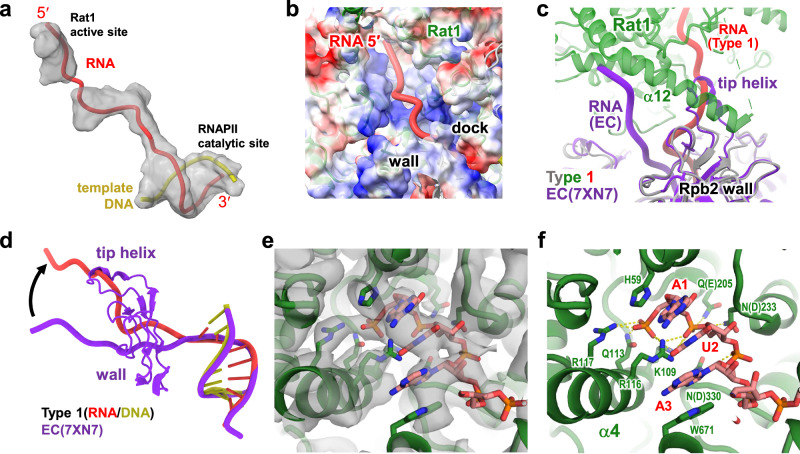
Fig. 3 RNA structure and path in the RNAPII pre-termination complex (type 1). a Cryo-EM map of the RNA in the type-1 complex. The cryo-EM map was segmented by ChimeraX65, and regions around the RNA and DNA/RNA hybrid are shown as transparent surfaces overlaid on the structural model. b RNA channel in the type-1 complex. A transparent surface of RNAPII and Rat1 (colored by electrostatic surface potentials) is overlaid on the cartoon model (colored as in Fig. 2c). c, d Comparing the RNA path at the RNAPII RNA exit. The structure of RNAPII EC (PDB 7XN7, purple) is superposed on the type-1 complex (colored as in Fig. 2c) by the Rpb1 subunit. e, f Structure of the Rat1 active site with the RNA 5´-end incorporated. e The cryo-EM map (transparent surfaces) is overlaid on the structural model (cartoon and stick models). f The side chains of the Rat1 residues interacting with the RNA are shown as stick models. Putative hydrogen bonds between Rat1 and RNA are shown as yellow dashed lines. The wild-type amino acids are shown in parentheses for the residues mutated in the Rat1 active site (residues 205, 233, and 330).
Fig. 3 RNA structure and path in the RNAPII pre-termination complex (type 1). a Cryo-EM map of the RNA in the type-1 complex. The cryo-EM map was segmented by ChimeraX65, and regions around the RNA and DNA/RNA hybrid are shown as transparent surfaces overlaid on the structural model. b RNA channel in the type-1 complex. A transparent surface of RNAPII and Rat1 (colored by electrostatic surface potentials) is overlaid on the cartoon model (colored as in Fig. 2c). c, d Comparing the RNA path at the RNAPII RNA exit. The structure of RNAPII EC (PDB 7XN7, purple) is superposed on the type-1 complex (colored as in Fig. 2c) by the Rpb1 subunit. e, f Structure of the Rat1 active site with the RNA 5´-end incorporated. e The cryo-EM map (transparent surfaces) is overlaid on the structural model (cartoon and stick models). f The side chains of the Rat1 residues interacting with the RNA are shown as stick models. Putative hydrogen bonds between Rat1 and RNA are shown as yellow dashed lines. The wild-type amino acids are shown in parentheses for the residues mutated in the Rat1 active site (residues 205, 233, and 330).CC BY-NC-ND · PubMed Central
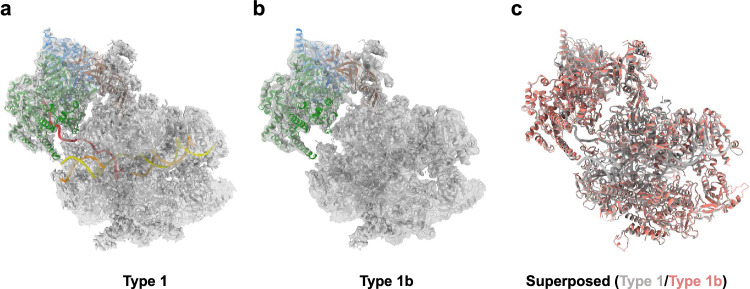
Fig. 4 Structure of RNAPII bound with Rat1-Rai1 at the RNA exit (type 1b). a, b Structures of the type-1 and type-1b complexes. Cryo-EM maps (white surfaces) are overlaid on the structural models (cartoon representation, colored as in Fig. 2). c Comparison of the type-1 and type-1b complexes. The structural models for the type-1 and type-1b complexes are superimposed by the Rpb1 subunit.
Fig. 4 Structure of RNAPII bound with Rat1-Rai1 at the RNA exit (type 1b). a, b Structures of the type-1 and type-1b complexes. Cryo-EM maps (white surfaces) are overlaid on the structural models (cartoon representation, colored as in Fig. 2). c Comparison of the type-1 and type-1b complexes. The structural models for the type-1 and type-1b complexes are superimposed by the Rpb1 subunit.CC BY-NC-ND · PubMed Central
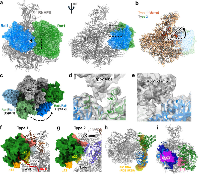
Fig. 5 Structure of Rat1-Rai1 bound within the RNAPII cleft (type 2). a Structure of Rat1-Rai1 bound within the RNAPII cleft (the type-2 complex). The structural model is displayed in two orientations, colored as in Fig. 2. The transparent surfaces of Rat1 and Rai1 are overlaid. b Comparison of the type-1 and type-2 complexes. The structural models for the type-1 (light orange) and type-2 complexes (colored as in (a)) are superimposed by the Rpb2 subunit. Structural models for RNAPII are shown in cartoon representation. The RNAPII clamp of the type-1 complex is colored red. c Comparison of the Rat1-Rai-1 binding sites in the type-1 and type-2 complexes. The structural models are superimposed by the Rpb2 subunit. d A close-up view of the interaction between the Rpb2 lobe and the Rat1 α20 helix. Cryo-EM map is overlaid on the structural model. e A close-up view of the interaction between the RNAPII clamp and the Rai1. Cryo-EM map is overlaid on the structural model. f, g Comparison of the Rat1 and RNAPII interfaces in the type-1 and type-2 complexes. RNAPII is shown as a cartoon representation, and Rat1 is shown as a surface representation. The Rat1 regions interacting with RNAPII in the type-1 complex are colored salmon (Rat1-Rpb7 interface, Fig. 2d), yellow (the α20 helix), and orange (the α12 helix), respectively. h Overlapping binding sites for Rat1-Rai1 and double-stranded DNA in the transcription pre-initiation complex (PIC). The structural model of the type-2 complex is shown in cartoon representation, colored as in (a). The structural model of RNAPII PIC (PDB 5FZ5) is superimposed on the type-2 complex by the Rpb2 subunit. The template DNA (yellow) and the non-template DNA (orange) of the PIC are shown in surface representation. i Overlapping binding sites for Rat1-Rai1 and elongation factors Spt4, Spt5, and Elf1. The structural model of the type-2 complex is shown in cartoon representation, colored as in (a). The structural model of RNAPII EC (PDB 7XN7) is superposed on the type-2 complex by the Rpb2 subunit. Elongation factors Spt4, Spt5, and Elf1 are shown in surface representation.
Fig. 5 Structure of Rat1-Rai1 bound within the RNAPII cleft (type 2). a Structure of Rat1-Rai1 bound within the RNAPII cleft (the type-2 complex). The structural model is displayed in two orientations, colored as in Fig. 2. The transparent surfaces of Rat1 and Rai1 are overlaid. b Comparison of the type-1 and type-2 complexes. The structural models for the type-1 (light orange) and type-2 complexes (colored as in (a)) are superimposed by the Rpb2 subunit. Structural models for RNAPII are shown in cartoon representation. The RNAPII clamp of the type-1 complex is colored red. c Comparison of the Rat1-Rai-1 binding sites in the type-1 and type-2 complexes. The structural models are superimposed by the Rpb2 subunit. d A close-up view of the interaction between the Rpb2 lobe and the Rat1 α20 helix. Cryo-EM map is overlaid on the structural model. e A close-up view of the interaction between the RNAPII clamp and the Rai1. Cryo-EM map is overlaid on the structural model. f, g Comparison of the Rat1 and RNAPII interfaces in the type-1 and type-2 complexes. RNAPII is shown as a cartoon representation, and Rat1 is shown as a surface representation. The Rat1 regions interacting with RNAPII in the type-1 complex are colored salmon (Rat1-Rpb7 interface, Fig. 2d), yellow (the α20 helix), and orange (the α12 helix), respectively. h Overlapping binding sites for Rat1-Rai1 and double-stranded DNA in the transcription pre-initiation complex (PIC). The structural model of the type-2 complex is shown in cartoon representation, colored as in (a). The structural model of RNAPII PIC (PDB 5FZ5) is superimposed on the type-2 complex by the Rpb2 subunit. The template DNA (yellow) and the non-template DNA (orange) of the PIC are shown in surface representation. i Overlapping binding sites for Rat1-Rai1 and elongation factors Spt4, Spt5, and Elf1. The structural model of the type-2 complex is shown in cartoon representation, colored as in (a). The structural model of RNAPII EC (PDB 7XN7) is superposed on the type-2 complex by the Rpb2 subunit. Elongation factors Spt4, Spt5, and Elf1 are shown in surface representation.CC BY-NC-ND · PubMed Central
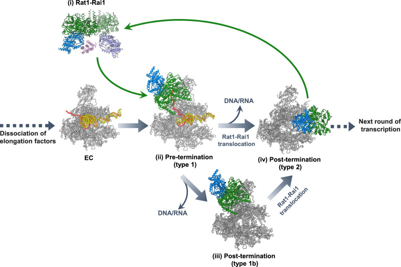
Fig. 6 Model of Rat1-dependent transcription termination. (i) Rat1-Rai1 exists as a tetramer in free form, and the tetramer holds one Rtt103 molecule. When recruited to transcribing RNAPII, the Rat1-Rai1 tetramer dissociates into a dimer. The Rat1-Rai1 binding to RNAPII requires the prior dissociation of Spt6 and Spt4/5 from RNAPII (except for the KOW5 domain of Spt5). (ii) Rat1 captures the monophosphorylated RNA 5´-end, which is generated by cleavage of mRNA precursor at the polyadenylation site and starts trimming of the RNA. When Rat1 trimmed the RNA to its length of 20–22 nt, Rat1-Rai1 forms a close contact with RNAPII. (iii) Further RNA trimming triggers bubble collapse and DNA/RNA release. (iv) After termination, Rat1-Rai1 is relocated to the empty DNA-binding site of RNAPII for occlusion. Alternatively, Rat1-Rai1 could move from the RNA-exit site (ii) to the RNAPII cleft (iv), to actively dislodge DNA/RNA from the RNAPII cleft. Rat1-Rai1 bound to the RNAPII cleft would protect RNAPII until it is recycled into a PIC for another round of transcription.
Fig. 6 Model of Rat1-dependent transcription termination. (i) Rat1-Rai1 exists as a tetramer in free form, and the tetramer holds one Rtt103 molecule. When recruited to transcribing RNAPII, the Rat1-Rai1 tetramer dissociates into a dimer. The Rat1-Rai1 binding to RNAPII requires the prior dissociation of Spt6 and Spt4/5 from RNAPII (except for the KOW5 domain of Spt5). (ii) Rat1 captures the monophosphorylated RNA 5´-end, which is generated by cleavage of mRNA precursor at the polyadenylation site and starts trimming of the RNA. When Rat1 trimmed the RNA to its length of 20–22 nt, Rat1-Rai1 forms a close contact with RNAPII. (iii) Further RNA trimming triggers bubble collapse and DNA/RNA release. (iv) After termination, Rat1-Rai1 is relocated to the empty DNA-binding site of RNAPII for occlusion. Alternatively, Rat1-Rai1 could move from the RNA-exit site (ii) to the RNAPII cleft (iv), to actively dislodge DNA/RNA from the RNAPII cleft. Rat1-Rai1 bound to the RNAPII cleft would protect RNAPII until it is recycled into a PIC for another round of transcription.CC BY-NC-ND · PubMed Central

Verification

Claims the paper makes

The claims and statistics of this paper haven’t been checked yet.

Run the check — free, no account needed. It reads the open-access text and checks the sources it cites.

A few free checks a day · papers already verified open instantly · results are public

Self-check

Percentages, sums and reported differences re-computed from the paper’s own numbers.

The self-check needs the paper’s full text.

Citations

Cites

Show all 1 references

Research works printed but not linked: 61

  1. Osman, S. & Cramer, P . Structural Biology of RNA Polymerase II Transcription: 20 Years On. Annu Rev Cell Dev Biol 36(2020). · DOI
  2. 10.1146/annurev-genet-110711-155440 · DOI
  3. 10.1038/nrg3662 · DOI · Paper page
  4. 10.1101/gad.824700 · DOI
  5. 10.1038/nsb0903-679 · DOI
  6. 10.1021/cr400071f · DOI
  7. 10.1101/gad.200303.112 · DOI
  8. 10.1016/j.jmb.2016.02.006 · DOI
  9. 10.1038/nrm.2017.10 · DOI · Paper page
  10. Mohamed, A.A. , Vazquez Nunez, R. & Vos, S.M . Structural advances in transcription elongation. Curr Opin Struct Biol 75(2022). · DOI
  11. Sekine, S. , Ehara, H. , Kujirai, T. & Kurumizaka, H . Structural perspectives on transcription in chromatin. Trends Cell Biol (2023). · DOI
  12. 10.1016/j.molcel.2022.12.021 · DOI
  13. 10.1016/j.tig.2020.05.008 · DOI · Paper page
  14. 10.1073/pnas.84.23.8306 · DOI · Paper page
  15. 10.1016/0968-0004(89)90132-1 · DOI
  16. 10.1101/gad.2.4.440 · DOI
  17. 10.1126/science.280.5361.298 · DOI
  18. 10.1016/j.ceb.2005.04.006 · DOI
  19. 10.1101/gad.17268411 · DOI
  20. 10.1038/nrm3943 · DOI
  21. Proudfoot, N . Transcriptional termination in mammals: Stopping the RNA polymerase II juggernaut. Science 352, aad9926 (2016). · DOI
  22. 10.1038/nature03035 · DOI
  23. 10.1016/j.molcel.2006.11.011 · DOI
  24. 10.1042/BST20130202 · DOI
  25. 10.1016/j.bbagrm.2013.03.005 · DOI
  26. 10.1074/jbc.270.27.16063 · DOI
  27. 10.1128/MCB.20.11.4006-4015.2000 · DOI
  28. 10.1038/nature03041 · DOI
  29. 10.1038/nsmb.1893 · DOI
  30. 10.1016/0092-8674(89)90126-8 · DOI
  31. 10.1016/J.MOLCEL.2023.08.007 · DOI
  32. 10.1101/gad.1409106 · DOI
  33. 10.1101/gad.332833.119 · DOI
  34. Ehara, H. , Kujirai, T. , Shirouzu, M. , Kurumizaka, H. & Sekine, S . Structural basis of nucleosome disassembly and reassembly by RNAPII elongation complex with FACT. Science 377, eabp9466 (2022). · DOI
  35. 10.1038/s41586-018-0440-4 · DOI · Paper page
  36. 10.1126/science.1059493 · DOI · Paper page
  37. 10.1038/nature07731 · DOI
  38. 10.1038/nature17970 · DOI
  39. 10.1038/nature17990 · DOI
  40. 10.1126/science.aan8552 · DOI
  41. 10.1126/science.aav8912 · DOI
  42. 10.1093/nar/gkq894 · DOI
  43. 10.1101/gad.463708 · DOI
  44. Murayama, Y. et al. Structural basis of the transcription termination factor Rho engagement with transcribing RNA polymerase from Thermus thermophilus. Sci Adv 9, eade7093 (2023). · DOI
  45. 10.1038/s41586-022-05658-1 · DOI
  46. 10.1007/s10969-014-9190-1 · DOI
  47. 10.1016/j.bbrc.2017.04.039 · DOI
  48. 10.1016/bs.mie.2016.04.012 · DOI
  49. 10.1016/j.jmb.2011.11.010 · DOI
  50. 10.1016/j.jsb.2015.08.008 · DOI
  51. 2019, Positive-unlabeled convolutional neural networks for particle picking in cryo-electron micrographs, Nature Methods, 16, 1153, 10.1038/s41592-019-0575-8 · DOI
  52. 2019, Real-time cryo-electron microscopy data preprocessing with Warp, Nat Methods, 16, 1146, 10.1038/s41592-019-0580-y · DOI
  53. Kimanius, D. et al. Data-driven regularisation lowers the size barrier of cryo-EM structure determination. bioRxiv (2023). · DOI
  54. 10.1002/jcc.20084 · DOI
  55. 10.1107/S0907444910007493 · DOI · Paper page
  56. 2018, ISOLDE: a physically realistic environment for model building into low-resolution electron-density maps, Acta Crystallogr D Struct Biol, 74, 519, 10.1107/S2059798318002425 · DOI
  57. 10.1107/S0907444912001308 · DOI · Paper page
  58. 10.1093/nar/gkm216 · DOI
  59. 10.1107/S0907444994003112 · DOI
  60. 10.1093/nar/22.22.4673 · DOI · Paper page
  61. P. Gouet , E. Courcelle , D. Stuart , F. Métoz , ESPript: analysis of multiple sequence alignments in PostScript. Bioinformatics (Oxford , England ) 15, (1999). · DOI

Metadata

Authors
Yanagisawa, T., Murayama, Y., Ehara, H., Goto, M., Aoki, M., & Sekine, S.-I.
Affiliations
RIKEN Center for Biosystems Dynamics Research (JP)
Corresponding author
Sekine, Shun-ichi
Venue
Nature Communications
Publisher
openRxiv
Published
2024-03-29
Volume
15
Issue
1
Pages
7854-7854
ISSN
2041-1723 (ISSN-L)
OpenAlex
W4402341346
Cited by
15
References
1
Citations by year
2026: 6 · 2025: 6 · 2024: 3
FWCI
2.04
Type
journal article
Language
en
Open access
Licence
cc-by-nc-nd
Locations
  • Nature Communications (publishedVersion, cc-by-nc-nd)
  • bioRxiv (Cold Spring Harbor Laboratory) (acceptedVersion, cc-by-nc-nd)
  • PubMed Central (publishedVersion, cc-by-nc-nd)
  • PubMed (publishedVersion)
  • RePEc: Research Papers in Economics (submittedVersion)
  • DOAJ (DOAJ: Directory of Open Access Journals) (submittedVersion)
Funders
  • Ministry of Education, Culture, Sports, Science and Technology (JP20H05906, JP24H00062, JP20H05690)
  • Japan Society for the Promotion of Science (JP 20H05906, 20H05690, 24H00062)
  • RIKEN
Topics
  • Life Sciences › Biochemistry, Genetics and Molecular Biology › Molecular Biology › Genomics and Chromatin Dynamics
  • Life Sciences › Biochemistry, Genetics and Molecular Biology › Molecular Biology › RNA Research and Splicing
  • Life Sciences › Biochemistry, Genetics and Molecular Biology › Molecular Biology › Nuclear Structure and Function
Keywords
RNA polymerase II, RAI1, 5'-3' exonuclease, Pichia pastoris, heterotetramer, cryo-EM structure
MeSH terms
Saccharomycetales, Exoribonucleases, Models, Molecular, Protein Binding, RNA, Messenger, Saccharomyces cerevisiae, Transcription, Genetic, Cryoelectron Microscopy, Saccharomyces cerevisiae Proteins, Transcription Termination, Genetic

About these checks

Registry, retraction, Cites and statistics results are computed, not judged, with one exception: a printed reference that no index lists is read once by a model before it counts as a citation error. Claims are extracted by a model and kept only when their quote appears verbatim in the text; each cited source is then checked, with a model’s help, for whether it says what the claim relies on. Those verdicts can be wrong — read the claim’s quote and the cited source before relying on one.

Statistics and arithmetic are recomputed, not judged.

Confidence bands are shown only for fields calibrated on our annotated test set. None is calibrated yet, so no band is shown.

What counts as a claim ›How we measure confidence ›How we check this ›

Are you an author, or do you see a problem with this page? Request a review

How to cite

Cite CoChat for the checks on this page, and the paper for its own findings.

This page
CoChat. (2026). Citation check: Structural basis of eukaryotic transcription termination by the Rat1 exonuclease complex. https://app.cochat.ai/papers/10.1101/2024.03.28.587100
The paper
Yanagisawa, T., Murayama, Y., Ehara, H., Goto, M., Aoki, M., & Sekine, S.-I. (2024). Structural basis of eukaryotic transcription termination by the Rat1 exonuclease complex. Nature Communications. https://doi.org/10.1101/2024.03.28.587100