

The DOI matches its Crossref record; 203 of the 203 references it cites were found in a registry or an index.
Human dendritic cell subsets: an update
Open accessAbstract
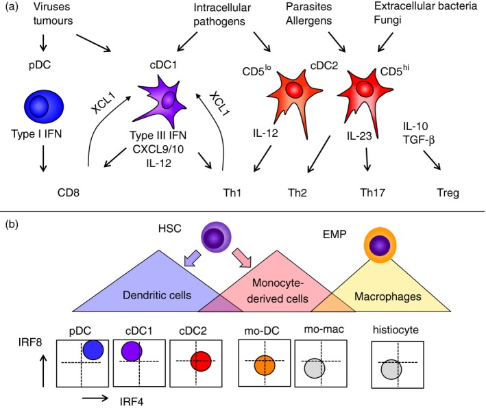
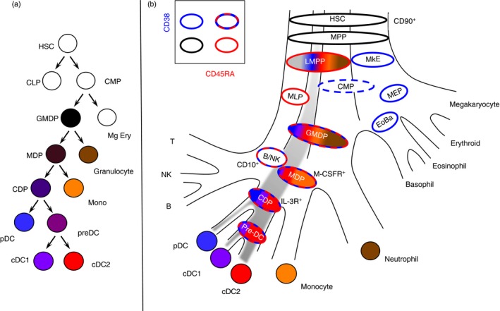
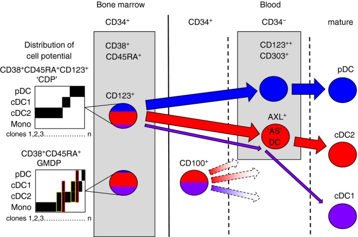
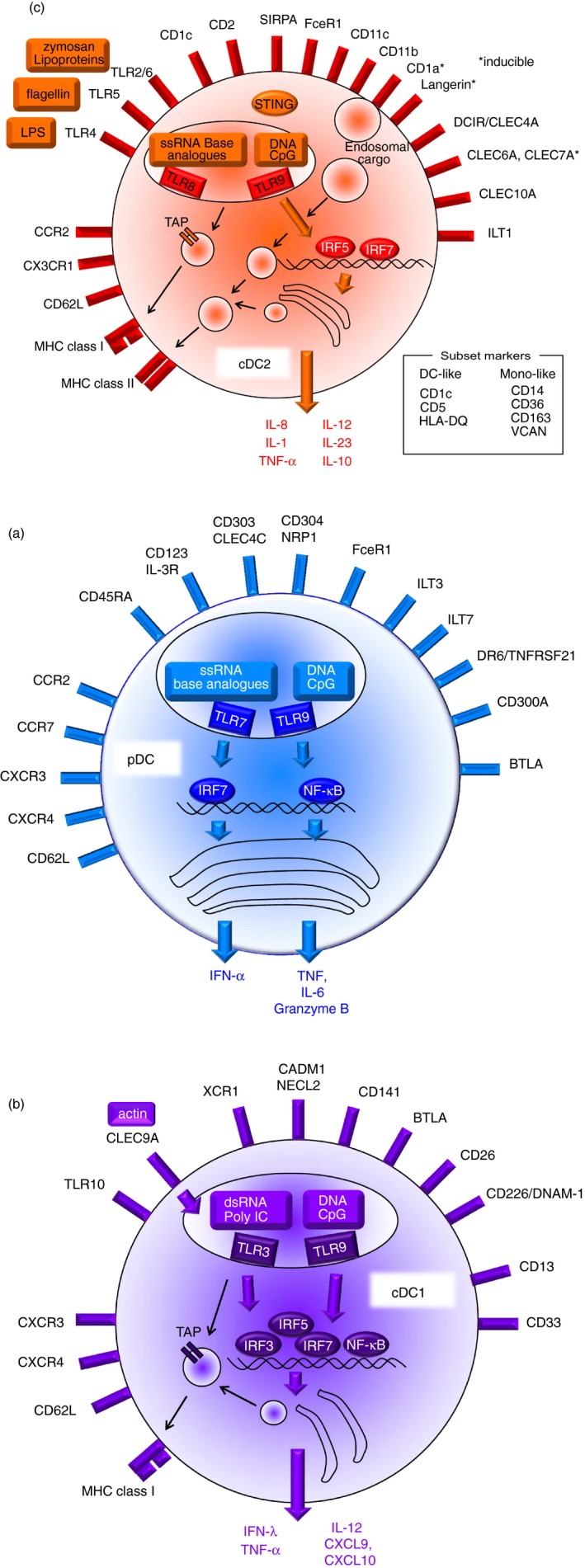
Dendritic cells (DC) are a class of bone-marrow-derived cells arising from lympho-myeloid haematopoiesis that form an essential interface between the innate sensing of pathogens and the activation of adaptive immunity. This task requires a wide range of mechanisms and responses, which are divided between three major DC subsets: plasmacytoid DC (pDC), myeloid/conventional DC1 (cDC1) and myeloid/conventional DC2 (cDC2). Each DC subset develops under the control of a specific repertoire of transcription factors involving differential levels of IRF8 and IRF4 in collaboration with PU.1, ID2, E2-2, ZEB2, KLF4, IKZF1 and BATF3. DC haematopoiesis is conserved between mammalian species and is distinct from monocyte development. Although monocytes can differentiate into DC, especially during inflammation, most quiescent tissues contain significant resident populations of DC lineage cells. An extended range of surface markers facilitates the identification of specific DC subsets although it remains difficult to dissociate cDC2 from monocyte-derived DC in some settings. Recent studies based on an increasing level of resolution of phenotype and gene expression have identified pre-DC in human blood and heterogeneity among cDC2. These advances facilitate the integration of mouse and human immunology, support efforts to unravel human DC function in vivo and continue to present new translational opportunities to medicine.
Overview
The claims and statistics of this paper haven’t been checked yet.
Run the check — free, no account needed. It reads the open-access text and checks the sources it cites.
Integrity signals
Registry
Cites
Statistics
Figures
Verification
Claims the paper makes
The claims and statistics of this paper haven’t been checked yet.
Run the check — free, no account needed. It reads the open-access text and checks the sources it cites.
Claims other papers make about this one
Each is checked against this paper’s own text: does it say what the citing paper says it does?
The source backs part of the claim, not all of it — or backs it for a different population, condition or comparison.
In the citing paper
“The three other proteins increased in abundance in TET2-CH were CD1C (a marker of conventional dendritic cells), CLEC4C (a marker of plasmacytoid dendritic cells) and CD86 (a marker of dendritic cells, monocytes and other antigen-presenting cells)51.”
From Rare variant associations with plasma protein levels in the UK Biobank (2023) · cites this paper as [51]
Experimental — accuracy not yet measuredIn the citing paper
“pDCs are instead characterized by a marked impairment of cytokine release in older people (27, 29).”
From Immunosenescence and Its Hallmarks: How to Oppose Aging Strategically? A Review of Potential Options for Therapeutic Intervention (2019) · cites this paper as [27]
Self-check
Percentages, sums and reported differences re-computed from the paper’s own numbers.
The self-check needs the paper’s full text.
Citations
Cites
Sallusto, F., & Lanzavecchia, A. (1994) · The Journal of Experimental Medicine · doi:10.1084/jem.179.4.1109 · Cited by 5,129
Simultaneous epitope and transcriptome measurement in single cells
Stoeckius, M., Hafemeister, C., Stephenson, W., Houck-Loomis, B., Chattopadhyay, P. K., Swerdlow, H., Satija, R., & Smibert, P. (2017) · Nature Methods · doi:10.1038/nmeth.4380 · Cited by 3,098
Nomenclature of monocytes and dendritic cells in blood
Ziegler‐Heitbrock, L., Ancuța, P., Crowe, S. M., Dalod, M., Grau, V., Hart, D. N. J., Leenen, P. J. M., Liu, Y.-J., MacPherson, G. G., Randolph, G. J., et al. (2010) · Blood · doi:10.1182/blood-2010-02-258558 · Cited by 2,436
Single-cell RNA-seq reveals new types of human blood dendritic cells, monocytes, and progenitors
Villani, A.-C., Satija, R., Reynolds, G., Sarkizova, S., Shekhar, K., Fletcher, J., Griesbeck, M., Butler, A., Zheng, S., Lazo, S., et al. (2017) · Science · doi:10.1126/science.aah4573 · Cited by 2,413
The Nature of the Principal Type 1 Interferon-Producing Cells in Human Blood
Siegal, F. P., Kadowaki, N., Shodell, M., Fitzgerald-Bocarsly, P. A., Shah, K., Ho, S., Antonenko, S., & Liu, Y.-J. (1999) · Science · doi:10.1126/science.284.5421.1835 · Cited by 2,293
IRF-7 is the master regulator of type-I interferon-dependent immune responses
Honda, K., Yanai, H., Negishi, H., Asagiri, M., Sato, M., Mizutani, T., Shimada, N., Ohba, Y., Takaoka, A., Yoshida, N., et al. (2005) · Nature · doi:10.1038/nature03464 · Cited by 2,229
Phosphorylation of innate immune adaptor proteins MAVS, STING, and TRIF induces IRF3 activation
Liu, S., Cai, X., Wu, J., Cong, Q., Chen, X., Li, T., Du, F., Ren, J., Wu, Y.-T., Grishin, N. V., et al. (2015) · Science · doi:10.1126/science.aaa2630 · Cited by 1,927
Dendritic cells, monocytes and macrophages: a unified nomenclature based on ontogeny
Guilliams, M., Ginhoux, F., Jakubzick, C., Naik, S. H., Onai, N., Schraml, B. U., Segura, É., Tussiwand, R., & Yona, S. (2014) · Nature reviews. Immunology · doi:10.1038/nri3712 · Cited by 1,923
Cella, M., Jarrossay, D., Facchetti, F., Alebardi, O., Nakajima, H., Lanzavecchia, A., & Colonna, M. (1999) · Nature Medicine · doi:10.1038/11360 · Cited by 1,734
Dzionek, A., Fuchs, A., Schmidt, P., Cremer, S., Zysk, M., Miltenyi, S., Buck, D., & Schmitz, J. (2000) · Journal of Immunology · doi:10.4049/jimmunol.165.11.6037 · Cited by 1,433
Cited by
Sorted by citations
Items 901–998 of 998
Langerin+ Dendritic Cells in Cutaneous Fibrosis: The TGF-β1 Signaling Axis
Lv, X., Xiang, C., Zheng, Y., Lv, X.-L., & Zhou, W.-X. (2025) · Clinical Cosmetic and Investigational Dermatology · doi:10.2147/ccid.s549199 · Cited by 5
“9,10 Functional Roles Langerin + LCs serve as immune sentinels in the epidermis, sampling antigens and migrating to lymph nodes to prime T cells.”
Immunopathogenesis of Sarcoidosis
Vagts, C. L., Ascoli, C., & Jacobson, J. R. (2025) · Seminars in Respiratory and Critical Care Medicine · doi:10.1055/a-2716-5737 · Cited by 3
“104 Collectively, these fi ndings indicate that DCs drive granuloma formation and maintenance through antigen presentation, co-stimulation, and continuous recruitment of immune cells.”
Immunological control of skin development: from homeostasis to developmental pathologies
Wei, Y., Gu, J., Zhao, Z., & Gu, B. (2025) · Frontiers in Immunology · doi:10.3389/fimmu.2025.1685633 · Cited by 4
“Their differentiation is dependent on transcription factors such as Irf8, Irf4, Id2, and Batf3 (74, 75).”
ALOX15: a key regulator in the pathogenesis and therapy of chronic rhinosinusitis with nasal polyps
Wang, Z., Qiu, X., Wang, Y., Wang, Z., Wang, B., Zhang, B., Yan, S., & Zhang, L. (2025) · International Immunopharmacology · doi:10.1016/j.intimp.2025.115994 · Cited by 3
Peng, S., Chen, Q., Chen, Z., Yao, M., Cai, Y., He, D., Cai, Y., Cheng, K., Li, J., Cai, H., et al. (2025) · Pancreatology · doi:10.1016/j.pan.2025.12.012 · Cited by 3
Dinh, T. T., Lordo, M. R., Zhang, A. Y., Goda, C., Shilo, N., Altynova, E. S., Ruesch, M., Kronen, P., Broughton, M. J., Jeremy, E. G., et al. (2025) · Leukemia · doi:10.1038/s41375-025-02829-7 · Cited by 4
“1 ILC3s are expanded in AML.”
Tang, S., Yokoo, T., Toshimitsu, T., Sashihara, T., Iwama, Y., Hachimura, S., & Hojo, K. (2026) · Journal of Functional Foods · doi:10.1016/j.jff.2026.107154 · Cited by 3
Virus-like particles: Bridging immunization and precision drug delivery
Gupta, J., Tiwari, P., & Kumar, R. (2026) · Gene · doi:10.1016/j.gene.2026.150208 · Cited by 3
Parvin, M., Alptekin, A., Kashif, S., Selina, F., Bushra, M. A., Syam, M., Rashid, M. H., Arnold, A., Liu, Y., Manicassamy, S., et al. (2026) · Cancers · doi:10.3390/cancers18101619 · Cited by 3
“Dendritic cells may also express mannose receptor/s under inflammatory skin disease conditions [59,60].”
Li, X., Jin, K., Fang, J., Ma, X., He, J., Lu, A., Jia, Z., & Shi, A. (2026) · Frontiers in Immunology · doi:10.3389/fimmu.2026.1835197 · Cited by 4
“DCs, key APCs, migrate through hepatic sinusoids and traf fi c to lymphoid tissues such as lymph nodes (70).”
WKYMVm Works by Targeting Immune Cells
Yang, Y., Zhao, J., Jiang, C., Zhang, Y., Han, M., & Liu, H. (2023) · Journal of Inflammation Research · doi:10.2147/jir.s390394 · Cited by 3
Wei, X., Tang, Y., Zheng, M., Xu, Y., & Wang, Z. (2024) · Frontiers in Medicine · doi:10.3389/fmed.2024.1391843 · Cited by 3
“DC, as the most functional antigen-presenting cells discovered so far, can induce the generation of specific cytotoxic T lymphocytes (25).”
BackgroundHan, S., & Ju, M. H. (2024) · Journal of Immunology Research · doi:10.1155/2024/4468145 · Cited by 3
“This study focused on fi ve cDC1-related genes (CD141, XCR1, CLEC9A, CADM1, and BTLA), frequently cited in related literature [11, 12, 13].”
BackgroundSia, J., D’Souza, C., Castle, R., Huang, Y., Yeang, H. X. A., Idrizi, R., Jana, M. K., Siva, S., Phillips, C., & Neeson, P. J. (2024) · Clinical and Translational Radiation Oncology · doi:10.1016/j.ctro.2024.100863 · Cited by 3
“These immune cell types share a monocytic link, in that monocytes can differentiate into DCs closely resembling MHC-II-high Type 2 cDCs [26], and mMDSCs are aberrant precursors of monocytes under inflammatory myelopoietic signalling [27].”
BackgroundSauerer, T., Bremm, F., Beenen, A. C., Heger, L., Dudziak, D., Bosch, N. C., Erdmann, M., Berking, C., Schaft, N., & Dörrie, J. (2025) · MedComm · doi:10.1002/mco2.70111 · Cited by 3
Decreased levels and function of dendritic cells in blood and airways predict COVID ‐19 severity
Österberg, B., Falck‐Jones, S., Vangeti, S., Åhlberg, E., Yu, M., Granja, D., Snik, M. E., Falck‐Jones, R., Barros, G. W. F., Charles, A., et al. (2025) · Clinical & Translational Immunology · doi:10.1002/cti2.70026 · Cited by 4
Dendritic Cells in Xenotransplantation: Shaping the Cellular Immune Response Toward Tolerance
Yung, G. P., Wakley, T., Kouklas, A., & Seebach, J. D. (2025) · Xenotransplantation · doi:10.1111/xen.70037 · Cited by 3
“The commitment and development of DC overlap with myeloid cells such as monocytes and macrophages, making it difficult to draw a clear distinction despite the use of transcription factors and surface markers [48].”
Ivy Leaf Dry Extract EA 575® Is a Potent Immunomodulator Acting on Dendritic Cells
Čolić, M. J., Tomić, S., Bekić, M., Dubovina, A., Häberlein, H., Rademaekers, A., Mašić, S., & Bokonjić, D. (2025) · Pharmaceutics · doi:10.3390/pharmaceutics17060773 · Cited by 3
Chen, X., Xie, Z., Wu, R., He, X., Qin, M., Wang, H., Bai, S., Chen, Z., He, C., Ou, Y., et al. (2025) · Advanced Materials · doi:10.1002/adma.202507743 · Cited by 3
Harnessing dendritic cells as immunological bridges to potentiate mRNA cancer vaccines
Wu, R., Li, H., Li, Z., Hao, K., & Tian, H. (2025) · Journal of Materials Chemistry B · doi:10.1039/d5tb00995b · Cited by 3
Seth, N., Abeykoon, J. P., Goyal, G., Go, R. S., Tessier, S., King, R. L., & Ravindran, A. (2025) · Cancers · doi:10.3390/cancers17122055 · Cited by 3
“…in cross-presenting antigens to CD8+ cytotoxic T-lymphocytes, playing a central role in antiviral and antitumor immunity. cDC2 cells, expressing CD1c and SIRP α , are adept at presenting antigens to CD4+ helper T-cells and directing T-cell polarization into Th1, Th2, or Th17 subsets [19,24].”
Lopez, B. S., Hurley, D. J., Giancola, S. C., Giguère, S., & Hart, K. A. (2020) · Veterinary Immunology and Immunopathology · doi:10.1016/j.vetimm.2020.110099 · Cited by 3
Sun, D., Lin, R., & Ouyang, Y. (2022) · Canadian Respiratory Journal · doi:10.1155/2022/6810745 · Cited by 3
“DC, derived from hematopoietic stem cells in the bone marrow and monocytes in peripheral blood [14], is the only antigen-presenting cell that can activate the proliferation and activation of original Tcells, which is positioned to perform a crucial role in the connection between specific and nonspecific immunity.”
BackgroundCai, L., Wang, Q., Chen, C., & Kang, P. (2022) · PubMed Central · doi:10.7507/1002-1892.202204106 · Cited by 3
Advances in understanding the role of dendritic cells in aplastic anaemia
Gao, M., Zhang, D., & Xu, R. (2023) · Scandinavian Journal of Immunology · doi:10.1111/sji.13265 · Cited by 3
Antigens and Antigen Presentation
Schroeder, H. W., & Rich, R. R. (2023) · Elsevier eBooks · doi:10.1016/b978-0-7020-8165-1.00006-x · Cited by 3
Immune heterogeneity in cardiovascular diseases from a single-cell perspective
Su, X., Wang, L., Ma, N., Yang, X., Liu, C., Yang, F., Li, J., Yi, X., & Xing, Y. (2023) · Frontiers in Cardiovascular Medicine · doi:10.3389/fcvm.2023.1057870 · Cited by 5
“Based on lineage, DCs are classi fi ed into three main populations: plasmacytoid (pDC) or conventional/myeloid DCs (cDC1 and cDC2) (63).”
Kandasamy, K., Johana, N. B., Tan, L. G., Tan, Y., Yeo, J., Yusof, N. N. B., Li, Z., Koh, J., Ginhoux, F., Chan, J. K. Y., et al. (2023) · Stem Cell Research & Therapy · doi:10.1186/s13287-023-03366-9 · Cited by 3
The Role of Innate Immune Cells in Allergen Immunotherapy
Jin, L., Li, W., & Zhu, R. (2023) · Current Treatment Options in Allergy · doi:10.1007/s40521-023-00337-6 · Cited by 3
Revolutionizing cancer treatment by boosting dendritic cell vaccine efficacy with graphene oxide
Mohamed, R., Ramli, M. M., Reshak, A. H., Osman, N. H., Aziz, M. Y., & Mansor, M. S. (2023) · Trends in Immunotherapy · doi:10.24294/ti.v7.i1.2039 · Cited by 3
Xu, H., Kang, J., Zhong, S., Chen, M., Hu, P., Ren, H., Zhou, Z., & Lei, Y. (2023) · BMC Immunology · doi:10.1186/s12865-023-00571-2 · Cited by 4
“Mature DCs stimulate T lymphocytes for activation and differentiation and then initiate immune responses through the expression of MHC, co-stimulatory molecules including CD80, CD83, CD86, various cluster of differentiation and the production of cytokines, including TNF-α, IL-10 and IL-12 [25, 26].”
BackgroundCui, L., Zhu, L., Chen, J., Li, C., Yu, Y., & Xu, S. (2023) · Genes · doi:10.3390/genes14101961 · Cited by 3
“Both migratory DCs and resident DCs can be further subdivided into cDC1s and cDC2s [46–49]. cDC1s, which depend on the transcription factors interferon regulatory factor 8 ( IRF-8 ) , inhibitor of DNA binding 2 ( Id2 ), and Batf3 for their development [50–52], selectively express the C-type lectin…”
BackgroundKang, C., Li, X., Liu, P., Liu, Y., Niu, Y., Zeng, X., Liu, J., Zhao, H., & Qiu, S. (2023) · Współczesna Onkologia · doi:10.5114/wo.2023.133741 · Cited by 3
“Dendritic cells are a class of bone marrow-derived cells arising from lympho-myeloid haematopoiesis that can be categorized into conventional DCs, plasmacytoid DCs, monocyte-derived DCs, and Langerhans cells [2, 16].”
BackgroundBrami, I., Zuckerman, T., Ram, R., Avni, B. R., Peretz, G., Ostrovsky, D. A., Lior, Y., Faour, C., McElvaney, O. F., McElvaney, N. G., et al. (2023) · International Journal of Molecular Sciences · doi:10.3390/ijms25010422 · Cited by 3
“For cytometric analysis, mDCs were defined as previously reported [47].”
MethodSamborska, I., Maievskyi, O., Podzihun, L., & Lavrynenko, V. (2024) · Wiadomości Lekarskie · doi:10.36740/wlek202401115 · Cited by 3
Tao, S., Tao, R., Busch, D. H., Widera, M., Schaal, H., & Drexler, I. (2019) · Frontiers in Immunology · doi:10.3389/fimmu.2019.02850 · Cited by 3
U-DCS: characterization of the first permanent human dendritic sarcoma cell line
Mellert, K., Benckendorff, J., Leithäuser, F., Zimmermann, K., Wiegand, P., Frascaroli, G., Buck, M., Malaisé, M., Hartmann, G., Barchet, W., et al. (2020) · Scientific Reports · doi:10.1038/s41598-020-77471-7 · Cited by 4
Cai, Y., Chen, M., Deng, Y., Liu, L., Lin, X., Lu, P., Guo, Y., & Han, M. (2022) · Current Medical Science · doi:10.1007/s11596-022-2532-5 · Cited by 3
Ibrahim, J. W., Garantziotis, S., & Savani, R. C. (2019) · Elsevier eBooks · doi:10.1016/b978-0-323-68353-1.00009-9 · Cited by 3
Dendritic cells’ characteristics in patients with treated systemic lupus erythematosus
Wardowska, A., Smoleńska, Ż., Lisowska, K. A., Zdrojewski, Z., & Pikuła, M. (2020) · Acta Biochimica Polonica · doi:10.18388/abp.2020_5416 · Cited by 3
“Both subpopulations share their antigen-presenting characteristics but differ in terms of localization, function, and phenotype (Collin & Bigley, 2018).”
BackgroundRodriguez, M., Porada, C. D., & Almeida-Porada, G. D. (2019) · Current Stem Cell Reports · doi:10.1007/s40778-019-00165-y · Cited by 3
Preface—Dendritic cells: Master regulators of innate and adaptive immunity
Lhuillier, C., & Galluzzi, L. (2019) · International review of cytology · doi:10.1016/s1937-6448(19)30095-4 · Cited by 3
Huang, Q., Wang, J., Shen, S., Wang, Y., Chen, Y., Wu, S., Xu, W., Chen, B., Lin, M., & Wu, J. (2021) · bioRxiv (Cold Spring Harbor Laboratory) · doi:10.1101/2021.02.23.432590 · Cited by 2
“Similar to the canonical categorization of DCs into three independent developmental lineages including conventional DCs (cDCs), plasmacytoid DCs (pDCs), and monocyte-derived DCs (moDCs) (26, 27), the mDC1 in our data expressed cDCs-associated genes like LAMP3 (28), and the pDC here expressed pDCs markers, such as IRF4…”
ResultLiang, Q., Wang, L., Xu, J., Lin, A., Wu, Y., Tao, Q., Zhang, B., Min, H., Song, S., & Gao, Q. (2023) · Cell Death Discovery · doi:10.1038/s41420-023-01518-7 · Cited by 2
Kumagai, Y. (2024) · PLoS Computational Biology · doi:10.1371/journal.pcbi.1012480 · Cited by 2
“However, CD1C and CLEC10A are markers for cDC2, while CLEC9A is a marker for cDC1 22 , implying that those DCs are mixed in the cluster.”
BackgroundZhao, C., Jia, B., Jiang, Y., Shike, H., Annageldiyev, C., Cioccio, J., Minagawa, K., Mineishi, S., Ehmann, W., Schell, T. D., et al. (2025) · Cancer Immunology Immunotherapy · doi:10.1007/s00262-025-03971-y · Cited by 2
“Compared with blood dendritic cells (DCs), MoDCs are less efficacious in polarizing the proliferating T lymphocytes [23, 24] which is likely due to the suboptimal conditions during the maturation of MoDCs [25, 26].”
Sun, L., Su, G., Li, Z.-H., & Xu, R. (2025) · Journal of Chemical & Engineering Data · doi:10.1021/acs.jced.5c00094 · Cited by 2
Trang, V. T. D., Mikkelsen, M. D., Christensen, M. D., Meier, S., Hunt, C. J., Holck, J., Hreggviðsson, G. Ó., Freysdóttir, J., Cao, H. T. T., Khánh, H. H. N., et al. (2025) · International Journal of Biological Macromolecules · doi:10.1016/j.ijbiomac.2025.145930 · Cited by 2
Production of mRNA-Loaded Dendritic Cell Cancer Vaccines
Hønnåshagen, K., Skoge, L. J., Sæbøe‐Larssen, S., Haakenstad, H., Kvalheim, G., & Sioud, M. (2025) · Methods in molecular biology · doi:10.1007/978-1-0716-4742-4_11 · Cited by 2
Purinergic Receptors in Dendritic Cells
Wang, M., Zhao, X., Hou, S., Wu, Z., & Yin, H. (2025) · Journal of Inflammation Research · doi:10.2147/jir.s549102 · Cited by 2
“21–23 Overall, the diverse functionalities and adaptability of DCs underscore their essential role in orchestrating immune responses and maintaining homeostasis within the immune system.”
Cuenca‐Escalona, J., Kramer, R., Marjalizo-Jimenez, C., Jorge, D.-A., Netea, M. G., Flórez‐Grau, G., de Vries, I. J. M., & Horrevorts, S. K. (2025) · Cellular Immunology · doi:10.1016/j.cellimm.2025.105060 · Cited by 2
Nanobiotechnology for dendritic cell-based cancer immunotherapy
Li, R. H., Yi, W., Qian, X., Hu, S., Shen, H., Yan, D., & Li, Y. (2026) · Cell Biomaterials · doi:10.1016/j.celbio.2026.100485 · Cited by 2
Clinical applications and challenges of CD40/CD40L signaling regulation in autoimmune diseases
Man, Y., Chen, X., Liu, Y., Zhang, B., Hu, J., & Hou, X. (2026) · Frontiers in Immunology · doi:10.3389/fimmu.2026.1830312 · Cited by 2
“Enrichment and phenotypic analyses of dendritic cell subsets isolated from human peripheral blood, tonsillar tissue, and dermis have de fi ned distinct populations in these compartments, and several of these, including epidermal Langerhans cells, have been shown to express functional CD40 (91 – 94).”
Kłosowicz, A., Pastuszczak, M., Dyduch, G., Englert, K., Łukasik, A., & Wojas‐Pelc, A. (2020) · Dermatologic Therapy · doi:10.1111/dth.14509 · Cited by 2
“DCs are divided into five major subsets: plasmacytoid DCs (pDCS), myeloid/conventional DC1 (cDC1), myeloid/conventional DC2 (cDC2), Langerhans cells (LCs) and monocyte-derived dendritic cells (mo-DCs).(12) Innate recognition of pathogens and the activation of adaptive immunity mediated by DCs require a wide range of m…”
BackgroundInfluential(2021) · Frontiers research topics · doi:10.3389/978-2-88966-718-5 · Cited by 3
Pi, Y., Li, Y.-F., Liang, R., Jian, X., Leng, J., & Zhang, L. (2022) · Central European Journal of Immunology · doi:10.5114/ceji.2022.117767 · Cited by 2
“As a heterogeneous population, human DCs include resting-state DCs, inflammatory DCs, and Langerhans cells [3].”
BackgroundBesson, S., Laurin, D., Chauvière, C., Thépaut, M., Kleman, J.-P., Pezet, M., Manches, O., Fieschi, F., Aspord, C., & Fender, P. (2022) · Biomedicines · doi:10.3390/biomedicines10112881 · Cited by 2
“Plasmacytoid DC are specialized to sense and respond to viruses, whereas conventional DC are more specialized in cross presentation via MHC class I to activate CD8+ T cells [21].”
BackgroundWu, X., & Xu, F. (2023) · Clinical and Translational Discovery · doi:10.1002/ctd2.173 · Cited by 2
Staal, A. H. J., Cortenbach, K. R. G., Gorris, M. A. J., van der Woude, L. L., Srinivas, M., Heijmen, R. H., Geuzebroek, G. S. C., Grewal, N., Hebeda, K. M., de Vries, I. J. M., et al. (2023) · Frontiers in Cardiovascular Medicine · doi:10.3389/fcvm.2023.1127685 · Cited by 2
Endotoxin-Tolerance Mimicking to Study TLR in Promotion of Tolerogenic DCs and Tr1 Cells
Scalisi, G., Ricciuti, D., & Manni, G. (2023) · Methods in molecular biology · doi:10.1007/978-1-0716-3366-3_5 · Cited by 2
Zhang, H., Liu, C., Zhai, X., Zhang, Q., Zhou, Y., Huang, H., Ding, M., Shi, Q., Liu, Y., Wang, H., et al. (2023) · International Immunopharmacology · doi:10.1016/j.intimp.2023.110907 · Cited by 2
Tao, Y., Li, M., & Liu, C. (2024) · IET Systems Biology · doi:10.1049/syb2.12109 · Cited by 3
“These cells are essential for initiating humoral and cellular immune responses, as well as for protecting against infectious diseases [52–54].”
Althobiti, M. M. (2024) · International Journal of Pharmaceutical Investigation · doi:10.5530/ijpi.20250002 · Cited by 2
scTrans: Sparse attention powers fast and accurate cell type annotation in single-cell RNA-seq data
Zou, Z., Liu, Y., Bai, Y., Luo, J., & Zhang, Z. (2025) · PLoS Computational Biology · doi:10.1371/journal.pcbi.1012904 · Cited by 2
Kikuchi, K. M., Wada, H., Saito, Y., Uesugi, T., Takahashi, K., Azuma, N., Mawatari, T., Nishimura, K., & Iwama, Y. (2025) · Journal of Functional Foods · doi:10.1016/j.jff.2025.106815 · Cited by 2
Watanabe, M., Yasunaga, J., Hussein, O., Tanaka, A., Shichijo, T., Izaki, M., Okamoto, Y., Komohara, Y., & Matsuoka, M. (2025) · Cancer Immunology Research · doi:10.1158/2326-6066.cir-24-0306 · Cited by 2
Tian, Y., Chen, W.-M., & Zhang, Y. (2025) · Medicine · doi:10.1097/md.0000000000044276 · Cited by 2
“CD11c + monocytes can differentiate into dendritic cells with potent antigen-presenting capabilities, [35] potentially enhancing immune recognition of premalignant plasma cells.”
Wang, Y., Liu, X., Bond, A. C., Oh, S., Ochiai, S., Larson, A. R., Qualls, A., Sun, I., Chopin, M., Gardner, J. M., et al. (2025) · Proceedings of the National Academy of Sciences · doi:10.1073/pnas.2508184122 · Cited by 2
Schloss, J. M., Ali, R., Babad, J., Guerrero‐Ros, I., Pongsachai, J., He, L.-Z., Keler, T., & DiLorenzo, T. P. (2019) · ImmunoHorizons · doi:10.4049/immunohorizons.1900014 · Cited by 2
“Although humans possess DCs with similar phenotypes to the murine subsets, they are not exactly parallel (16).”
BackgroundAllergic Disease - New Developments in Diagnosis and Therapy
Özdemır, Ö. (2022) · IntechOpen eBooks · doi:10.5772/intechopen.102204 · Cited by 2
Food Allergies: New Challenges of Our Civilization
Васильевич, K. В., Черевко, Н. А., Koshkarova, N. S., & Klimov, A. (2022) · IntechOpen eBooks · doi:10.5772/intechopen.106627 · Cited by 2
Features of the microenvironment of oncourological tumors
Molchanov, O., Maistrenko, D. N., Гранов, Д. А., Lisitsin, I. Y., & Romanov, A. A. (2023) · Urology reports (St - Petersburg) · doi:10.17816/uroved112576 · Cited by 2
Current status in cellular-based therapies for prevention and treatment of COVID-19
Hattab, D., Amer, M. F. A., Gazzali, A. M., Chuah, L., & Bakhtiar, A. (2023) · Critical Reviews in Clinical Laboratory Sciences · doi:10.1080/10408363.2023.2177605 · Cited by 3
“pDCs are distributed in the lung’s parenchyma, airways, and alveolar septa, and they are the primary source of type I IFN involved in the viral response [24,25].”
BackgroundInfluentialGulubova, M. V., Aleksandrova, E., Yovchev, Y., Chonov, D., Chilingirov, P., & Vlaykova, T. I. (2023) · Biotechnology & Biotechnological Equipment · doi:10.1080/13102818.2023.2266517 · Cited by 2
Lamendour, L., Gilotin, M., Deluce, N. K.-N., Lakhrif, Z., Meley, D., Anne Poupon, B., Laboute, T., di Tommaso, A., Pin, J., Mulleman, D., et al. (2024) · Frontiers in Immunology · doi:10.3389/fimmu.2024.1369117 · Cited by 2
“Hence, pDCs were identi fi ed by CD123 + /CD303 + , cDC1 cells by CD11c + /CD141 + , and cDC2 cells by CD11c + /CD1c + labeling in the HLA-DR gate (30, 31).”
BackgroundRole of Dendritic Cells in Cancer Immunotherapy
Das, B., Sethy, C., Bhal, S., Acharya, S. S., Kundu, C. N., & Das, B. (2024) · Springer eBooks · doi:10.1007/978-981-97-7022-9_6 · Cited by 2
Antigen Presentation to Lymphocytes
Muharemagic, D., Scott, W., & Mishto, M. (2019) · Encyclopedia of Life Sciences · doi:10.1002/9780470015902.a0001227.pub3 · Cited by 2
A specialist antigen storage compartment in dendritic cells to sustain cross‐presentation
Cerovic, V., & Milling, S. W. F. (2021) · Immunology · doi:10.1111/imm.13422 · Cited by 2
“In particular, a subset of DCs referred to as DC1 [5] have been shown to have a propensity for efficient cross- presentation, which appear to be evolutionarily conserved across multiple species [6].”
BackgroundQiu, G., Meng, L. M., Xie, J., Feng, H., Sun, S., Gao, C., Zhong, S., Xu, X., Kang, B., Xu, H., et al. (2022) · Research Square · doi:10.21203/rs.3.rs-1250384/v1 · Cited by 2
IgG derived dendritic cells can induce production of IL-17 by T cells in multiple sclerosisPreprint
Pournasrolla, N., Ahmadi, E., Siroos, S. B., Nourizadeh, M., Harirchian, M. H., & Izad, M. H. (2022) · bioRxiv (Cold Spring Harbor Laboratory) · doi:10.1101/2022.08.15.503963 · Cited by 2
“Although the exact role of mo-DCs has not defined yet, it is widely accepted that mo-DCs have a synergic with cDCs in response to inflammation or infection (9, 12).”
BackgroundSolvin, Å. Ø., Chawla, K., Jenssen, M., Olsen, L. C., Furberg, A., Danielsen, K., Saunes, M., Hveem, K., Sætrom, P., & Løset, M. (2023) · medRxiv · doi:10.1101/2023.11.03.23298021 · Cited by 3
“FAM129C , suggested as a transcriptional marker of plasmacytoid dendritic cells (pDCs) 29 was the most dysregulated (log 2 FC 3.08) in PP skin with PASI 10. pDCs have been recognized in the development of psoriasis 30 and our results lends further weight to their roles increased disease activity.”
ResultInfluentialSingle-cell genetics identifies cell-type-specific effector genes across complex traits and diseasesPreprint
Henry, A., Senabouth, A., Tyebally, M., Bowen, B., Allen, P. C., Spenceley, E., Sagi-Zsigmond, E., McCloy, R. A., Cuomo, A. S. E., Fan, J., et al. (2025) · medRxiv · doi:10.1101/2025.08.28.25334614 · Cited by 3
Distinct Immune Homeostasis Remodeling Patterns after HLA-matched and Haploidentical TransplantationPreprint
Huang, X.-J., Guo, H. L., Guo, L., Wang, B., Jiang, X., Wu, Z., Mo, X., Sun, Y., Zhang, Y., Wang, Z.-D., et al. (2023) · Research Square · doi:10.21203/rs.3.rs-3599648/v1 · Cited by 2
M2 Macrophages in the Integument and in the Musculoskeletal System
Rőszer, T. (2020) · Progress in inflammation research · doi:10.1007/978-3-030-50480-9_7 · Cited by 1
The Serotonergic System in Hematopoiesis and Hematopoietic Disorders
Côté, F., & Coman, T. (2021) · The Receptors · doi:10.1007/978-3-030-55920-5_4 · Cited by 1
Therapeutic dendritic cell cancer vaccines in hematologic malignancies
Bindal, P., Rosenblatt, J. M., & Avigan, D. (2021) · ImmunoMedicine · doi:10.1002/imed.1022 · Cited by 1
The potential of cord blood to replenish young immune cells against cancer
Li, X., Cheng, L., Li, J., & Ren, R. (2021) · Aging and Cancer · doi:10.1002/aac2.12032 · Cited by 1
Issabekova, A., Zhumabekova, M., Zhunussova, M., & Ogay, V. B. (2021) · Russian Open Medical Journal · doi:10.15275/rusomj.2021.0209 · Cited by 1
“cDC2s have been reported to present antigens through MHC class II and activate the expansion of CD4 T cells [9, 10].”
BackgroundIsolation and Identification of Dendritic Cell Subsets from Human and Mouse Tumors
Rocca, Y., Voissière, A., Valladeau‐Guilemond, J., & Bendriss‐Vermare, N. (2023) · Methods in molecular biology · doi:10.1007/978-1-0716-2938-3_14 · Cited by 1
Liu, X., Qi, Y., Hu, F., Shu, K., & Lei, T. (2022) · Oncology and Translational Medicine · doi:10.1007/s10330-022-0582-2 · Cited by 1
Fang, C.-H., Cheng, Y. F., Lin, S., Lai, W.-Y., Liao, L.-R., Chiu, Y., & Lee, J. (2024) · BMC Cancer · doi:10.1186/s12885-024-12707-7 · Cited by 1
“IFN-Is (comprising IFN-α, β, ε, κ, and ω) trigger monocytes differentiating into DCs and promote DC antigen-presenting activity in cross-presentation [40, 41].”
BackgroundThe Role of Sex Hormones and Gender Differences in the Immunity of Cancer
Cosci, I., Fiore, P. D., D, S. M. M., & Ferlin, A. (2024) · Springer eBooks · doi:10.1007/978-3-030-80962-1_377-1 · Cited by 1
The Function of B and T Lymphocyte Attenuator and Its Role in Transplantation
Ma, K., Han, H., Bao, Y., Chen, R., Yang, Y., & Shao, W. (2025) · Apmis · doi:10.1111/apm.70012 · Cited by 1
Naito, T., Morikawa, M., Ayaka, M. A. K. I., KATO-NAGAOKA, N., HAGIHIRA, Y., IWATA, A., WATANABE, O., Shida, K., Matsumoto, S., & Hori, T. (2025) · Bioscience of Microbiota Food and Health · doi:10.12938/bmfh.2025-004 · Cited by 2
“Plasmacytoid dendritic cells detect and combat viral infections by producing type I and type III interferons and activating CD4 + and CD8 + T cells in response to influenza virus [10, 12, 13].”
Progress in the development of cancer vaccines for lung cancer utilizing dendritic cells (Review)
Hu, H., Chen, W.-J., Sun, C., & Xie, J. (2025) · Oncology Letters · doi:10.3892/ol.2025.15044 · Cited by 1
“The results indicated that the efficacy of the dual‑gene‑modified DC vaccine against LLC was greater than that of the respective single‑gene‑modified DC vaccines, potentially due to its enhanced ability to modulate the immune response of the TME by targeting CAFs.”
InfluentialMadrid, D. M. D. C., Gu, W., Karim, S. J. I., Lowke, M. T., Kelleher, A. M., Warren, W. C., & Driver, J. P. (2025) · The Journal of Immunology · doi:10.1093/jimmun/vkaf032 · Cited by 3
Weber, S., Wei, X., Chen, G., Yankouskaya, K., Yin, D., Allabauer, I., Jobst‐Schwan, T., Wiesener, M. S., Schiffer, M., Dudziak, D., et al. (2025) · Clinical & Experimental Immunology · doi:10.1093/cei/uxaf026 · Cited by 1
Xu, H., Shen, P., Zhao, Z., Xing, Y., Yi, T., Yang, C., Gao, H., Guo, H., Zhao, M., Zhang, S., et al. (2025) · Alcohol · doi:10.1016/j.alcohol.2025.05.006 · Cited by 1
Metadata
- Authors
- Collin, M., & Bigley, V.
- Affiliations
- Newcastle upon Tyne Hospitals NHS Foundation Trust (GB)
- Corresponding author
- Collin, Matthew
- Venue
- Immunology
- Publisher
- Wiley
- Published
- 2018-02-27
- Volume
- 154
- Issue
- 1
- Pages
- 3-20
- ISSN
- 0019-2805 (ISSN-L)
- 1365-2567
- PMC
- PMC5904714
- OpenAlex
- W2781617771
- Cited by
- 1,301
- References
- 203
- Citations by year
- 2026: 69 · 2025: 169 · 2024: 167 · 2023: 191 · 2022: 237 · 2021: 198 · 2020: 155 · 2019: 87 · 2018: 27
- FWCI
- 11.83
- Type
- journal article
- Language
- en
- Open access
- hybrid
- doi.org
- Licence
- Locations
- Immunology (publishedVersion, cc-by)
- Europe PMC (PubMed Central) (publishedVersion, cc-by)
- PubMed Central (publishedVersion, cc-by)
- PubMed (publishedVersion)
- Newcastle University ePrints (Newcastle Univesity) (cc-by)
- Funders
- Cancer Research UK (C30484/A21025)
- Wellcome Trust (101155/Z/13/Z)
- Topics
- Life Sciences › Immunology and Microbiology › Immunology › Immunotherapy and Immune Responses
- Life Sciences › Immunology and Microbiology › Immunology › Immune Response and Inflammation
- Life Sciences › Immunology and Microbiology › Immunology › T-cell and B-cell Immunology
- Keywords
- dendritic cells, dendritic cell subsets, plasmacytoid dendritic cells, myeloid dendritic cells, DC1, DC2, transcription factors, IRF8, IRF4, PU.1, monocyte-derived dendritic cells, cell surface markers, innate immunity, adaptive immunity
- MeSH terms
- Kruppel-Like Factor 4, Cell Communication, Cell Differentiation, Dendritic Cells, Humans, Immunity, Phenotype, Signal Transduction, Biomarkers, Immunophenotyping
About these checks
Registry, retraction, Cites and statistics results are computed, not judged, with one exception: a printed reference that no index lists is read once by a model before it counts as a citation error. Claims are extracted by a model and kept only when their quote appears verbatim in the text; each cited source is then checked, with a model’s help, for whether it says what the claim relies on. Those verdicts can be wrong — read the claim’s quote and the cited source before relying on one.
Statistics and arithmetic are recomputed, not judged.
Confidence bands are shown only for fields calibrated on our annotated test set. None is calibrated yet, so no band is shown.
What counts as a claim ›How we measure confidence ›How we check this ›
Are you an author, or do you see a problem with this page? Request a review
