

The DOI matches its Crossref record; 203 of the 203 references it cites were found in a registry or an index.
Human dendritic cell subsets: an update
Open accessAbstract
Dendritic cells (DC) are a class of bone-marrow-derived cells arising from lympho-myeloid haematopoiesis that form an essential interface between the innate sensing of pathogens and the activation of adaptive immunity. This task requires a wide range of mechanisms and responses, which are divided between three major DC subsets: plasmacytoid DC (pDC), myeloid/conventional DC1 (cDC1) and myeloid/conventional DC2 (cDC2). Each DC subset develops under the control of a specific repertoire of transcription factors involving differential levels of IRF8 and IRF4 in collaboration with PU.1, ID2, E2-2, ZEB2, KLF4, IKZF1 and BATF3. DC haematopoiesis is conserved between mammalian species and is distinct from monocyte development. Although monocytes can differentiate into DC, especially during inflammation, most quiescent tissues contain significant resident populations of DC lineage cells. An extended range of surface markers facilitates the identification of specific DC subsets although it remains difficult to dissociate cDC2 from monocyte-derived DC in some settings. Recent studies based on an increasing level of resolution of phenotype and gene expression have identified pre-DC in human blood and heterogeneity among cDC2. These advances facilitate the integration of mouse and human immunology, support efforts to unravel human DC function in vivo and continue to present new translational opportunities to medicine.
Overview
The claims and statistics of this paper haven’t been checked yet.
Run the check — free, no account needed. It reads the open-access text and checks the sources it cites.
Integrity signals
Registry
Cites
Statistics
Figures
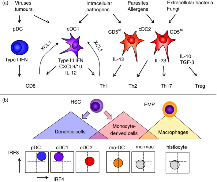
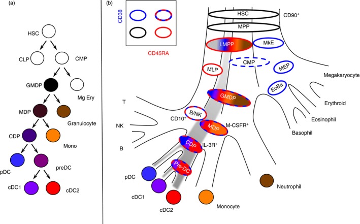
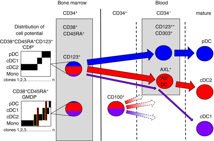
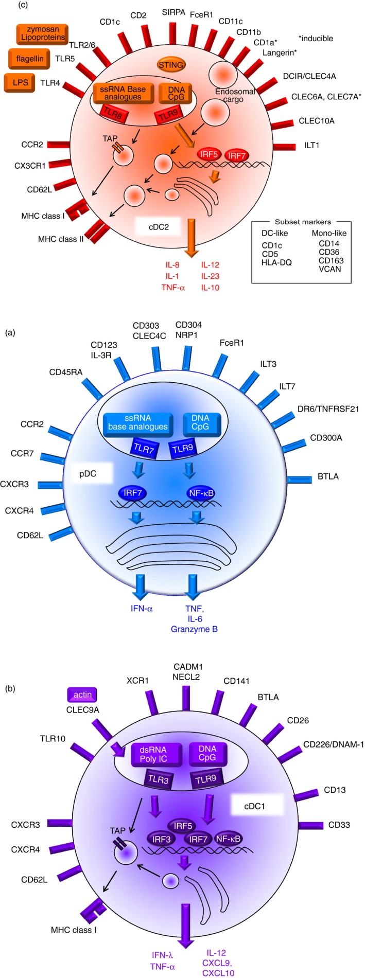
Verification
Claims the paper makes
The claims and statistics of this paper haven’t been checked yet.
Run the check — free, no account needed. It reads the open-access text and checks the sources it cites.
Claims other papers make about this one
Each is checked against this paper’s own text: does it say what the citing paper says it does?
The source backs part of the claim, not all of it — or backs it for a different population, condition or comparison.
In the citing paper
“The three other proteins increased in abundance in TET2-CH were CD1C (a marker of conventional dendritic cells), CLEC4C (a marker of plasmacytoid dendritic cells) and CD86 (a marker of dendritic cells, monocytes and other antigen-presenting cells)51.”
From Rare variant associations with plasma protein levels in the UK Biobank (2023) · cites this paper as [51]
Experimental — accuracy not yet measuredIn the citing paper
“pDCs are instead characterized by a marked impairment of cytokine release in older people (27, 29).”
From Immunosenescence and Its Hallmarks: How to Oppose Aging Strategically? A Review of Potential Options for Therapeutic Intervention (2019) · cites this paper as [27]
Self-check
Percentages, sums and reported differences re-computed from the paper’s own numbers.
The self-check needs the paper’s full text.
Citations
Cites
Sallusto, F., & Lanzavecchia, A. (1994) · The Journal of Experimental Medicine · doi:10.1084/jem.179.4.1109 · Cited by 5,129
Simultaneous epitope and transcriptome measurement in single cells
Stoeckius, M., Hafemeister, C., Stephenson, W., Houck-Loomis, B., Chattopadhyay, P. K., Swerdlow, H., Satija, R., & Smibert, P. (2017) · Nature Methods · doi:10.1038/nmeth.4380 · Cited by 3,098
Nomenclature of monocytes and dendritic cells in blood
Ziegler‐Heitbrock, L., Ancuța, P., Crowe, S. M., Dalod, M., Grau, V., Hart, D. N. J., Leenen, P. J. M., Liu, Y.-J., MacPherson, G. G., Randolph, G. J., et al. (2010) · Blood · doi:10.1182/blood-2010-02-258558 · Cited by 2,436
Single-cell RNA-seq reveals new types of human blood dendritic cells, monocytes, and progenitors
Villani, A.-C., Satija, R., Reynolds, G., Sarkizova, S., Shekhar, K., Fletcher, J., Griesbeck, M., Butler, A., Zheng, S., Lazo, S., et al. (2017) · Science · doi:10.1126/science.aah4573 · Cited by 2,413
The Nature of the Principal Type 1 Interferon-Producing Cells in Human Blood
Siegal, F. P., Kadowaki, N., Shodell, M., Fitzgerald-Bocarsly, P. A., Shah, K., Ho, S., Antonenko, S., & Liu, Y.-J. (1999) · Science · doi:10.1126/science.284.5421.1835 · Cited by 2,293
IRF-7 is the master regulator of type-I interferon-dependent immune responses
Honda, K., Yanai, H., Negishi, H., Asagiri, M., Sato, M., Mizutani, T., Shimada, N., Ohba, Y., Takaoka, A., Yoshida, N., et al. (2005) · Nature · doi:10.1038/nature03464 · Cited by 2,229
Phosphorylation of innate immune adaptor proteins MAVS, STING, and TRIF induces IRF3 activation
Liu, S., Cai, X., Wu, J., Cong, Q., Chen, X., Li, T., Du, F., Ren, J., Wu, Y.-T., Grishin, N. V., et al. (2015) · Science · doi:10.1126/science.aaa2630 · Cited by 1,927
Dendritic cells, monocytes and macrophages: a unified nomenclature based on ontogeny
Guilliams, M., Ginhoux, F., Jakubzick, C., Naik, S. H., Onai, N., Schraml, B. U., Segura, É., Tussiwand, R., & Yona, S. (2014) · Nature reviews. Immunology · doi:10.1038/nri3712 · Cited by 1,923
Cella, M., Jarrossay, D., Facchetti, F., Alebardi, O., Nakajima, H., Lanzavecchia, A., & Colonna, M. (1999) · Nature Medicine · doi:10.1038/11360 · Cited by 1,734
Dzionek, A., Fuchs, A., Schmidt, P., Cremer, S., Zysk, M., Miltenyi, S., Buck, D., & Schmitz, J. (2000) · Journal of Immunology · doi:10.4049/jimmunol.165.11.6037 · Cited by 1,433
Cited by
Sorted by citations
Items 601–700 of 998
Aghamajidi, A., Farhangnia, P., Pashangzadeh, S., Damavandi, A. R., & Jafari, R. (2022) · Cancer Cell International · doi:10.1186/s12935-022-02727-3 · Cited by 12
“Although, DCs mediate antigen trafficking and stimulation of CD8+ T-cell responses, however, TAR-DCs exhibited immunosuppressive properties by low expression of costimulatory molecules and high expression of regulatory molecules.”
BackgroundInfluentialAghamiri, S. S., Puniya, B. L., Amin, R., & Helikar, T. (2023) · Frontiers in Immunology · doi:10.3389/fimmu.2023.1112985 · Cited by 12
Role of dendritic cell‑derived exosomes in allergic rhinitis (Review)
Kang, C., He, H., Liu, P., Liu, Y., Li, X., Zhang, J., Hong, R., Zeng, X., Zhao, H., Liu, J., et al. (2023) · International Journal of Molecular Medicine · doi:10.3892/ijmm.2023.5320 · Cited by 13
“This sequential invagination of the plasma membrane eventually leads to the development of (20). dcs, dendritic cells; HSC, hematopoietic stem cell; CMP, common myeloid progenitor; GMDP, granulocyte‑macrophage DC progenitor; MDP, macro‑ phage DC progenitor; CDP, common DC precursor; CLP, common…”
BackgroundLin, Q. X. X., Rajagopalan, D., Gamage, A. M., Tan, L. M., Venkatesh, P. N., Chan, W. O. Y., Kumar, D., Agrawal, R., Chen, Y., Fong, S., et al. (2024) · Nature Communications · doi:10.1038/s41467-023-44524-0 · Cited by 17
Development of innate and adaptive immunity to RSV in young children
Parsons, E. L., Kim, J. S., & Malloy, A. M. W. (2024) · Cellular Immunology · doi:10.1016/j.cellimm.2024.104824 · Cited by 17
Cell-specific gene networks and drivers in rheumatoid arthritis synovial tissues
Pélissier, A., Laragione, T., Gulko, P. S., & Martínez, M. R. (2024) · Frontiers in Immunology · doi:10.3389/fimmu.2024.1428773 · Cited by 11
Du, J., Fang, L., Dong, K., & Zhou, Z. (2024) · Journal of Affective Disorders · doi:10.1016/j.jad.2024.10.050 · Cited by 12
Dendritic cells in triple-negative breast cancer: From pathophysiology to therapeutic applications
Sebastião, A. I., Simões, G., Oliveira, F., Mateus, D., Falcão, A. C., Carrascal, M. A., Gomes, C. M. F., Neves, B. M., & Cruz, M. T. (2025) · Cancer Treatment Reviews · doi:10.1016/j.ctrv.2025.102884 · Cited by 14
Yin, J., Ye, Y., Gao, Y., Xu, Q., Su, M., Sun, S., Xu, W., Fu, Q., Wang, A., & Hu, S. (2025) · International Journal of Molecular Sciences · doi:10.3390/ijms26052269 · Cited by 12
“Dendritic cells (DCs) are the most efficient and specialized antigen-presenting cells (APCs) in the innate immune system and are capable of initiating adaptive immune responses [65].”
Biomarkers in immunology: Their impact on immune function and response
Kaushik, D., Xu, B., & Kumar, M. (2025) · Advances in Biomarker Sciences and Technology · doi:10.1016/j.abst.2025.03.001 · Cited by 14
Microbial-induced trained immunity for cancer immunotherapy
Vuscan, P., Kischkel, B., Joosten, L. A. B., & Netea, M. G. (2025) · Pharmacological Reviews · doi:10.1016/j.pharmr.2025.100074 · Cited by 14
Regulation of allergic inflammation by dendritic cells
Chow, T. G., & Gill, M. A. (2019) · Current Opinion in Allergy and Clinical Immunology · doi:10.1097/aci.0000000000000603 · Cited by 12
“Although many of the subsets participate in both antigen presentation to T cells and signaling to coordinate immune responses, differences exist in their predominant functions, recently reviewed here [2], which will be discussed when pertinent throughout this review.”
BackgroundWei, B. M., Hanlon, D. J., Khalil, D., Han, P., Tatsuno, K., Sobolev, O., & Edelson, R. L. (2020) · PubMed · Cited by 13
Prenatal Development and Function of Human Mononuclear Phagocytes
Miah, M., Goh, I., & Haniffa, M. A. (2021) · Frontiers in Cell and Developmental Biology · doi:10.3389/fcell.2021.649937 · Cited by 13
Singh, A., Das, D., Kurra, S., Arava, S. K., Gupta, S., & Sharma, A. (2021) · Cytokine · doi:10.1016/j.cyto.2021.155598 · Cited by 12
Dendritic Cells and Antiphospholipid Syndrome: An Updated Systematic Review
Tang, K., Chen, H., Chen, T., Bracci, N. R., & Lin, C. (2021) · Life · doi:10.3390/life11080801 · Cited by 12
“DCs can be divided into three major subsets: conventional DCs, DCs derived from monocytes, and plasmacytoid DCs [16].”
BackgroundInhibition of renalase drives tumour rejection by promoting T cell activation
Guo, X., Jessel, S., Qu, R., Kluger, Y., Tian-min, C., Hollander, L. L., Safirstein, R. L., Nelson, B., Cha, C. H., Bosenberg, M., et al. (2022) · European Journal of Cancer · doi:10.1016/j.ejca.2022.01.002 · Cited by 12
Chemically engineered glycan-modified cancer vaccines to mobilize skin dendritic cells
Duinkerken, S., Li, R. J. E., van Haften, F. J., de Gruijl, T. D., Chiodo, F., Schetters, S. T. T., & van Kooyk, Y. (2019) · Current Opinion in Chemical Biology · doi:10.1016/j.cbpa.2019.10.001 · Cited by 12
Immune activation of the monocyte-derived dendritic cells using patients own circulating tumor cells
Kološtová, K., Pospíšilová, E., Matkowski, R., Szelachowska, J., & Bobek, V. (2022) · Cancer Immunology Immunotherapy · doi:10.1007/s00262-022-03189-2 · Cited by 12
Buffa, D. J., O’Neil, T. R., Vine, E. E., Sarkawt, L., van Dijk, F. A. W., Dong, O. A., Nasr, N., Cunningham, A. L., Bertram, K. M., & Harman, A. N. (2025) · PLoS Pathogens · doi:10.1371/journal.ppat.1013490 · Cited by 13
Targeting FGL2 in glioma immunosuppression and malignant progression
Ma, X., Zhu, H., Cheng, L., Chen, X., Shu, K., & Zhang, S. (2022) · Frontiers in Oncology · doi:10.3389/fonc.2022.1004700 · Cited by 11
Modulation by luminal factors on the functions and migration of intestinal innate immunity
Higashiyama, M., Miura, S., & Hokari, R. (2023) · Frontiers in Immunology · doi:10.3389/fimmu.2023.1113467 · Cited by 12
Colombo, S. A. P., Brown, S. L., Hepworth, M. R., Hankinson, J., Granato, F., Kitchen, S. J., Hussell, T., Simpson, A., Cook, P. C., & MacDonald, A. S. (2023) · Discovery Immunology · doi:10.1093/discim/kyad009 · Cited by 12
“The C-type lectin receptor CD206 (mannose receptor) is a pattern recognition receptor (PRR) associated with signalling by type-2 cytokines, particularly IL-4 and IL-13 [38], and used as a moDC marker on CD1a+ cells [37].”
BackgroundGorodilova, A. V., Kitaeva, K. V., Filin, I. Y., Mayasin, Y. P., Kharisova, C. B., Issa, S. S., Solovyeva, V. V., & Rizvanov, A. A. (2023) · Current Issues in Molecular Biology · doi:10.3390/cimb45100509 · Cited by 12
Tolerogenic dendritic cells in radiation-induced lung injury
Liu, B., Wang, Y., Han, G., & Zhu, M. (2024) · Frontiers in Immunology · doi:10.3389/fimmu.2023.1323676 · Cited by 11
“These cells are known to secrete IL-12, type I, and III interferons (IFNs), and are believed to facilitate Th1-assisted T-cells and natural killer responses (72, 73).”
BackgroundExploiting Leishmania—Primed Dendritic Cells as Potential Immunomodulators of Canine Immune Response
Valério-Bolas, A., Meunier, M., Palma-Marques, J., Rodrigues, A., Santos, A., Nunes, T., Ferreira, R. R. F., Armada, A., Alves, J. C., Antunes, W., et al. (2024) · Cells · doi:10.3390/cells13050445 · Cited by 12
“In addition to being important mediators between innate and adaptive immunity, DCs are also recognized for their highly efficient phagocytic potential [55].”
BackgroundThe pleiotropic CLEC10A: implications for harnessing this receptor in the tumor microenvironment
van der Meijs, N. L., Travecedo, M. A., Marcelo, F., & van Vliet, S. J. (2024) · Expert Opinion on Therapeutic Targets · doi:10.1080/14728222.2024.2374743 · Cited by 11
“The subset-defining markers and general functions of cDC2 and DC3 are reviewed in more detail elsewhere [62,63].”
The role of sialoglycans in modulating dendritic cell function and tumour immunity
Silva, Z., Soares, C. O., Barbosa, M., Palma, A. S., Marcelo, F., & Videira, P. A. (2024) · Seminars in Immunology · doi:10.1016/j.smim.2024.101900 · Cited by 11
Human genital dendritic cell heterogeneity confers differential rapid response to HIV-1 exposure
Parthasarathy, S., de Lara, L. M., Carrillo-Salinas, F. J., Werner, A., Borchers, A., Iyer, V. V., Vogell, A. B., Fortier, J. M., Wira, C. R., & Rodríguez‐García, M. (2024) · Frontiers in Immunology · doi:10.3389/fimmu.2024.1472656 · Cited by 11
“Characterization and delineation of DC subsets is classically dependent on surface protein expression patterns (34).”
BackgroundYang, C., Qu, J., Wu, J., Cai, S., Liu, W., Deng, Y., Meng, Y., Zheng, L., Zhang, L., Wang, L., et al. (2024) · Clinical and Translational Medicine · doi:10.1002/ctm2.70091 · Cited by 11
Song, W., Chen, X., Haiying, W., & Rahimian, N. (2024) · European journal of medical research · doi:10.1186/s40001-024-02230-7 · Cited by 14
Shu, Y., Li, S., Du, Y., & Zheng, X. (2025) · Frontiers in Immunology · doi:10.3389/fimmu.2025.1611976 · Cited by 11
“Human DCs are broadly categorized into three main types, including monocyte-derived DCs (moDCs), plasmacytoid DCs (pDCs), and conventional DCs (cDCs) (6).”
Imai, J., Ohashi, S., & Sakai, T. (2020) · Pharmaceutics · doi:10.3390/pharmaceutics12020153 · Cited by 12
“Most of these DC classifications have been examined in mice, strongly indicating that experimental confirmation is required for DC vaccines [31,33].”
BackgroundInfluentialLiu, Y., Jin, Z., Huang, X., Che, Y.-C., & Liu, Q. (2020) · Frontiers in Oncology · doi:10.3389/fonc.2020.553536 · Cited by 11
“DCs promote immunity or tolerance by sampling and presenting antigens to T cells and by providing immunomodulatory signals through cell–cell contacts and cytokines (120, 121).”
Background3. Immune responses against African swine fever virus infection
Montoya, M., Franzoni, G., Pérez-Núñez, D., Revilla, Y., Galindo, I., Alonso, C., Netherton, C. L., & Blohm, U. (2021) · Wageningen Academic Publishers eBooks · doi:10.3920/978-90-8686-910-7_3 · Cited by 11
“…amounts of type I interferon following viral stimulation (Collin and Bigley , 2018), whereas cDC1 are specialised in priming a Th1 response through IL-12 secretion and antigen presentation via MHC class I to CD8 + T cells, and cDC2 can stimulate Th2 and Th17 responses (Collin and Bigley , 2018).”
InfluentialPeil, J., Bock, F., Kiefer, F., Schmidt, R., Heindl, L. M., Cursiefen, C., & Schlereth, S. L. (2022) · International Journal of Molecular Sciences · doi:10.3390/ijms23031478 · Cited by 12
“moDCs originate from monocytes and appear in inflammation, cancer, or infection [203,237,247].”
BackgroundInfluentialDendritic cells metabolism: a strategic path to improve antitumoral DC vaccination
Currivan, E., Finlay, D. K., & Moreira, D. (2022) · Clinical & Experimental Immunology · doi:10.1093/cei/uxac048 · Cited by 11
Different Roles of Dendritic Cells for Chronic Rhinosinusitis Treatment According to Phenotype
Tai, J., Kwak, J., Han, M. S., & Kim, T. H. (2022) · International Journal of Molecular Sciences · doi:10.3390/ijms23148032 · Cited by 11
“During infection, they express many receptors of cDC2, such as BDCA1, CD1a, CD11c, CD14, and CD172a [36].”
BackgroundSebastião, A. I., Mateus, D., Carrascal, M. A., Sousa, C., Cortes, L., Bachmann, M. F., do Carmo, A., Matos, A. M., Sales, M. G. F., & Cruz, M. T. (2023) · Pharmaceutics · doi:10.3390/pharmaceutics15030825 · Cited by 11
“At the end of the replication cycle, SARS-CoV-2 induces cellular pyroptosis and triggers the release of both danger-associated molecular patterns (DAMPs) and pathogen-associated molecular patterns (PAMPs).”
BackgroundInfluentialWang, H., Xu, L., Wu, Z., & Chen, X. (2020) · Fish & Shellfish Immunology · doi:10.1016/j.fsi.2020.04.026 · Cited by 11
Menzner, A.-K., Rottmar, T., Völkl, S., Bosch, J. J., Mougiakakos, D., Mackensen, A., & Resheq, Y. J. (2021) · Cancers · doi:10.3390/cancers13030461 · Cited by 11
“In this study, we show that CAT-DCs fail to upregulate common DC-maturation-markers such as CD1a, CD1c, CD83, CD49d, and HLA-DR [39].”
BackgroundInfluentialDevelopment of an Inflammatory CD14+ Dendritic Cell Subset in Humanized Mice
Iwabuchi, R., Ide, K., Terahara, K., Wagatsuma, R., Iwaki, R., Matsunaga, H., Tsunetsugu‐Yokota, Y., Takeyama, H., & Takahashi, Y. (2021) · Frontiers in Immunology · doi:10.3389/fimmu.2021.643040 · Cited by 11
Therapeutic Cancer Vaccine Therapy for Acute Myeloid Leukemia
Wu, M., Wang, S., Chen, J., Zhou, L., Guo, Z., & Li, Y. (2021) · Immunotherapy · doi:10.2217/imt-2020-0277 · Cited by 11
Doyle, C. M., Vine, E. E., Bertram, K. M., Baharlou, H., Rhodes, J. W., Dervish, S., Gosselink, M. P., Re, A. M. D., Collins, G. P., Reza, F., et al. (2021) · Frontiers in Immunology · doi:10.3389/fimmu.2021.727952 · Cited by 11
Dang, D., Liu, Y., Zhou, Q., Li, H., Wang, Y., & Wu, H. M. (2021) · Infection Genetics and Evolution · doi:10.1016/j.meegid.2021.105121 · Cited by 11
Dhindsa, R. S., Burren, O. S., Sun, B. B., Prins, B. P., Matelska, D., Wheeler, E., Mitchell, J., Oerton, E., Hristova, V. A., Smith, K. R., et al. (2022) · bioRxiv (Cold Spring Harbor Laboratory) · doi:10.1101/2022.10.09.511476 · Cited by 11
Tandem repeat variation shapes immune cell type-specific gene expression
Tanudisastro, H. A., Cuomo, A. S. E., Weisburd, B., Welland, M. J., Spenceley, E., Franklin, M., Xue, A., Huang, H. L., Bowen, B., Fan, J., et al. (2026) · Nature · doi:10.1101/2024.11.02.621562 · Cited by 14
“The effect sizes of type-2 conventional dendritic cells (cDC2) and monocytes showed strong concordance, consistent with cDC2 sharing the most markers with monocytes amongst all dendritic cell (DC) subtypes 37 .”
Recent advances in the immunomodulation mechanism of mesenchymal stem cell therapy in liver diseases
Liu, P., Yao, L., Hu, X., Wang, Z., Xiong, Z., & Jiang, Y. (2023) · Journal of Gastroenterology and Hepatology · doi:10.1111/jgh.16247 · Cited by 10
Research progress on mitochondria regulating tumor immunity
Li, J., Xu, P., & Chen, S. (2024) · Journal of Zhejiang University (Medical Sciences) · doi:10.3724/zdxbyxb-2023-0484 · Cited by 11
Poženel, P., Zajc, K., & Švajger, U. (2024) · Journal of Translational Medicine · doi:10.1186/s12967-024-05368-4 · Cited by 10
“Dendritic cells (DCs) act at the central stage of our immunological network with the most important antigen-presenting cell (APC) function of the immune cell repertoire [1].”
BackgroundBuilding a human lung from pluripotent stem cells to model respiratory viral infections
Turner, D. L., Amoozadeh, S., Baric, H., Stanley, E. G., & Werder, R. B. (2024) · Respiratory Research · doi:10.1186/s12931-024-02912-0 · Cited by 11
“Similar classification has occurred with respect to human DC populations (CD141 + cDC1, CD1c + cDC2, moDCs and pDCs) [202– 205]; however, studies describing the role of these subsets during infections with human-specific respiratory pathogens are limited.”
ResultDendritic cell-based immunotherapy in non-small cell lung cancer: a comprehensive critical review
de Oliveira, J. B., Silva, S. B., Fernandes, I. L., Batah, S. S., Herrera, A. J. R., Cetlin, A. A., & Fabro, A. T. (2024) · Frontiers in Immunology · doi:10.3389/fimmu.2024.1376704 · Cited by 11
“Although human DC subsets have traditionally been recognized through a limited selection of markers, such as CD1c + , CD141 + , and CD14 + (22), this simplistic phenotypical representation fails to capture the complexity of DC identity in various physiological processes and tumoral…”
BackgroundBooth, C. A. G., Bouyssou, J., Togami, K., Armand, O., Rivas, H. G., Yan, K., Rice, S., Cheng, S., Lachtara, E. M., Bourquin, J., et al. (2024) · JCI Insight · doi:10.1172/jci.insight.183889 · Cited by 10
“Globally, the mutational landscape of BPDCN shows similarities to both myeloid and lymphoid malignancies (4), which may reflect the lympho-myeloid ontogeny of pDCs (16, 17).”
Tertiary lymphoid structures in colorectal cancer - organization and immune cell interactions
Gulubova, M. V. (2024) · American journal of clinical and experimental immunology. · doi:10.62347/gryy2849 · Cited by 10
Laminarins and their derivatives affect dendritic cell activation and their crosstalk with T cells
Christensen, M. D., Allahgholi, L., Dobruchowska, J. M., Moenaert, A., Guðmundsson, H., Friðjónsson, Ó. H., Karlsson, E. N., Hreggviðsson, G. Ó., & Freysdóttir, J. (2025) · International Journal of Biological Macromolecules · doi:10.1016/j.ijbiomac.2025.141287 · Cited by 10
Pathophysiology of bone remodelling cycle: Role of immune system and lipids
Parolini, C. (2025) · Biochemical Pharmacology · doi:10.1016/j.bcp.2025.116844 · Cited by 10
Colorectal Organoids: Models, Imaging, Omics, Therapy, Immunology, and Ethics
Taglieri, M., Gregorio, L. D., Matis, S., Uras, C. R. M., Ardy, M., Casati, S., Marchese, M., Poggi, A. D., Raffaghello, L., & Benelli, R. (2025) · Cells · doi:10.3390/cells14060457 · Cited by 12
“Recently, it has been shown that conventional DC type 2 (DC2) relevant for the induction of T helper cell responses [223] can shift to a CD14+ population with impaired T cell-activating capabilities when co-cultured with CRC organoids [224].”
Elahi, Z., Jameson, V. J., Sakkas, M., Butcher, S., Mintern, J. D., Radford, K. J., & Wells, C. A. (2025) · Journal of Leukocyte Biology · doi:10.1093/jleuko/qiaf045 · Cited by 10
Sun, F., Gao, X., Li, T., Zhao, X., & Zhu, Y. (2025) · Biochimica et Biophysica Acta (BBA) - Reviews on Cancer · doi:10.1016/j.bbcan.2025.189350 · Cited by 13
Ju, X., Putz, E. M., Liu, Y., Yuan, D., Sun, G., Koda, S., Fu, Z., Shao, S., Tong, C., Deng, B., et al. (2025) · Nature Communications · doi:10.1038/s41467-025-60922-y · Cited by 10
“Theenzyme indoleamine 2,3-dioxygenase 1 (IDO1) and arginase expressed in DCs deplete the essential amino acids L-tryptophan and arginine in T cells and generate immune suppressive metabolites 7 .”
Hawkins, A. N., Determann, B. F., Nelson, B. N., & Wozniak, K. L. (2021) · Frontiers in Immunology · doi:10.3389/fimmu.2021.722500 · Cited by 11
Zuo, H., van Lierop, M.-J. C., Kaspers, J., Bos, R., Reurs, A. W., Sarkar, S., Konry, T., Kamermans, A., Kooij, G., de Vries, H. E., et al. (2021) · Cells · doi:10.3390/cells10113233 · Cited by 10
“Dendritic cells possess this cross-presenting activity, but one type of DC, the myeloid/conventional DC type 1 (cDC1), seems superior in this [46,47], and these cells are found in relatively high quantities in LNs [48].”
Solhaug, M. B., Schreurs, O., Schenck, K., Blix, I. J. S., & Bækkevold, E. S. (2021) · European Journal Of Oral Sciences · doi:10.1111/eos.12835 · Cited by 10
Han, C., Yan, Y., Yan, L., Meng, G.-X., Yang, C., Liu, H., Ding, Z., Dong, Z., Hong, J., Chen, Z., et al. (2022) · Journal of Cancer Research and Clinical Oncology · doi:10.1007/s00432-022-04008-y · Cited by 10
“The phenomenon is probably attributable to that DCs are antigen-presenting cells in the host body (Collin and Bigley 2018), and it is more efficient to administer DC plus antigens directly to bypass the recognition step and the loss of antigens.”
BackgroundscESI: evolutionary sparse imputation for single-cell transcriptomes from nearest neighbor cells
Liu, Q., Luo, X., Li, J., & Wang, G. (2022) · Briefings in Bioinformatics · doi:10.1093/bib/bbac144 · Cited by 10
Chen, J., Xing, Q.-L., Yang, H., Yang, F., Luo, Y., Kong, W., & Wang, Y. (2022) · Scientific Reports · doi:10.1038/s41598-022-13818-6 · Cited by 10
Lin, C., Yang, H., Zhao, W., & Wang, W. (2022) · Biochemical and Biophysical Research Communications · doi:10.1016/j.bbrc.2022.06.037 · Cited by 10
Carenza, C., Franzese, S., Castagna, A., Terzoli, S., Simonelli, M., Persico, P., Bello, L., Nibali, M. C., Pessina, F., Kunderfranco, P., et al. (2023) · Frontiers in Immunology · doi:10.3389/fimmu.2022.1074762 · Cited by 11
Daniel, S. K., Sullivan, K. M., Dickerson, L. K., van den Bijgaart, R. J. E., Utria, A. F., Labadie, K. P., Kenerson, H. L., Jiang, X., Smythe, K. S., Campbell, J. S., et al. (2024) · Scientific Reports · doi:10.1038/s41598-024-55593-6 · Cited by 10
“2C) 44–46 .”
Monasterio, G., Guevara, J., Ibarra, J. P., Castillo, F. A., Díaz-Zúñiga, J. A., Álvarez, C., Cafferata, E. A., & Vernal, R. (2018) · Clinical Oral Investigations · doi:10.1007/s00784-018-2641-5 · Cited by 10
Tethering Innate Surface Receptors on Dendritic Cells: A New Avenue for Immune Tolerance Induction?
Lamendour, L., Deluce, N. K.-N., Mouline, C. C., Gouilleux‐Gruart, V., & Velge-Roussel, F. (2020) · International Journal of Molecular Sciences · doi:10.3390/ijms21155259 · Cited by 10
“They are identified by their expression of CD123, CD4, MHCII, CD303 (BDCA2), CD304 (BDCA4), and CD45RA (Table 1) [8].”
BackgroundComprehensive Phenotyping of Dendritic Cells in Cancer Patients by Flow Cytometry
Carenza, C., Franzese, S., Calcaterra, F., Mavilio, D., & Bella, S. D. (2020) · Cytometry Part A · doi:10.1002/cyto.a.24245 · Cited by 10
Dendritic cell responses to Plasmodium falciparum in a malaria-endemic setting
Turner, T. C., Arama, C., Ongoïba, A., Doumbo, S., Doumtabé, D., Kayentao, K., Skinner, J., Li, S., Traoré, B., Crompton, P. D., et al. (2021) · Malaria Journal · doi:10.1186/s12936-020-03533-w · Cited by 10
Sarchioto, M., Howe, F. A., Dumitriu, I. E., Morgante, F., Stern, J. S., Edwards, M. J., & Martino, D. (2021) · European Journal of Neurology · doi:10.1111/ene.14837 · Cited by 10
“Among DC subsets, MDC1s have a specific capability to present antigens via major histocompatibility complex class II to activate naïve CD4+ T cells, and to promote T- helper 1 responses [11].”
BackgroundInfluentialChatterjee, G., Sriram, H., Ghogale, S., Deshpande, N., Khanka, T., Girase, K., Verma, S., Arolkar, G., Dasgupta, N., Narula, G., et al. (2021) · British Journal of Haematology · doi:10.1111/bjh.17801 · Cited by 10
Zheng, L., Zhao, F., Ru, J., Liu, L., Wang, Z., Wang, N., Shu, X., Wei, Z., & Guo, H. (2021) · Viruses · doi:10.3390/v13112158 · Cited by 10
“Unlike DCs of human and mice [37,38], porcine DCs selectively express TLR3, TLR5, TLR7, TLR9, and TLR10, which were significantly higher in the inactivated TGEV vaccine with the MSN group.”
BackgroundDong, Y. W., Li, Y., Yao, Y., & Song, Q. (2022) · Frontiers in Oncology · doi:10.3389/fonc.2022.897323 · Cited by 10
Differential IRF8 Requirement Defines Two Pathways of Dendritic Cell Development in HumansPreprint
Cytlak, U., Resteu, A., Pagan, S., Green, K., Milne, P., Maisuria, S., McDonald, D., Hulme, G., Filby, A. J., Carpenter, B., et al. (2019) · SSRN Electronic Journal · doi:10.2139/ssrn.3365028 · Cited by 10
Mengistu, D. T., Curtis, J. L., & Freeman, C. M. (2024) · Trends in Immunology · doi:10.1016/j.it.2024.04.010 · Cited by 9
Dendritic cells in Parkinson's disease: Regulatory role and therapeutic potential
Mula, A. R. A., Yuan, X., & Lu, J. (2024) · European Journal of Pharmacology · doi:10.1016/j.ejphar.2024.176690 · Cited by 9
Jiang, Y., Bei, W.-X., Li, W., Huang, Y., He, S., Zhu, X., Zheng, L., Xia, W., Dong, S., Liu, Q., et al. (2024) · Clinical and Translational Medicine · doi:10.1002/ctm2.70061 · Cited by 10
“35,36 40 These phenomena were similar to the results observed in renal cell carcinoma in a recent study.”
ResultImmunopathogenesis and immunotherapy of diabetes-associated periodontitis
Li, S., Li, S., Meng, L., Gao, R., Liu, H., & Li, M. (2025) · Clinical Oral Investigations · doi:10.1007/s00784-024-06141-z · Cited by 9
“Under normal conditions, immature DCs exhibit high migratory capacity and can efficiently uptake, process, and deliver antigens; mature DCs can effectively activate the initial T cells and mainly perform the initiation, regulation, and maintenance of the immune response [32].”
Dumolard, L., Gerster, T., Chuffart, F., Decaens, T., Hilleret, M., Larrat, S., Saas, P., Jouvin‐Marche, E., Durantel, D., Marche, P. N., et al. (2025) · Hepatology Communications · doi:10.1097/hc9.0000000000000625 · Cited by 9
Roshal, M., & Gao, Q. (2025) · American Journal of Clinical Pathology · doi:10.1093/ajcp/aqae187 · Cited by 10
Boller, A.-L., Ruland, T., Dantas, R. L., Michels, S., Dannlowski, U., Scheu, S., Baune, B. T., Culmsee, C., & Alferink, J. (2025) · Brain Behavior and Immunity · doi:10.1016/j.bbi.2025.07.004 · Cited by 10
“…and transcription factor expression: conventional DCs (cDCs) and plasmacytoid DCs (pDCs). cDCs are further divided into cDC1, which are specialized in antigen cross-presentation to CD8⁺ T cells, and cDC2, which primarily activate CD4⁺ T cells to support adaptive immunity (Collin & Bigley, 2018).”
Human cDC1s display constitutive activation of the UPR sensor IRE1
García-González, P., Fernández, D., Gutiérrez, D., Parra-Cordero, M., & Osorio, F. (2022) · European Journal of Immunology · doi:10.1002/eji.202149774 · Cited by 7
Zheng, F. Y., Zhang, W., Yang, B., & Chen, M. (2022) · Annals of Translational Medicine · doi:10.21037/atm-22-5351 · Cited by 10
Mathieu, E., Marquant, Q., Chain, F., Bouguyon, E., Saint‐Criq, V., Goffic, R. L., Descamps, D., Langella, P., Tompkins, T. A., Binda, S., et al. (2023) · Nutrients · doi:10.3390/nu15020263 · Cited by 8
Zhao, M., Zhang, Q., Song, Z., Lei, H., Li, J., Peng, F., & Lin, S. (2023) · Translational Cancer Research · doi:10.21037/tcr-23-83 · Cited by 9
Moon, H., Eccles, J. D., Kim, S.-J., Kim, K.-H., Kim, Y.-M., Rehman, J., Lee, H., Kanabar, P. N., Christman, J. W., Ackerman, S. J., et al. (2023) · Journal of Allergy and Clinical Immunology · doi:10.1016/j.jaci.2023.07.016 · Cited by 9
Zhao, F., Gu, W., Li, L., Qu, Z., Xu, M.-Y., Liu, K., Zhang, F., Liu, H., Xu, J., & Yin, H.-Y. (2023) · International Immunopharmacology · doi:10.1016/j.intimp.2023.110771 · Cited by 9
Highlighting the Role of DC-NK Cell Interplay in Immunobiology and Immunotherapy
Calmeiro, J., Carrascal, M. A., Gomes, C. M. F., Falcão, A. C., Cruz, M. T., & Neves, B. M. (2018) · InTech eBooks · doi:10.5772/intechopen.78804 · Cited by 9
“There are three major subpopulations of DCs: plasmacytoid DCs (pDCs) characterized by the expression of CD123, CD303, CD304, FCER1, ILT3, and ILT7; classical DCs 1 (cDC1) that express CD141, CLEC9A, XCR1, and CADM1; and classical DCs 2 expressing CD1c, CD11c, CD11b, CD2, FCER1, SIRPA, ILT1, and CLEC4A [2, 22, 23].”
BackgroundMurray, S. M., Zhang, Y., Douek, D. C., & Sékaly, R. (2020) · Frontiers in Immunology · doi:10.3389/fimmu.2020.00261 · Cited by 9
Tembhare, P. R., Sriram, H., Chatterjee, G., Khanka, T., Gokarn, A. G., Mirgh, S. P., Rajendra, A., Chaturvedi, A., Ghogale, S., Deshpande, N., et al. (2021) · Immunology and Cell Biology · doi:10.1111/imcb.12504 · Cited by 10
“They recognize viral antigens through Toll-like receptors, migrate to lymphoid organs and initiate a specific immune response.(29) An acute viral infection induces DC activation, followed by migration of DCs from peripheral blood to the tissue with infection (such as lungs), leading to depletion in the circulating DC…”
BackgroundAmodio, G., Mandelli, A., Curto, R., Rancoita, P. M. V., Stabilini, A., Bonfanti, R., Pellegrin, M. D., Bosi, E., Serio, C. D., Battaglia, M., et al. (2021) · Frontiers in Immunology · doi:10.3389/fimmu.2021.750162 · Cited by 9
Chan, A. C. L., Liu, Y., Devlin, S. M., Gao, Q., Baik, J., Sigler, A. M., Londono, D., Arcila, M. E., Levine, R. L., Doǧan, A., et al. (2022) · HemaSphere · doi:10.1097/hs9.0000000000000685 · Cited by 9
Du, Y., Hertoghs, N., Duffy, F. J., Carnes, J., McDermott, S. M., Neal, M., Schwedhelm, K. V., McElrath, M., Rosa, S. D., Aitchison, J., et al. (2021) · bioRxiv · doi:10.1371/journal.ppat.1010282 · Cited by 10
“250 Because different types of DCs can activate and skew different T-helper cell responses, we also 251 assessed the proportion of Th1 and Th2 CD4+ T cells (Collin and Bigley, 2018) and observed 252 greater proportions of Th2 CD4 T cells in NP, and a trend towards more Th1 cells in P subjects 253 (Fig.”
BackgroundZhu, B., Sun, Y., Wei, X., Zhou, H., Cao, J., Li, C., & Wu, N. (2022) · Journal of Healthcare Engineering · doi:10.1155/2022/1964081 · Cited by 9
“Dendritic cells (DCs) activate T cells to become cytotoxic T lymphocytes (CTLs) and initiate a primary immune response [7].”
BackgroundThe Tropomyosin Family as Novel Biomarkers in Relation to Poor Prognosis in Glioma
Huang, K., Wang, H., Xu, J., Xu, R.-M., Liu, Z., Li, Y., & Xu, Z. (2022) · Biology · doi:10.3390/biology11081115 · Cited by 9
“Th2 and Th17 infiltration were expected to contribute to the immune suppression type of DCs, as shown in previous studies [36].”
ResultQiu, G., Zhong, S., Xie, J., Feng, H., Sun, S., Gao, C., Xu, X., Kang, B., Xu, H., Zhao, C. G., et al. (2022) · Journal of Immunology Research · doi:10.1155/2022/9634073 · Cited by 9
“DCs form an essential interface between the innate sensing of pathogens and the activation of adaptive immunity [6].”
Metadata
- Authors
- Collin, M., & Bigley, V.
- Affiliations
- Newcastle upon Tyne Hospitals NHS Foundation Trust (GB)
- Corresponding author
- Collin, Matthew
- Venue
- Immunology
- Publisher
- Wiley
- Published
- 2018-02-27
- Volume
- 154
- Issue
- 1
- Pages
- 3-20
- ISSN
- 0019-2805 (ISSN-L)
- 1365-2567
- PMC
- PMC5904714
- OpenAlex
- W2781617771
- Cited by
- 1,301
- References
- 203
- Citations by year
- 2026: 69 · 2025: 169 · 2024: 167 · 2023: 191 · 2022: 237 · 2021: 198 · 2020: 155 · 2019: 87 · 2018: 27
- FWCI
- 11.83
- Type
- journal article
- Language
- en
- Open access
- hybrid
- doi.org
- Licence
- Locations
- Immunology (publishedVersion, cc-by)
- Europe PMC (PubMed Central) (publishedVersion, cc-by)
- PubMed Central (publishedVersion, cc-by)
- PubMed (publishedVersion)
- Newcastle University ePrints (Newcastle Univesity) (cc-by)
- Funders
- Cancer Research UK (C30484/A21025)
- Wellcome Trust (101155/Z/13/Z)
- Topics
- Life Sciences › Immunology and Microbiology › Immunology › Immunotherapy and Immune Responses
- Life Sciences › Immunology and Microbiology › Immunology › Immune Response and Inflammation
- Life Sciences › Immunology and Microbiology › Immunology › T-cell and B-cell Immunology
- Keywords
- dendritic cells, dendritic cell subsets, plasmacytoid dendritic cells, myeloid dendritic cells, DC1, DC2, transcription factors, IRF8, IRF4, PU.1, monocyte-derived dendritic cells, cell surface markers, innate immunity, adaptive immunity
- MeSH terms
- Kruppel-Like Factor 4, Cell Communication, Cell Differentiation, Dendritic Cells, Humans, Immunity, Phenotype, Signal Transduction, Biomarkers, Immunophenotyping
About these checks
Registry, retraction, Cites and statistics results are computed, not judged, with one exception: a printed reference that no index lists is read once by a model before it counts as a citation error. Claims are extracted by a model and kept only when their quote appears verbatim in the text; each cited source is then checked, with a model’s help, for whether it says what the claim relies on. Those verdicts can be wrong — read the claim’s quote and the cited source before relying on one.
Statistics and arithmetic are recomputed, not judged.
Confidence bands are shown only for fields calibrated on our annotated test set. None is calibrated yet, so no band is shown.
What counts as a claim ›How we measure confidence ›How we check this ›
Are you an author, or do you see a problem with this page? Request a review
