

The DOI matches its Crossref record; 71 of the 72 references it cites were found in a registry or an index.
Context conditioning and extinction in humans: differential contribution of the hippocampus, amygdala and prefrontal cortex
Open accessAbstract
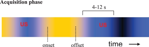
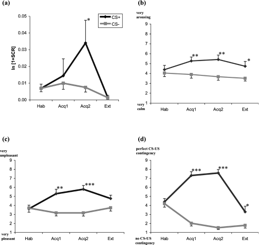
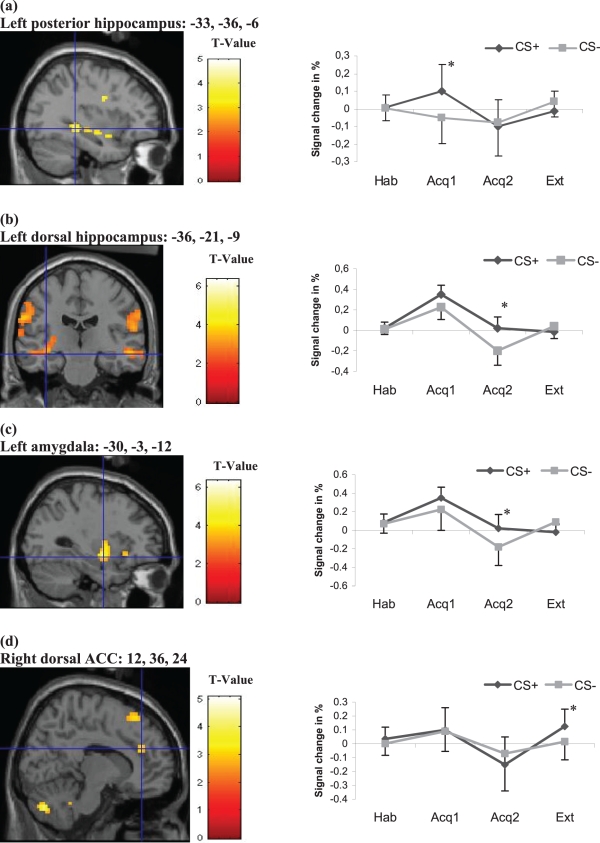
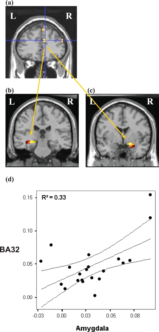
Functional magnetic resonance imaging was used to investigate the role of the hippocampus, amygdala and medial prefrontal cortex (mPFC) in a contextual conditioning and extinction paradigm provoking anxiety. Twenty-one healthy persons participated in a differential context conditioning procedure with two different background colours as contexts. During acquisition increased activity to the conditioned stimulus (CS+) relative to the CS- was found in the left hippocampus and anterior cingulate cortex (ACC). The amygdala, insula and inferior frontal cortex were differentially active during late acquisition. Extinction was accompanied by enhanced activation to CS+ vs. CS- in the dorsal anterior cingulate cortex (dACC). The results are in accordance with animal studies and provide evidence for the important role of the hippocampus in contextual learning in humans. Connectivity analyses revealed correlated activity between the left posterior hippocampus and dACC (BA32) during early acquisition and the dACC, left posterior hippocampus and right amygdala during extinction. These data are consistent with theoretical models that propose an inhibitory effect of the mPFC on the amygdala. The interaction of the mPFC with the hippocampus may reflect the context-specificity of extinction learning.
Overview
The claims and statistics of this paper haven’t been checked yet.
Run the check — free, no account needed. It reads the open-access text and checks the sources it cites.
Integrity signals
Registry
Cites
Statistics
Figures
Verification
Claims the paper makes
The claims and statistics of this paper haven’t been checked yet.
Run the check — free, no account needed. It reads the open-access text and checks the sources it cites.
Claims other papers make about this one
Each is checked against this paper’s own text: does it say what the citing paper says it does?
The cited source says what the claim attributes to it.
In the citing paper
“Connectivity analyses have demonstrated functional coupling between the mPFC and amygdala during extinction learning [107], and vmPFC and amygdala during extinction recall [89, 108].”
From Prefrontal cortex, amygdala, and threat processing: implications for PTSD (2022) · cites this paper as [107]
The cited source says what the claim attributes to it.
In the citing paper
“Importantly, functional connectivity analyses also suggest coupling of the PFC and hippocampus during contextual extinction learning [107] and recall [89].”
From Prefrontal cortex, amygdala, and threat processing: implications for PTSD (2022) · cites this paper as [107]
The cited source says what the claim attributes to it.
In the citing paper
“The dACC is a region previously identified as important for both extinction learning (Lang et al., 2009; Maier et al., 2012) and emotion regulation through cognitive reappraisal (Ochsner et al., 2002, 2004), and a region previously shown to be hypoactivated during emotion regulation in GAD (Blair et al., 2012).”
From Neural correlates of emotion acceptance vs worry or suppression in generalized anxiety disorder (2017) · cites this paper as Lang et al., 2009
The cited source says what the claim attributes to it.
In the citing paper
“Increased late-phase dACC activation in the acceptance condition was found in regions that directly overlap with those reported in the extinction of contextual fear, and in particular the early stages of within-session extinction (Lang et al., 2009; Etkin et al., 2011).”
From Neural correlates of emotion acceptance vs worry or suppression in generalized anxiety disorder (2017) · cites this paper as Lang et al., 2009
The cited source says what the claim attributes to it.
In the citing paper
“Although an emphasis has been placed on the role of subgenual and rostral ACC in the modulation of amygdala activation during extinction of cue conditioned fear in studies of human fear conditioning (Delgado et al., 2008; Milad et al., 2007; Phelps et al., 2004), studies examining the extinction of contextual fear have implicated a larger role for the dACC (Lang et al., 2009; Sehlmeyer et al., 201”
From Neural correlates of emotion acceptance vs worry or suppression in generalized anxiety disorder (2017) · cites this paper as Lang et al., 2009
Self-check
Percentages, sums and reported differences re-computed from the paper’s own numbers.
The self-check needs the paper’s full text.
Citations
Cites
Controlling the False Discovery Rate: A Practical and Powerful Approach to Multiple Testing
Benjamini, Y., & Hochberg, Y. (1995) · Journal of the Royal Statistical Society Series B: Statistical Methodology · doi:10.1111/j.2517-6161.1995.tb02031.x · Cited by 109,482
Tzourio-Mazoyer, N., Landeau, B., Papathanassiou, D., Crivello, F., Etard, O., Delcroix, N., Mazoyer, B., & Joliot, M. (2002) · NeuroImage · doi:10.1006/nimg.2001.0978 · Cited by 16,319
LeDoux, J. E. (2000) · Annual Review of Neuroscience · doi:10.1146/annurev.neuro.23.1.155 · Cited by 8,444
Rescorla, R. A. (1972) · Medical Entomology and Zoology · Cited by 7,268
Differential contribution of amygdala and hippocampus to cued and contextual fear conditioning.
Phillips, R. G., & LeDoux, J. E. (1992) · Behavioral Neuroscience · doi:10.1037/0735-7044.106.2.274 · Cited by 3,292
Interoception: the sense of the physiological condition of the body
(Bud) Craig, A. (2003) · Current Opinion in Neurobiology · doi:10.1016/s0959-4388(03)00090-4 · Cited by 2,556
Modality-Specific Retrograde Amnesia of Fear
Kim, J. J., & Fanselow, M. S. (1992) · Science · doi:10.1126/science.1585183 · Cited by 2,483
Neurons in medial prefrontal cortex signal memory for fear extinction
Milad, M. R., & Quirk, G. J. (2002) · Nature · doi:10.1038/nature01138 · Cited by 1,836 · cited 1×
“Our data are in line with animal data and human neuroimaging studies providing evidence that the mPFC regulates the expression of fear and anxiety by inhibiting the amygdala (Milad & Quirk, 2002; Maren & Quirk, 2004; Sotres-Bayon et al., 2004; Milad et al., 2006).”
Phelps, E. A., Delgado, M. R., Nearing, K. I., & LeDoux, J. E. (2004) · Neuron · doi:10.1016/j.neuron.2004.08.042 · Cited by 1,753
Publication Recommendations for Electrodermal Measurements
Fowles, D. C., Christie, M. J., Edelberg, R., Grings, W. W., Lykken, D. T., & Venables, P. H. (1981) · Psychophysiology · doi:10.1111/j.1469-8986.1981.tb03024.x · Cited by 1,700
Research works printed but not linked: 1
- Rescorla R.A., 1972, Classical conditioning II: current theory and research, 64
Cited by
Sorted by citations
Items 101–171 of 171
Ebrahimi, C., Koch, S., Friedel, E., Crespo, I., Fydrich, T., Ströhle, A., Heinz, A., & Schlagenhauf, F. (2017) · Neurobiology of Learning and Memory · doi:10.1016/j.nlm.2017.05.008 · Cited by 19
Contingency awareness as a prerequisite for differential contextual fear conditioning
Baeuchl, C., Hoppstädter, M., Meyer, P., & Flor, H. (2018) · Cognitive Affective & Behavioral Neuroscience · doi:10.3758/s13415-018-00666-z · Cited by 20
Oxytocin differentially modulates pavlovian cue and context fear acquisition
Cavalli, J., Ruttorf, M., Pahi, M. R., Zidda, F., Flor, H., & Nees, F. (2017) · Social Cognitive and Affective Neuroscience · doi:10.1093/scan/nsx028 · Cited by 17
Zuj, D. V., Palmer, M. A., Malhi, G. S., Bryant, R. A., & Felmingham, K. L. (2018) · Behaviour Research and Therapy · doi:10.1016/j.brat.2018.08.005 · Cited by 17
Functional MRI Studies of the Hippocampus
Nees, F., & Pohlack, S. T. (2014) · Monographs in clinical neuroscience/Frontiers of neurology and neuroscience/Monographs in neural sciences · doi:10.1159/000356427 · Cited by 17
Abnormal fear circuits activities correlated to physical symptoms in somatic anxiety patients
Chen, Y., Wu, Y., Mu, J., Qiu, B., Wang, K., & Tian, Y. (2020) · Journal of Affective Disorders · doi:10.1016/j.jad.2020.05.036 · Cited by 15
The Neurocircuitry of Fear and PTSD
VanElzakker, M. B., Staples-Bradley, L. K., & Shin, L. M. (2017) · Springer eBooks · doi:10.1007/978-1-4939-7148-0_10 · Cited by 19
Namkung, H., Thomas, K. L., Hall, J., & Sawa, A. (2021) · Neuroscience & Biobehavioral Reviews · doi:10.1016/j.neubiorev.2021.12.025 · Cited by 17
Converging evidence for an impact of a functionalNOSgene variation on anxiety-related processes
Kuhn, M., Haaker, J., Glotzbach-Schoon, E., Schümann, D., Andreatta, M., Mechias, M.-L., Raczka, K. A., Gartmann, N., Büchel, C., Mühlberger, A., et al. (2016) · Social Cognitive and Affective Neuroscience · doi:10.1093/scan/nsv151 · Cited by 16
Markett, S., Montag, C., & Reuter, M. (2016) · Neuroimaging Personality, Social Cognition, and Character · doi:10.1016/b978-0-12-800935-2.00005-1 · Cited by 15
van Hoof, M.-J., Riem, M. M. E., Garrett, A. S., van der Wee, N. J. A., van IJzendoorn, M. H., & Vermeiren, R. R. J. M. (2019) · European Journal of Psychotraumatology · doi:10.1080/20008198.2019.1583525 · Cited by 14
Memory and Posttraumatic Stress Disorder
Flor, H., & Wessa, M. (2010) · Zeitschrift für Psychologie · doi:10.1027/0044-3409/a000012 · Cited by 14
Haim‐Nachum, S., & Levy‐Gigi, E. (2019) · Cognition · doi:10.1016/j.cognition.2019.104021 · Cited by 13
Martynova, O., Tetereva, A., Balaev, V. V., Portnova, G. V., Ushakov, V., & Ivanitsky, A. M. (2020) · International Journal of Psychophysiology · doi:10.1016/j.ijpsycho.2020.01.002 · Cited by 16
Scott, J. A., Goodrich‐Hunsaker, N. J., Kalish, K., Lee, A., Hunsaker, M. R., Schumann, C. M., Carmichael, O. T., & Simon, T. J. (2016) · Journal of Psychiatry and Neuroscience · doi:10.1503/jpn.140299 · Cited by 13
Pahi, M. R., Winkelmann, T., Pohlack, S. T., Cavalli, J., Nees, F., & Flor, H. (2019) · Brain Structure and Function · doi:10.1007/s00429-019-01887-9 · Cited by 13
Implicit Learning in Transient Global Amnesia and the Role of Stress
Nees, F., Griebe, M., Ebert, A., Ruttorf, M., Gerber, B. R., Wolf, O. T., Schad, L. R., Gass, A., & Szabo, K. (2016) · Frontiers in Behavioral Neuroscience · doi:10.3389/fnbeh.2016.00222 · Cited by 12
Cortico-limbic connectivity changes following fear extinction and relationships with trait anxiety
Belleau, E. L., Pedersen, W. S., Miskovich, T. A., Helmstetter, F. J., & Larson, C. L. (2018) · Social Cognitive and Affective Neuroscience · doi:10.1093/scan/nsy073 · Cited by 11
Neural correlates of sensory preconditioning: A preliminary fMRI investigation
Yu, T., Lang, S., Birbaumer, N., & Kotchoubey, B. (2013) · Human Brain Mapping · doi:10.1002/hbm.22253 · Cited by 11
Xue, L., Li, Z. D., Chen, Z. X., Wang, X. G., Shi, Y., & Zhao, H. (2013) · Neuroscience · doi:10.1016/j.neuroscience.2013.12.014 · Cited by 11
Simon, R. A., Barazanji, N., Jones, M. P., Bednarska, O., Icenhour, A., Engström, M., Hamilton, J. P., Keita, Å. V., & Walter, S. (2021) · Scientific Reports · doi:10.1038/s41598-020-80873-2 · Cited by 11
Ma, Y., Jiao, F., Batsikadze, G., Yavari, F., & Nitsche, M. A. (2024) · Brain Stimulation · doi:10.1016/j.brs.2024.07.004 · Cited by 9
Mattera, A., Cavallo, A., Granato, G., Baldassarre, G., & Pagani, M. (2022) · Frontiers in Psychology · doi:10.3389/fpsyg.2022.944838 · Cited by 12
Tracking explicit and implicit long-lasting traces of fearful memories in humans
Packard, P. A., Rodríguez‐Fornells, A., Stein, L. M., Nicolás, B., & Fuentemilla, L. (2014) · Neurobiology of Learning and Memory · doi:10.1016/j.nlm.2014.09.004 · Cited by 11
Intensity Dependence of Auditory Evoked Potentials in Primary Dysmenorrhea
Zhang, B., Xu, Y., He, W., Wang, J., Chai, H., Shen, C., Zhu, Q., & Wang, W. (2017) · Journal of Pain · doi:10.1016/j.jpain.2017.06.009 · Cited by 9
Kim, S. M., & Han, D. H. (2021) · Journal of Psychosomatic Research · doi:10.1016/j.jpsychores.2021.110534 · Cited by 8
Chang, W.-L., & Hen, R. (2024) · Advances in neurobiology · doi:10.1007/978-3-031-62983-9_10 · Cited by 8
Chang, W.-L., Tegang, K., Samuels, B. A., Saxe, M., Wichmann, J., David, D. J. P., Mendez‐David, I., Augustin, A., Fischer, H., Golling, S., et al. (2024) · Biological Psychiatry Global Open Science · doi:10.1016/j.bpsgos.2024.100419 · Cited by 10
Retigabine calms seizure-induced behavior following status epilepticus
Slomko, A. M., Naseer, Z., Ali, S. S., Wongvravit, J. P., & Friedman, L. K. (2014) · Epilepsy & Behavior · doi:10.1016/j.yebeh.2014.06.010 · Cited by 8
Levy‐Gigi, E., Haim‐Nachum, S., Hall, J. M., Crouse, J. J., Winwood-Smith, R., Lewis, S. J. G., & Moustafa, A. A. (2018) · Neuroscience Letters · doi:10.1016/j.neulet.2018.11.006 · Cited by 7
Nees, F., Pohlack, S. T., Grimm, O., Winkelmann, T., Zidda, F., & Flor, H. (2019) · Cortex · doi:10.1016/j.cortex.2019.08.020 · Cited by 9
Siehl, S., Wicking, M., Pohlack, S. T., Winkelmann, T., Zidda, F., Steiger-White, F., Nees, F., & Flor, H. (2023) · Psychological Medicine · doi:10.1017/s0033291722003695 · Cited by 6
Where Sounds Occur Matters: Context Effects Influence Processing of Salient Vocalisations
Azhari, A., Rigo, P., Bornstein, M. H., & Esposito, G. (2020) · Brain Sciences · doi:10.3390/brainsci10070429 · Cited by 5
Principles of extinction learning of nonaversive experience
Uengoer, M., Lissek, S., Tegenthoff, M., Manahan‐Vaughan, D., & Lachnit, H. (2020) · e-Neuroforum · doi:10.1515/nf-2020-0013 · Cited by 5
Stegmann, Y., Andreatta, M., Pauli, P., Keil, A., & Wieser, M. J. (2022) · Psychophysiology · doi:10.1111/psyp.14229 · Cited by 5
From Gut Feeling to Gray Matter: Mapping Defecation Urgency in Irritable Bowel Syndrome
Barazanji, N., Sjödahl, J., Orell, G., Evripidou, M., Adjeiwaah, M., Icenhour, A., Lundberg, P., & Walter, S. (2026) · Gastroenterology · doi:10.1053/j.gastro.2026.01.032 · Cited by 4
Hoglund, B. K., Carfagno, V. F., Olive, M. F., & Leyrer‐Jackson, J. M. (2022) · International review of neurobiology · doi:10.1016/bs.irn.2022.10.004 · Cited by 4
The Cognitive Neuroscience of Fear Learning
Stjepanović, D., & LaBar, K. S. (2018) · Wiley eBooks · doi:10.1002/9781119170174.epcn102 · Cited by 4
MouseCircuits.org: An online repository to guide the circuit era of disordered affectPreprint
Anderson, K. R., & Dumitriu, D. (2020) · bioRxiv (Cold Spring Harbor Laboratory) · doi:10.1101/2020.02.16.951608 · Cited by 3
Stem Cell Therapy for Post-Traumatic Stress Disorder: A Novel Therapeutic Approach
Gala, D., Gurusamy, V., Patel, K., Damodar, S., Swaminath, G., & Ullal, G. (2021) · Diseases · doi:10.3390/diseases9040077 · Cited by 4
Chapter 12. Anxiety, Obsessive-Compulsive, and Trauma-Related Disorders
Conroy, M. L., Maruca-Sullivan, P. E., Lane, C. E., & Cook, J. M. (2022) · American Psychiatric Association Publishing eBooks · doi:10.1176/appi.books.9781615375196.ds12 · Cited by 3
Cognición social contexto-dependiente y redes frontotemporo-insulares
Sedeño, L., Moya, Á., Baker, P., & Ibáñez, A. M. (2013) · International Journal of Social Psychology Revista de Psicología Social · doi:10.1174/021347413807719085 · Cited by 5
Ridderbusch, I. C., Yang, Y., Weber, H., Reif, A., Herterich, S., Ströhle, A., Pfleiderer, B., Arolt, V., Wïttchen, H., Lueken, U., et al. (2020) · NeuroImage Clinical · doi:10.1016/j.nicl.2020.102268 · Cited by 3
Elemental and configural representation of a conditioned context
Genheimer, H., Pauli, P., & Andreatta, M. (2024) · Behavioural Brain Research · doi:10.1016/j.bbr.2024.115119 · Cited by 2
The Neural Substrates of Fear Extinction
Lueken, U., & Maslowski, N. I. (2012) · Springer eBooks · doi:10.1007/978-1-4614-3342-2_5 · Cited by 3
Demori, I., & Burlando, B. (2026) · International Journal of Molecular Sciences · doi:10.3390/ijms27146384 · Cited by 1
Posttraumatic Stress Disorder: From Neurobiology to Cycles of Violence
McCullough, K. M., & Ressler, K. J. (2018) · Springer eBooks · doi:10.1007/978-3-030-00503-0_3 · Cited by 1
Facing Fear: An Adaptive Response
Southwick, S. M., & Charney, D. S. (2018) · Cambridge University Press eBooks · doi:10.1017/9781108349246.004 · Cited by 1
Nostadt, A., Nitsche, M. A., Tegenthoff, M., & Lissek, S. (2023) · Scientific Reports · doi:10.1038/s41598-023-47704-6 · Cited by 1
The Search for Neurophysiological Mechanisms of Configural Learning
Chernyshev, B., Ushakov, V. L., & Poznyak, L. A. (2024) · Neuroscience and Behavioral Physiology · doi:10.1007/s11055-024-01723-6 · Cited by 1
Serotonin modulates learning content-specific neuroplasticity of functional brain networksPreprint
Klöbl, M., Seiger, R., Vanicek, T., Handschuh, P. A., Reed, M. B., Spurny‐Dworak, B., Ritter, V., Godbersen, G. M., Gryglewski, G., Kraus, C., et al. (2021) · bioRxiv (Cold Spring Harbor Laboratory) · doi:10.1101/2021.01.15.426779 · Cited by 1
The anterior insular cortex associates temporally discontiguous stimuli during threat learning
Namkung, H., Lockhart, S., de Chabot, J., Guttman, L., Isehak, I., Kwon, H.-B., & Sawa, A. (2021) · doi:10.1101/2021.10.19.465048 · Cited by 1
The role of kinesthetic and visuospatial cues in pain-induced movement avoidancePreprint
Fuchs, X., & Heed, T. (2023) · bioRxiv (Cold Spring Harbor Laboratory) · doi:10.1101/2023.12.19.572163 · Cited by 1
Chang, W.-L., Tegang, K., Samuels, B. A., Saxe, M., Wichmann, J., David, D. J. P., Mendez‐David, I., Augustin, A., Fischer, H., Golling, S., et al. (2024) · bioRxiv (Cold Spring Harbor Laboratory) · doi:10.1101/2024.02.01.578406 · Cited by 1
Southwick, S. M., & Charney, D. S. (2012) · Cambridge University Press eBooks · doi:10.1017/cbo9781139013857.003
Regulação emocional pela atenção: um estudo de neuroimagem por ressonância magnética funcional
Sanchez, T. A. (2009) · Digital Library of Theses and Dissertations (Universidade de São Paulo) · doi:10.11606/t.59.2009.tde-29062010-154952
Role of D1 receptor antagonism in contextual fear learning and memory
Heath, F. (2015) · Nottingham ePrints (University of Nottingham)
Sex differences in extinction to negative stimuli
Sun, N., Lu, H., & Qu, C. (2018) · Medicine · doi:10.1097/md.0000000000010503
Neurobiology and Neuroimaging of PTSD
Milad, M. R., & Moore, K. N. (2018) · Oxford University Press eBooks · doi:10.1093/med/9780190457136.003.0015 · Cited by 1
Cavalcante, K. M. H. (2019) · Scientific Electronic Archives · doi:10.36560/1242019871
Sakamoto, I. M. (2020) · Scholar Commons (University of South Carolina)
Transcranial Magnetic Stimulation in the Treatment of Anxiety Disorders
Lorenzo, G. D., Jannini, T. B., Longo, L., Rossi, R., Siracusano, A., & Dell’Osso, B. M. (2020) · Springer eBooks · doi:10.1007/978-3-030-43356-7_13
Canadian College of Neuropsychopharmacology 2015 award winners
(2016) · Journal of Psychiatry and Neuroscience · doi:10.1503/jpn.160054
Friedman, L. K., Kahen, B., Velı́šek, L., & Velı́šková, J. (2022) · Brain Research · doi:10.1016/j.brainres.2022.147849
Neuronale Korrelate von Placeboeffekt, Furchtextinktion und willentlicher Emotionsregulation
Geier, K. (2014) · eDiss (Georg-August-Universität Göttingen) · doi:10.53846/goediss-4626
(2018) · Elsevier eBooks · doi:10.1016/b978-0-12-811351-6.00026-7
Search for neurophysiological mechanisms of configurational learning
Chernyshev, B., Ushakov, V., & Poznyak, L. A. (2024) · Журнал высшей нервной деятельности им И П Павлова · doi:10.31857/s0044467724020028
Puberty, sex, and fear extinction retention: A neuroimaging study in youth
Bhargava, S., Zundel, C. G., Shampine, M. M., Ely, S. L., Carpenter, C., Losiowski, J., Chanamolu, S., Jande, J., Tamimi, R., Sharma, K., et al. (2025) · Developmental Cognitive Neuroscience · doi:10.1016/j.dcn.2025.101595 · Cited by 1
Ågren, T. (2012) · KTH Publication Database DiVA (KTH Royal Institute of Technology)
Metadata
- Authors
- Lang, S., Kroll, A., Lipinski, S. J., Wessa, M., Ridder, S., Christmann, C., Schad, L. R., & Flor, H.
- Affiliations
- Heidelberg University (DE)
- Venue
- European Journal of Neuroscience
- Publisher
- Wiley
- Published
- 2009-02
- Volume
- 29
- Issue
- 4
- Pages
- 823-832
- ISSN
- 0953-816X (ISSN-L)
- 1460-9568
- PMC
- PMC2695154
- OpenAlex
- W1991878909
- Cited by
- 171
- References
- 72
- Citations by year
- 2026: 2 · 2025: 1 · 2024: 8 · 2023: 3 · 2022: 7 · 2021: 8 · 2020: 10 · 2019: 11 · 2018: 18 · 2017: 12 · 2016: 15 · 2015: 11 · 2014: 16 · 2013: 16 · 2012: 10
- FWCI
- 5.32
- Type
- journal article
- Language
- en
- Open access
- green
- www.ncbi.nlm.nih.gov
- Locations
- European Journal of Neuroscience (publishedVersion)
- PubMed Central (publishedVersion)
- PubMed (publishedVersion)
- Funders
- Deutsche Forschungsgemeinschaft
- Topics
- Life Sciences › Neuroscience › Cognitive Neuroscience › Memory and Neural Mechanisms
- Life Sciences › Neuroscience › Cognitive Neuroscience › Neural and Behavioral Psychology Studies
- Life Sciences › Neuroscience › Cognitive Neuroscience › Memory Processes and Influences
- Keywords
- contextual conditioning, extinction learning, hippocampus, amygdala, medial prefrontal cortex, anterior cingulate cortex, anxiety, contextual learning, hippocampal-amygdala interaction, neuronal connectivity
- MeSH terms
- Adult, Amygdala, Cerebral Cortex, Conditioning, Psychological, Cues, Electric Stimulation, Extinction, Psychological, Female, Galvanic Skin Response, Hippocampus, Humans, Magnetic Resonance Imaging, Male, Neural Pathways, Photic Stimulation, Regression Analysis, Prefrontal Cortex, Young Adult
About these checks
Registry, retraction, Cites and statistics results are computed, not judged, with one exception: a printed reference that no index lists is read once by a model before it counts as a citation error. Claims are extracted by a model and kept only when their quote appears verbatim in the text; each cited source is then checked, with a model’s help, for whether it says what the claim relies on. Those verdicts can be wrong — read the claim’s quote and the cited source before relying on one.
Statistics and arithmetic are recomputed, not judged.
Confidence bands are shown only for fields calibrated on our annotated test set. None is calibrated yet, so no band is shown.
What counts as a claim ›How we measure confidence ›How we check this ›
Are you an author, or do you see a problem with this page? Request a review
