

The DOI matches its Crossref record; 71 of the 72 references it cites were found in a registry or an index.
Context conditioning and extinction in humans: differential contribution of the hippocampus, amygdala and prefrontal cortex
Open accessAbstract
Functional magnetic resonance imaging was used to investigate the role of the hippocampus, amygdala and medial prefrontal cortex (mPFC) in a contextual conditioning and extinction paradigm provoking anxiety. Twenty-one healthy persons participated in a differential context conditioning procedure with two different background colours as contexts. During acquisition increased activity to the conditioned stimulus (CS+) relative to the CS- was found in the left hippocampus and anterior cingulate cortex (ACC). The amygdala, insula and inferior frontal cortex were differentially active during late acquisition. Extinction was accompanied by enhanced activation to CS+ vs. CS- in the dorsal anterior cingulate cortex (dACC). The results are in accordance with animal studies and provide evidence for the important role of the hippocampus in contextual learning in humans. Connectivity analyses revealed correlated activity between the left posterior hippocampus and dACC (BA32) during early acquisition and the dACC, left posterior hippocampus and right amygdala during extinction. These data are consistent with theoretical models that propose an inhibitory effect of the mPFC on the amygdala. The interaction of the mPFC with the hippocampus may reflect the context-specificity of extinction learning.
Overview
The claims and statistics of this paper haven’t been checked yet.
Run the check — free, no account needed. It reads the open-access text and checks the sources it cites.
Integrity signals
Registry
Cites
Statistics
Figures
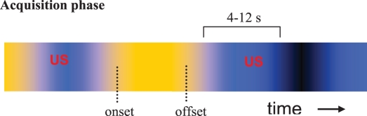
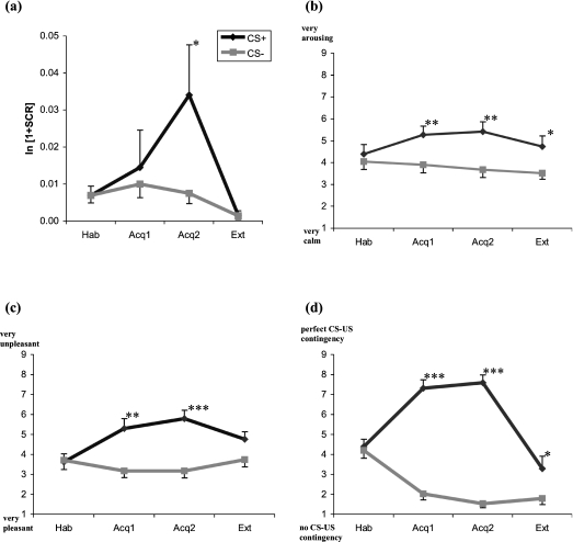
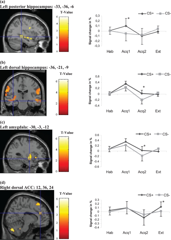
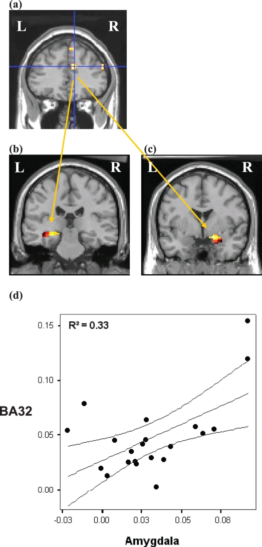
Verification
Claims the paper makes
The claims and statistics of this paper haven’t been checked yet.
Run the check — free, no account needed. It reads the open-access text and checks the sources it cites.
Claims other papers make about this one
Each is checked against this paper’s own text: does it say what the citing paper says it does?
The cited source says what the claim attributes to it.
In the citing paper
“Connectivity analyses have demonstrated functional coupling between the mPFC and amygdala during extinction learning [107], and vmPFC and amygdala during extinction recall [89, 108].”
From Prefrontal cortex, amygdala, and threat processing: implications for PTSD (2022) · cites this paper as [107]
The cited source says what the claim attributes to it.
In the citing paper
“Importantly, functional connectivity analyses also suggest coupling of the PFC and hippocampus during contextual extinction learning [107] and recall [89].”
From Prefrontal cortex, amygdala, and threat processing: implications for PTSD (2022) · cites this paper as [107]
The cited source says what the claim attributes to it.
In the citing paper
“The dACC is a region previously identified as important for both extinction learning (Lang et al., 2009; Maier et al., 2012) and emotion regulation through cognitive reappraisal (Ochsner et al., 2002, 2004), and a region previously shown to be hypoactivated during emotion regulation in GAD (Blair et al., 2012).”
From Neural correlates of emotion acceptance vs worry or suppression in generalized anxiety disorder (2017) · cites this paper as Lang et al., 2009
The cited source says what the claim attributes to it.
In the citing paper
“Increased late-phase dACC activation in the acceptance condition was found in regions that directly overlap with those reported in the extinction of contextual fear, and in particular the early stages of within-session extinction (Lang et al., 2009; Etkin et al., 2011).”
From Neural correlates of emotion acceptance vs worry or suppression in generalized anxiety disorder (2017) · cites this paper as Lang et al., 2009
The cited source says what the claim attributes to it.
In the citing paper
“Although an emphasis has been placed on the role of subgenual and rostral ACC in the modulation of amygdala activation during extinction of cue conditioned fear in studies of human fear conditioning (Delgado et al., 2008; Milad et al., 2007; Phelps et al., 2004), studies examining the extinction of contextual fear have implicated a larger role for the dACC (Lang et al., 2009; Sehlmeyer et al., 201”
From Neural correlates of emotion acceptance vs worry or suppression in generalized anxiety disorder (2017) · cites this paper as Lang et al., 2009
Self-check
Percentages, sums and reported differences re-computed from the paper’s own numbers.
The self-check needs the paper’s full text.
Citations
Cites
Controlling the False Discovery Rate: A Practical and Powerful Approach to Multiple Testing
Benjamini, Y., & Hochberg, Y. (1995) · Journal of the Royal Statistical Society Series B: Statistical Methodology · doi:10.1111/j.2517-6161.1995.tb02031.x · Cited by 109,482
Tzourio-Mazoyer, N., Landeau, B., Papathanassiou, D., Crivello, F., Etard, O., Delcroix, N., Mazoyer, B., & Joliot, M. (2002) · NeuroImage · doi:10.1006/nimg.2001.0978 · Cited by 16,319
LeDoux, J. E. (2000) · Annual Review of Neuroscience · doi:10.1146/annurev.neuro.23.1.155 · Cited by 8,444
Rescorla, R. A. (1972) · Medical Entomology and Zoology · Cited by 7,268
Differential contribution of amygdala and hippocampus to cued and contextual fear conditioning.
Phillips, R. G., & LeDoux, J. E. (1992) · Behavioral Neuroscience · doi:10.1037/0735-7044.106.2.274 · Cited by 3,292
Interoception: the sense of the physiological condition of the body
(Bud) Craig, A. (2003) · Current Opinion in Neurobiology · doi:10.1016/s0959-4388(03)00090-4 · Cited by 2,556
Modality-Specific Retrograde Amnesia of Fear
Kim, J. J., & Fanselow, M. S. (1992) · Science · doi:10.1126/science.1585183 · Cited by 2,483
Neurons in medial prefrontal cortex signal memory for fear extinction
Milad, M. R., & Quirk, G. J. (2002) · Nature · doi:10.1038/nature01138 · Cited by 1,836 · cited 1×
“Our data are in line with animal data and human neuroimaging studies providing evidence that the mPFC regulates the expression of fear and anxiety by inhibiting the amygdala (Milad & Quirk, 2002; Maren & Quirk, 2004; Sotres-Bayon et al., 2004; Milad et al., 2006).”
Phelps, E. A., Delgado, M. R., Nearing, K. I., & LeDoux, J. E. (2004) · Neuron · doi:10.1016/j.neuron.2004.08.042 · Cited by 1,753
Publication Recommendations for Electrodermal Measurements
Fowles, D. C., Christie, M. J., Edelberg, R., Grings, W. W., Lykken, D. T., & Venables, P. H. (1981) · Psychophysiology · doi:10.1111/j.1469-8986.1981.tb03024.x · Cited by 1,700
Research works printed but not linked: 1
- Rescorla R.A., 1972, Classical conditioning II: current theory and research, 64
Cited by
Sorted by citations
The Neurocircuitry of Fear, Stress, and Anxiety Disorders
Shin, L. M., & Liberzon, I. (2010) · Neuropsychopharmacology · doi:10.1038/npp.2009.83 · Cited by 2,083
The contextual brain: implications for fear conditioning, extinction and psychopathology
Maren, S., Phan, K. L., & Liberzon, I. (2013) · Nature Reviews Neuroscience · doi:10.1038/nrn3492 · Cited by 1,547
Fear Extinction as a Model for Translational Neuroscience: Ten Years of Progress
Milad, M. R., & Quirk, G. J. (2012) · Annual Review of Psychology · doi:10.1146/annurev.psych.121208.131631 · Cited by 1,466
Long-axis specialization of the human hippocampus
Poppenk, J., Evensmoen, H. R., Moscovitch, M., & Nadel, L. (2013) · Trends in Cognitive Sciences · doi:10.1016/j.tics.2013.03.005 · Cited by 1,100
Post-Traumatic Stress Disorder
Shalev, A. Y., Liberzon, I., & Marmar, C. R. (2017) · New England Journal of Medicine · doi:10.1056/nejmra1612499 · Cited by 852
Pain and the brain: specificity and plasticity of the brain in clinical chronic pain
Apkarian, V. A., Hashmi, J. A., & Baliki, M. N. (2011) · Pain · doi:10.1016/j.pain.2010.11.010 · Cited by 782
Functional connectivity mapping of the human precuneus by resting state fMRI
Zhang, S., & Li, C. R. (2011) · NeuroImage · doi:10.1016/j.neuroimage.2011.11.023 · Cited by 574
Craske, M. G., Rauch, S. L., Ursano, R., Prenoveau, J., Pine, D. S., & Zinbarg, R. E. (2009) · Depression and Anxiety · doi:10.1002/da.20633 · Cited by 529
From Pavlov to PTSD: The extinction of conditioned fear in rodents, humans, and anxiety disorders
VanElzakker, M. B., Kathryn Dahlgren, M., Caroline Davis, F., Dubois, S., & Shin, L. M. (2014) · Neurobiology of Learning and Memory · doi:10.1016/j.nlm.2013.11.014 · Cited by 482
Context Processing and the Neurobiology of Post-Traumatic Stress Disorder
Liberzon, I., & Abelson, J. L. (2016) · Neuron · doi:10.1016/j.neuron.2016.09.039 · Cited by 489
Izquierdo, I., Furini, C. R. G., & Myskiw, J. C. (2016) · Physiological Reviews · doi:10.1152/physrev.00018.2015 · Cited by 453
Fear is only as deep as the mind allows
Diekhof, E. K., Geier, K., Falkai, P., & Gruber, O. (2011) · NeuroImage · doi:10.1016/j.neuroimage.2011.05.073 · Cited by 413
Prefrontal cortex, amygdala, and threat processing: implications for PTSD
Alexandra Kredlow, M., Fenster, R. J., Laurent, E. S., Ressler, K. J., & Phelps, E. A. (2022) · Neuropsychopharmacology · doi:10.1038/s41386-021-01155-7 · Cited by 346
“Connectivity analyses have demonstrated functional coupling between the mPFC and amygdala during extinction learning [107], and vmPFC and amygdala during extinction recall [89, 108].”
Contextual social cognition and the behavioral variant of frontotemporal dementia
Ibáñez, A. M., & Manes, F. F. (2012) · Neurology · doi:10.1212/wnl.0b013e3182518375 · Cited by 337
Fear-Conditioning Mechanisms Associated with Trait Vulnerability to Anxiety in Humans
Indovina, I., Robbins, T. W., Núñez-Elizalde, A. O., Dunn, B. D., & Bishop, S. J. (2011) · Neuron · doi:10.1016/j.neuron.2010.12.034 · Cited by 310
Resilience: The Science of Mastering Life's Greatest Challenges
Southwick, S. M., & Charney, D. S. (2012) · Cited by 212
Montagrin, A., Saiote, C., & Schiller, D. (2017) · Hippocampus · doi:10.1002/hipo.22797 · Cited by 211
Kim, J. H., & Richardson, R. (2010) · Biological Psychiatry · doi:10.1016/j.biopsych.2009.09.003 · Cited by 189
Craske, M. G., Rauch, S. L., Ursano, R. J., Prenoveau, J. M., Pine, D. S., & Zinbarg, R. E. (2011) · FOCUS The Journal of Lifelong Learning in Psychiatry · doi:10.1176/foc.9.3.foc369 · Cited by 219
McDonald, A. J., & Mott, D. D. (2016) · Journal of Neuroscience Research · doi:10.1002/jnr.23709 · Cited by 189
Neural correlates of trait anxiety in fear extinction
Sehlmeyer, C., Dannlowski, U., Schöning, S., Kugel, H., Pyka, M., Pfleiderer, B., Zwitserlood, P., Schiffbauer, H., Heindel, W., Arolt, V., et al. (2011) · Psychological Medicine · doi:10.1017/s0033291710001248 · Cited by 165
Effect of multiple context exposure on renewal in spider phobia
Shiban, Y., Pauli, P., & Mühlberger, A. (2013) · Behaviour Research and Therapy · doi:10.1016/j.brat.2012.10.007 · Cited by 162
MRI findings in posttraumatic stress disorder
Kunimatsu, A., Yasaka, K., Akai, H., Kunimatsu, N., & Abe, O. (2019) · Journal of Magnetic Resonance Imaging · doi:10.1002/jmri.26929 · Cited by 173
Fear conditioning and extinction across development: Evidence from human studies and animal models
Shechner, T., Hong, M., Britton, J. C., Pine, D. S., & Fox, N. A. (2014) · Biological Psychology · doi:10.1016/j.biopsycho.2014.04.001 · Cited by 170
Empathy and contextual social cognition
Melloni, M., López, V., & Ibáñez, A. M. (2013) · Cognitive Affective & Behavioral Neuroscience · doi:10.3758/s13415-013-0205-3 · Cited by 147
Prefrontal neuronal circuits of contextual fear conditioning
Rozeske, R. R., Valério, S., Chaudun, F., & Herry, C. (2014) · Genes Brain & Behavior · doi:10.1111/gbb.12181 · Cited by 158
The centrality of fear extinction in linking risk factors to PTSD: A narrative review
Zuj, D. V., Palmer, M. A., Lommen, M. J. J., & Felmingham, K. L. (2016) · Neuroscience & Biobehavioral Reviews · doi:10.1016/j.neubiorev.2016.07.014 · Cited by 153
A neurobiological enquiry into the origins of our experience of the sublime and beautiful
Ishizu, T. (2014) · Frontiers in Human Neuroscience · doi:10.3389/fnhum.2014.00891 · Cited by 115
Unconditioned responses and functional fear networks in human classical conditioning
Linnman, C., Rougemont-Bücking, A., Beucke, J. C., Zeffiro, T. A., & Milad, M. R. (2011) · Behavioural Brain Research · doi:10.1016/j.bbr.2011.02.045 · Cited by 99
The Neurocircuitry of Fear, Stress, and Anxiety Disorders
Shin, L. M., & Liberzon, I. (2011) · FOCUS The Journal of Lifelong Learning in Psychiatry · doi:10.1176/foc.9.3.foc311 · Cited by 105
Siehl, S., King, J. A., Burgess, N., Flor, H., & Nees, F. (2018) · NeuroImage Clinical · doi:10.1016/j.nicl.2018.05.013 · Cited by 104
Wang, T., Guan, R.-L., Liu, M., Shen, X., Chen, J. Y., Zhao, M., & Luo, W. (2015) · Molecular Neurobiology · doi:10.1007/s12035-015-9312-1 · Cited by 93
Yao, H., Liu, Y., Zhou, B., Zhang, Z., An, N., Wang, P., Wang, L., Zhang, X., & Jiang, T. (2013) · European Journal of Radiology · doi:10.1016/j.ejrad.2013.03.019 · Cited by 93
Lonsdorf, T. B., Haaker, J., & Kalisch, R. (2014) · Social Cognitive and Affective Neuroscience · doi:10.1093/scan/nsu018 · Cited by 89
Molecular Specificity of Multiple Hippocampal Processes Governing Fear Extinction
Radulović, J., & Tronson, N. C. (2010) · Reviews in the Neurosciences · doi:10.1515/revneuro.2010.21.1.1 · Cited by 83
Levy-Gigi, E., Szabo, C., Richter-Levin, G., & Kéri, S. (2015) · Neuropsychology · doi:10.1037/neu0000131 · Cited by 85
Zuj, D. V., & Norrholm, S. D. (2018) · Progress in Neuro-Psychopharmacology and Biological Psychiatry · doi:10.1016/j.pnpbp.2018.08.014 · Cited by 89
Egorova, N., Gollub, R. L., & Kong, J. (2015) · NeuroImage Clinical · doi:10.1016/j.nicl.2015.09.012 · Cited by 82
Morriss, J., Gell, M., & van Reekum, C. M. (2018) · Neuroscience & Biobehavioral Reviews · doi:10.1016/j.neubiorev.2018.12.013 · Cited by 76
Balemans, M. C. M., Kasri, N. N., Kopanitsa, M. V., Afinowi, N. O., Ramakers, G. J. A., Peters, T. A., Beynon, A. J., Janssen, S. M., van Summeren, R. C. J., Eeftens, J. M., et al. (2012) · Human Molecular Genetics · doi:10.1093/hmg/dds490 · Cited by 78
Initial and sustained brain responses to contextual conditioned anxiety in humans
Andreatta, M., Glotzbach-Schoon, E., Mühlberger, A., Schulz, S. M., Wiemer, J., & Pauli, P. (2015) · Cortex · doi:10.1016/j.cortex.2014.09.014 · Cited by 73
Activation of the ventral striatum during aversive contextual conditioning in humans
Pohlack, S. T., Nees, F., Ruttorf, M., Schad, L. R., & Flor, H. (2012) · Biological Psychology · doi:10.1016/j.biopsycho.2012.04.004 · Cited by 73
Hippocampal but not amygdalar volume affects contextual fear conditioning in humans
Pohlack, S. T., Nees, F., Liebscher, C., Cacciaglia, R., Diener, S. J., Ridder, S., Woermann, F. G., & Flor, H. (2011) · Human Brain Mapping · doi:10.1002/hbm.21224 · Cited by 75
Circuit dysregulation and circuit-based treatments in posttraumatic stress disorder
Sheynin, J., & Liberzon, I. (2017) · Neuroscience Letters · doi:10.1016/j.neulet.2016.11.014 · Cited by 68
Cortisol modifies extinction learning of recently acquired fear in men
Merz, C. J., Hermann, A., Stark, R. M., & Wolf, O. T. (2013) · Social Cognitive and Affective Neuroscience · doi:10.1093/scan/nst137 · Cited by 69
Steiger-White, F., Nees, F., Wicking, M., Lang, S., & Flor, H. (2015) · International Journal of Psychophysiology · doi:10.1016/j.ijpsycho.2015.06.009 · Cited by 71
Feng, P., Feng, T., Chen, Z., & Lei, X. (2013) · Social Cognitive and Affective Neuroscience · doi:10.1093/scan/nst170 · Cited by 66
Raber, J., Arzy, S., Bertolus, J. B., Depue, B., Haas, H. E., Hofmann, S. G., Kangas, M., Kensinger, E., Lowry, C. A., Marusak, H. A., et al. (2019) · Neuroscience & Biobehavioral Reviews · doi:10.1016/j.neubiorev.2019.03.015 · Cited by 62
Social anxiety modulates amygdala activation during social conditioning
Pejic, T., Hermann, A., Vaitl, D., & Stark, R. M. (2011) · Social Cognitive and Affective Neuroscience · doi:10.1093/scan/nsr095 · Cited by 61
Neural correlates of emotion acceptance vs worry or suppression in generalized anxiety disorder
Ellard, K. K., Barlow, D. H., Whitfield-Gabrieli, S., Gabrieli, J. D. E., & Deckersbach, T. (2017) · Social Cognitive and Affective Neuroscience · doi:10.1093/scan/nsx025 · Cited by 59
“The dACC is a region previously identified as important for both extinction learning (Lang et al., 2009; Maier et al., 2012)”
InfluentialGlotzbach, E., Ewald, H., Andreatta, M., Pauli, P., & Mühlberger, A. (2012) · Cognition & Emotion · doi:10.1080/02699931.2012.656581 · Cited by 54
Fani, N., King, T. Z., Brewster, R. C., Srivastava, A., Stevens, J. S., Glover, E. M., Norrholm, S., Bradley, B., Ressler, K., & Jovanović, T. (2014) · Cortex; a journal devoted to the study of the nervous system and behavior · doi:10.1016/j.cortex.2014.11.006 · Cited by 62
Enhanced discrimination between threatening and safe contexts in high-anxious individuals
Glotzbach-Schoon, E., Tadda, R., Andreatta, M., Tröger, C., Ewald, H., Grillon, C., Pauli, P., & Mühlberger, A. (2013) · Biological Psychology · doi:10.1016/j.biopsycho.2013.01.011 · Cited by 57
Lissek, S., Glaubitz, B., Uengoer, M., & Tegenthoff, M. (2013) · NeuroImage · doi:10.1016/j.neuroimage.2013.05.025 · Cited by 60
Pohlack, S. T., Nees, F., Ruttorf, M., Cacciaglia, R., Winkelmann, T., Schad, L. R., Witt, S. H., Rietschel, M., & Flor, H. (2015) · Biological Psychiatry · doi:10.1016/j.biopsych.2014.12.018 · Cited by 50
Neural circuits for the adaptive regulation of fear and extinction memory
Plas, S. L., Tuna, T., Bayer, H., Juliano, V. A. L., Sweck, S. O., Pérez, A. D. A., Hassell, J. E., & Maren, S. (2024) · Frontiers in Behavioral Neuroscience · doi:10.3389/fnbeh.2024.1352797 · Cited by 55
Icenhour, A., Kattoor, J., Benson, S., Boekstegers, A., Schlamann, M. U., Merz, C. J., Forsting, M., & Elsenbruch, S. (2015) · Human Brain Mapping · doi:10.1002/hbm.22837 · Cited by 47
Mechanisms behind the Development of Chronic Low Back Pain and Its Neurodegenerative Features
Mosabbir, A. (2022) · Life · doi:10.3390/life13010084 · Cited by 61
Revealing Context-Specific Conditioned Fear Memories with Full Immersion Virtual Reality
Huff, N. C., Hernandez, J. A., Fecteau, M. E., Zielinski, D. J., Brady, R. B., & LaBar, K. S. (2011) · Frontiers in Behavioral Neuroscience · doi:10.3389/fnbeh.2011.00075 · Cited by 45
Wieser, M. J., Reicherts, P., Juravle, G., & von Leupoldt, A. (2016) · NeuroImage · doi:10.1016/j.neuroimage.2016.06.026 · Cited by 42
Hippocampal volume and declarative memory function in combat-related PTSD
Woodward, S. H., Kaloupek, D. G., Grande, L. J., Stegman, W. K., Kutter, C. J., LESKIN, L., Prestel, R. S., Schaer, M., Reiss, A. L., & D, S. E. M. (2009) · Journal of the International Neuropsychological Society · doi:10.1017/s1355617709990476 · Cited by 43
Default mode network connectivity of fear- and anxiety-related cue and context conditioning
Zidda, F., Andoh, J., Pohlack, S. T., Winkelmann, T., Dinu-Biringer, R., Cavalli, J., Ruttorf, M., Nees, F., & Flor, H. (2017) · NeuroImage · doi:10.1016/j.neuroimage.2017.10.024 · Cited by 44
The hidden price of repeated traumatic exposure
Levy‐Gigi, E., & Richter‐Levin, G. (2014) · Stress · doi:10.3109/10253890.2014.923397 · Cited by 44
Dossi, G., Delvecchio, G., Prunas, C., Soares, J. C., & Brambilla, P. (2020) · Frontiers in Psychiatry · doi:10.3389/fpsyt.2020.00176 · Cited by 41
Neural structures and mechanisms involved in scene recognition: A review and interpretation
Sewards, T. V. (2010) · Neuropsychologia · doi:10.1016/j.neuropsychologia.2010.11.018 · Cited by 40
Levy‐Gigi, E., Richter‐Levin, G., & Kéri, S. (2014) · Frontiers in Behavioral Neuroscience · doi:10.3389/fnbeh.2014.00281 · Cited by 38
Adaptive contextualization: a new role for the default mode network in affective learning
Marstaller, L., Burianová, H., & Reutens, D. C. (2017) · Human Brain Mapping · doi:10.1002/hbm.23442 · Cited by 37
Lecei, A., & van Winkel, R. (2019) · Neuroscience & Biobehavioral Reviews · doi:10.1016/j.neubiorev.2019.11.010 · Cited by 45
Straube, B., Lueken, U., Jansen, A., Konrad, C., Gloster, A. T., Gerlach, A. L., Ströhle, A., Wittmann, A., Pfleiderer, B., Gauggel, S., et al. (2014) · Psychotherapy and Psychosomatics · doi:10.1159/000359955 · Cited by 40
Contextual fear conditioning in humans using feature-identical contexts
Baeuchl, C., Meyer, P., Hoppstädter, M., Diener, C., & Flor, H. (2015) · Neurobiology of Learning and Memory · doi:10.1016/j.nlm.2015.03.001 · Cited by 38
Torregrossa, M. M., Gordon, J., & Taylor, J. R. (2013) · Journal of Neuroscience · doi:10.1523/jneurosci.0489-13.2013 · Cited by 35
The Neurobiology of Panic: A Chronic Stress Disorder
Goddard, A. W. (2017) · Chronic Stress · doi:10.1177/2470547017736038 · Cited by 39
A mechanism-oriented approach to psychopathology: The role of Pavlovian conditioning
Nees, F., Heinrich, A., & Flor, H. (2015) · International Journal of Psychophysiology · doi:10.1016/j.ijpsycho.2015.05.005 · Cited by 36
Ney, L. J., Vicario, C. M., Nitsche, M. A., & Felmingham, K. L. (2020) · Neurobiology of Learning and Memory · doi:10.1016/j.nlm.2020.107356 · Cited by 33
Escitalopram modulates learning content-specific neuroplasticity of functional brain networks
Klöbl, M., Seiger, R., Vanicek, T., Handschuh, P. A., Reed, M. B., Spurny‐Dworak, B., Ritter, V., Godbersen, G. M., Gryglewski, G., Kraus, C., et al. (2021) · NeuroImage · doi:10.1016/j.neuroimage.2021.118829 · Cited by 30
André, M. A. ̈. E., Wolf, O. T., & Manahan‐Vaughan, D. (2015) · Frontiers in Behavioral Neuroscience · doi:10.3389/fnbeh.2015.00125 · Cited by 29
Altered functional connectivity density in patients with herpes zoster and postherpetic neuralgia
Hong, S., Gu, L., Zhou, F., Liu, J., Huang, M., Jiang, J. P., He, L., Gong, H., & Zeng, X. (2018) · Journal of Pain Research · doi:10.2147/jpr.s154314 · Cited by 27
Neural measures associated with configural threat acquisition
Stout, D. M., Glenn, D. E., Acheson, D. T., Spadoni, A. D., Risbrough, V. B., & Simmons, A. N. (2018) · Neurobiology of Learning and Memory · doi:10.1016/j.nlm.2018.03.012 · Cited by 26
Feng, T., Feng, P., & Chen, Z. (2013) · Brain Research · doi:10.1016/j.brainres.2013.05.039 · Cited by 27
Lehner, M., Wisłowska-Stanek, A., Taracha, E., Maciejak, P., Szyndler, J., Skórzewska, A., Turzyńska, D., Sobolewska, A., Hamed, A., Bidziński, A., et al. (2009) · Neurobiology of Learning and Memory · doi:10.1016/j.nlm.2009.07.002 · Cited by 27
Learning, memory and brain plasticity in posttraumatic stress disorder: Context matters
Flor, H., & Nees, F. (2014) · Restorative Neurology and Neuroscience · doi:10.3233/rnn-139013 · Cited by 29
Estradiol and progesterone in female reward-learning, addiction, and therapeutic interventions
Hilz, E. N., & Lee, H. J. (2022) · Frontiers in Neuroendocrinology · doi:10.1016/j.yfrne.2022.101043 · Cited by 29
Imaging of Posttraumatic Stress Disorder
Kamiya, K., & Abe, O. (2019) · Neuroimaging Clinics of North America · doi:10.1016/j.nic.2019.09.010 · Cited by 29
André, M. A. E., Güntürkün, O., & Manahan‐Vaughan, D. (2014) · Hippocampus · doi:10.1002/hipo.22359 · Cited by 25
Direct Neuronal Glucose Uptake Is Required for Contextual Fear Acquisition in the Dorsal Hippocampus
Kong, L., Zhao, Y., Zhou, W., Yu, H., Teng, S.-W., Guo, Q., Chen, Z.-Y., & Wang, Y. (2017) · Frontiers in Molecular Neuroscience · doi:10.3389/fnmol.2017.00388 · Cited by 27
Classical conditioning in borderline personality disorder: an fMRI study
Krause-Utz, A., Keibel-Mauchnik, J., Ebner-Priemer, U., Bohus, M., & Schmahl, C. (2016) · European Archives of Psychiatry and Clinical Neuroscience · doi:10.1007/s00406-015-0593-1 · Cited by 24
Controllability and hippocampal activation during pain expectation in fibromyalgia syndrome
González‐Roldán, A. M., Bomba, I. C., Diesch, E., Montoya, P., Flor, H., & Kamping, S. (2016) · Biological Psychology · doi:10.1016/j.biopsycho.2016.09.007 · Cited by 27
Myskiw, J. D. C., Fiorenza, N. G., Izquierdo, L. A., & Izquierdo, I. (2010) · Neurobiology of Learning and Memory · doi:10.1016/j.nlm.2010.06.007 · Cited by 23
Glenn, D. E., Risbrough, V. B., Simmons, A. N., Acheson, D. T., & Stout, D. M. (2017) · Current topics in behavioral neurosciences · doi:10.1007/7854_2017_30 · Cited by 23
Guhn, A., Dresler, T., Hahn, T., Mühlberger, A., Ströhle, A., Deckert, J., & Herrmann, M. J. (2012) · Neuropsychobiology · doi:10.1159/000337002 · Cited by 22
Characterizing the neural circuitry associated with configural threat learning
Stout, D. M., Glenn, D. E., Acheson, D. T., Simmons, A. N., & Risbrough, V. B. (2019) · Brain Research · doi:10.1016/j.brainres.2019.06.003 · Cited by 22
Music models aberrant rule decoding and reward valuation in dementia
Clark, C. N., Golden, H. L., McCallion, O., Nicholas, J. M., Cohen, M. H., Slattery, C. F., Paterson, R. W., Fletcher, P. D., Mummery, C. J., Rohrer, J. D., et al. (2017) · Social Cognitive and Affective Neuroscience · doi:10.1093/scan/nsx140 · Cited by 21
Context conditioning in virtual reality as a model for pathological anxiety
Glotzbach-Schoon, E., Andreatta, M., Mühlberger, A., & Pauli, P. (2013) · e-Neuroforum · doi:10.1007/s13295-013-0047-z · Cited by 25
Andreatta, M., Neueder, D., Glotzbach-Schoon, E., Mühlberger, A., & Pauli, P. (2016) · Learning & Memory · doi:10.1101/lm.044073.116 · Cited by 24
Goodman, A. M., Harnett, N. G., Wheelock, M. D., Hurst, D. R., Orem, T. R., Gossett, E. W., Dunaway, C. A., Mrug, S., & Knight, D. C. (2018) · NeuroImage · doi:10.1016/j.neuroimage.2018.03.030 · Cited by 20
A Review of Preclinical Studies to Understand Fear During Adolescence
Kim, J. H., & Ganella, D. E. (2015) · Australian Psychologist · doi:10.1111/ap.12066 · Cited by 20
Neural circuitry changes associated with increasing self-efficacy in Posttraumatic Stress Disorder
Titcombe, R. F., Chen, J., Rahman, N., Kouri, N. A., Qian, M., Li, M., Bryant, R. A., Marmar, C. R., & Brown, A. D. (2018) · Journal of Psychiatric Research · doi:10.1016/j.jpsychires.2018.06.009 · Cited by 21
Fonteyne, R., Vervliet, B., Hermans, D., Baeyens, F., & Vansteenwegen, D. (2010) · Biological Psychology · doi:10.1016/j.biopsycho.2010.08.007 · Cited by 19
Lissek, S., Golisch, A., Glaubitz, B., & Tegenthoff, M. (2016) · Brain Imaging and Behavior · doi:10.1007/s11682-016-9662-y · Cited by 16
Human BDNF rs6265 polymorphism as a mediator for the generalization of contextual anxiety
Andreatta, M., Neueder, D., Genheimer, H., Schiele, M. A., Schartner, C., Deckert, J., Domschke, K., Reif, A., Wieser, M. J., & Pauli, P. (2018) · Journal of Neuroscience Research · doi:10.1002/jnr.24345 · Cited by 21
Metadata
- Authors
- Lang, S., Kroll, A., Lipinski, S. J., Wessa, M., Ridder, S., Christmann, C., Schad, L. R., & Flor, H.
- Affiliations
- Heidelberg University (DE)
- Venue
- European Journal of Neuroscience
- Publisher
- Wiley
- Published
- 2009-02
- Volume
- 29
- Issue
- 4
- Pages
- 823-832
- ISSN
- 0953-816X (ISSN-L)
- 1460-9568
- PMC
- PMC2695154
- OpenAlex
- W1991878909
- Cited by
- 171
- References
- 72
- Citations by year
- 2026: 2 · 2025: 1 · 2024: 8 · 2023: 3 · 2022: 7 · 2021: 8 · 2020: 10 · 2019: 11 · 2018: 18 · 2017: 12 · 2016: 15 · 2015: 11 · 2014: 16 · 2013: 16 · 2012: 10
- FWCI
- 5.32
- Type
- journal article
- Language
- en
- Open access
- green
- www.ncbi.nlm.nih.gov
- Locations
- European Journal of Neuroscience (publishedVersion)
- PubMed Central (publishedVersion)
- PubMed (publishedVersion)
- Funders
- Deutsche Forschungsgemeinschaft
- Topics
- Life Sciences › Neuroscience › Cognitive Neuroscience › Memory and Neural Mechanisms
- Life Sciences › Neuroscience › Cognitive Neuroscience › Neural and Behavioral Psychology Studies
- Life Sciences › Neuroscience › Cognitive Neuroscience › Memory Processes and Influences
- Keywords
- contextual conditioning, extinction learning, hippocampus, amygdala, medial prefrontal cortex, anterior cingulate cortex, anxiety, contextual learning, hippocampal-amygdala interaction, neuronal connectivity
- MeSH terms
- Adult, Amygdala, Cerebral Cortex, Conditioning, Psychological, Cues, Electric Stimulation, Extinction, Psychological, Female, Galvanic Skin Response, Hippocampus, Humans, Magnetic Resonance Imaging, Male, Neural Pathways, Photic Stimulation, Regression Analysis, Prefrontal Cortex, Young Adult
About these checks
Registry, retraction, Cites and statistics results are computed, not judged, with one exception: a printed reference that no index lists is read once by a model before it counts as a citation error. Claims are extracted by a model and kept only when their quote appears verbatim in the text; each cited source is then checked, with a model’s help, for whether it says what the claim relies on. Those verdicts can be wrong — read the claim’s quote and the cited source before relying on one.
Statistics and arithmetic are recomputed, not judged.
Confidence bands are shown only for fields calibrated on our annotated test set. None is calibrated yet, so no band is shown.
What counts as a claim ›How we measure confidence ›How we check this ›
Are you an author, or do you see a problem with this page? Request a review
