

The DOI matches its Crossref record; 52 of the 52 references it cites were found in a registry or an index.
Cysteine residues exposed on protein surfaces are the dominant intramitochondrial thiol and may protect against oxidative damage
Open accessAbstract
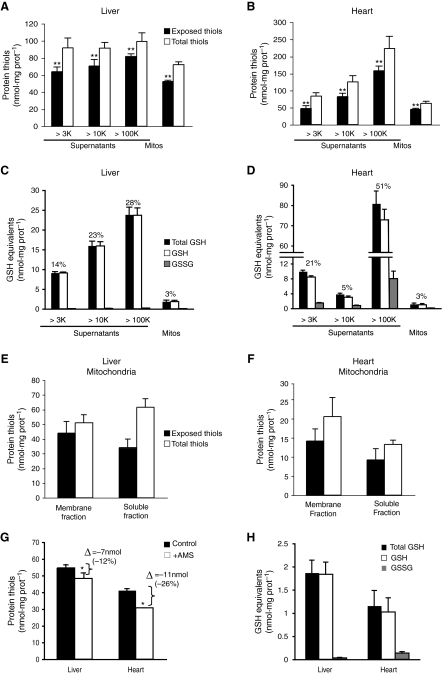
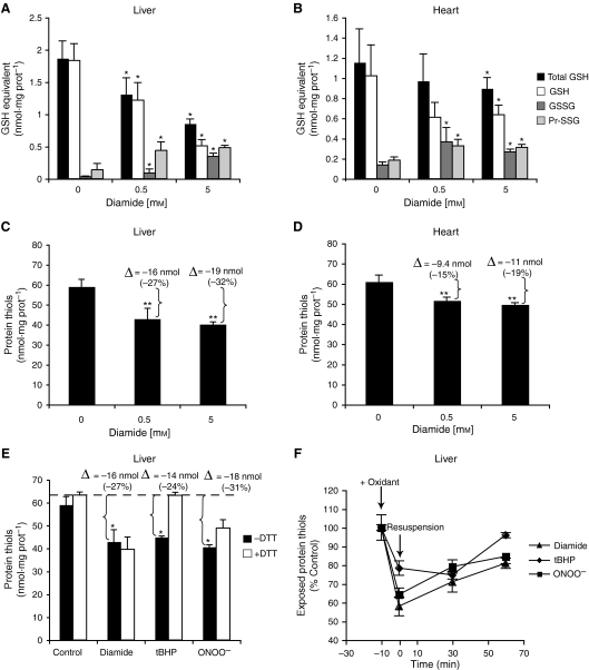
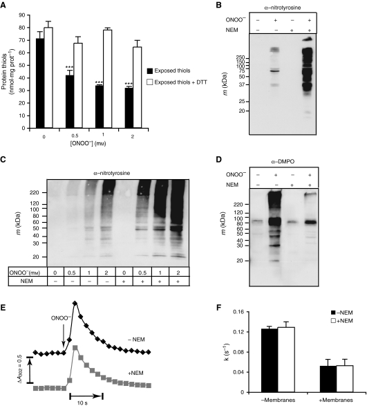
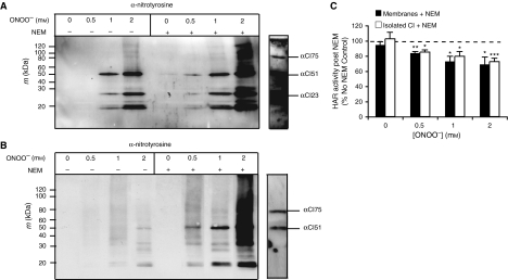
Cysteine plays a number of important roles in protecting the cell from oxidative damage through its thiol functional group. These defensive functions are generally considered to be carried out by the low molecular weight thiol glutathione and by cysteine residues in the active sites of proteins such as thioredoxin and peroxiredoxin. In addition, there are thiols exposed on protein surfaces that are not directly involved with protein function, although they can interact with the intracellular environment. In the present study, in subcellular fractions prepared from rat liver or heart, we show that the quantitatively dominant free thiols are those of cysteine residues exposed on protein surfaces and not those carried by glutathione. Within the mitochondrial matrix, the concentration of exposed protein thiols is 60–90 m m, which is approximately 26‐fold higher than the glutathione concentration in that compartment. This suggests that exposed protein thiols are of greater importance than glutathione for nonenzyme catalysed reactions of thiols with reactive oxygen and nitrogen species and with electrophiles within the cell. One such antioxidant role for exposed protein thiols may be to prevent protein oxidative damage. In the present study, in mitochondrial membranes and in complex I, we show that exposed protein thiols protect against tyrosine nitration and protein dysfunction caused by peroxynitrite. Therefore, exposed protein thiols are the dominant free thiol within the cell and may play a critical role in intracellular antioxidant defences against oxidative damage.
Overview
The claims and statistics of this paper haven’t been checked yet.
Run the check — free, no account needed. It reads the open-access text and checks the sources it cites.
Integrity signals
Registry
Cites
Statistics
Figures
Verification
Claims the paper makes
The claims and statistics of this paper haven’t been checked yet.
Run the check — free, no account needed. It reads the open-access text and checks the sources it cites.
Claims other papers make about this one
Each is checked against this paper’s own text: does it say what the citing paper says it does?
In the citing paper
“thiol residues are as capable as deprotonated amines of mediating nucleophilic attack, if not more so, and are found in large numbers in the mitochondrial matrix ((Requejo et al. 2010)).”
From The Mitochondrial Acylome Emerges: Proteomics, Regulation by Sirtuins, and Metabolic and Disease Implications (2018) · cites this paper as Requejo et al. 2010
Self-check
Percentages, sums and reported differences re-computed from the paper’s own numbers.
The self-check needs the paper’s full text.
Citations
Cites
Measurement of protein using bicinchoninic acid
Smith, P. K., Krohn, R. I., Hermanson, G. T., Mallia, A. K., Gartner, F. H., Provenzano, M. D., Fujimoto, E. K., Goeke, N. M., Olson, B. J., & Klenk, D. C. (1985) · Analytical Biochemistry · doi:10.1016/0003-2697(85)90442-7 · Cited by 18,482
How mitochondria produce reactive oxygen species
Murphy, M. P. (2009) · Biochemical Journal · doi:10.1042/bj20081386 · Cited by 8,207
Self-citationBeckman, J. S., Beckman, T. W., Chen, J., Marshall, P. A., & Freeman, B. A. (1990) · Proceedings of the National Academy of Sciences · doi:10.1073/pnas.87.4.1620 · Cited by 7,029
Schafer, F. Q., & Buettner, G. R. (2001) · Free Radical Biology and Medicine · doi:10.1016/s0891-5849(01)00480-4 · Cited by 4,587
Reconciling the chemistry and biology of reactive oxygen species
Winterbourn, C. C. (2008) · Nature Chemical Biology · doi:10.1038/nchembio.85 · Cited by 2,504
Peroxynitrite oxidation of sulfhydryls.
Radí, R., Beckman, J. S., Bush, K., & Freeman, B. Α. (1991) · Journal of Biological Chemistry · doi:10.1016/s0021-9258(20)64313-7 · Cited by 2,492
Peroxynitrite: biochemistry, pathophysiology and development of therapeutics
Szabó, C., Ischiropoulos, H., & Radi, R. (2007) · Nature Reviews Drug Discovery · doi:10.1038/nrd2222 · Cited by 2,134
A new collection method for the evaluation of nasal mucus proteins
Ruocco, L. A., Fattori, Romanelli, Martelloni, L., Casani, Samolewska, & Rezzonico (1998) · Clinical & Experimental Allergy · doi:10.1046/j.1365-2222.1998.00312.x · Cited by 1,296
Extensive Nitration of Protein Tyrosines in Human Atherosclerosis Detected by Immunohistochemistry
Beckman, J. S., Ye, Y. Z., Anderson, P. G., Chen, J., Accavitti, M. A., Tarpey, M. M., & White, C. R. (1994) · Biological Chemistry Hoppe-Seyler · doi:10.1515/bchm3.1994.375.2.81 · Cited by 1,122
Reactivity of biologically important thiol compounds with superoxide and hydrogen peroxide
Winterbourn, C. C., & Metodiewa, D. (1999) · Free Radical Biology and Medicine · doi:10.1016/s0891-5849(99)00051-9 · Cited by 799
Molecular and Cellular Aspects of Thiol–Disulfide Exchange
Gilbert, H. (1990) · Advances in enzymology and related areas of molecular biology/Advances in enzymology and related subjects · doi:10.1002/9780470123096.ch2 · Cited by 770
Glutathione: Toxicological Implications
Reed, D. J. (1990) · The Annual Review of Pharmacology and Toxicology · doi:10.1146/annurev.pa.30.040190.003131 · Cited by 756
Levonen, A., Landar, A. L., Ramachandran, A., CEASER, E. K., Dickinson, D. A., Zanoni, G., Morrow, J. D., & Darley-Usmar, V. M. (2004) · Biochemical Journal · doi:10.1042/bj20031049 · Cited by 569
Methionine in proteins defends against oxidative stress
Luo, S., & Levine, R. L. (2008) · The FASEB Journal · doi:10.1096/fj.08-118414 · Cited by 462
Quantifying the global cellular thiol–disulfide status
Hansen, R. E., Roth, D., & Winther, J. R. (2009) · Proceedings of the National Academy of Sciences · doi:10.1073/pnas.0812149106 · Cited by 437
Beer, S. M., Taylor, E. R., Brown, S. E., Dahm, C. C., Costa, N. J., Runswick, M. J., & Murphy, M. P. (2004) · Journal of Biological Chemistry · doi:10.1074/jbc.m408011200 · Cited by 404
Self-citationProtein Sulfhydryls and Their Role in the Antioxidant Function of Protein S-Thiolation
Thomas, J. A., Poland, B. W., & Honzatko, R. B. (1995) · Archives of Biochemistry and Biophysics · doi:10.1006/abbi.1995.1261 · Cited by 399
Reversible Glutathionylation of Complex I Increases Mitochondrial Superoxide Formation
Taylor, E. R., Hurrell, F., Shannon, R. J., Lin, T., Hirst, J., & Murphy, M. P. (2003) · Journal of Biological Chemistry · doi:10.1074/jbc.m209359200 · Cited by 389
Self-citationSuperoxide as an intracellular radical sink
Winterbourn, C. C. (1993) · Free Radical Biology and Medicine · doi:10.1016/0891-5849(93)90512-s · Cited by 364
The chemistry of pernitrites. Part I. Kinetics of decomposition of pernitrous acid
Hughes, M., & Nicklin, H. G. (1968) · Journal of the Chemical Society A Inorganic Physical Theoretical · doi:10.1039/j19680000450 · Cited by 356
Methionine residues may protect proteins from critical oxidative damage
Levine, R. L., Berlett, B. S., Moskovitz, J., Mosoni, L., & Stadtman, E. R. (1999) · Mechanisms of Ageing and Development · doi:10.1016/s0047-6374(98)00152-3 · Cited by 338
Cloning and Expression of a Novel Human Glutaredoxin (Grx2) with Mitochondrial and Nuclear Isoforms
Lundberg, M., Johansson, C., Chandra, J., Enoksson, M., Jacobsson, G., Lundström-Ljung, J., Johansson, M., & Holmgren, A. (2001) · Journal of Biological Chemistry · doi:10.1074/jbc.m011605200 · Cited by 335
Peroxynitrite rapidly permeates phospholipid membranes
Marla, S. S., Lee, J., & Groves, J. T. (1997) · Proceedings of the National Academy of Sciences · doi:10.1073/pnas.94.26.14243 · Cited by 310
A precise method for the determination of whole blood and plasma sulfhydryl groups
Ellman, G. L., & Lysko, H. (1979) · Analytical Biochemistry · doi:10.1016/s0003-2697(79)80122-0 · Cited by 268
Identification and Characterization of a New Mammalian Glutaredoxin (Thioltransferase), Grx2
Gladyshev, V. N., Liu, A., Novoselov, S. V., Krysan, K., Sun, Q., Kryukov, V. M., Kryukov, G. V., & Lou, M. F. (2001) · Journal of Biological Chemistry · doi:10.1074/jbc.m100020200 · Cited by 234
Ford, E., Hughes, M., & Wardman, P. (2002) · Free Radical Biology and Medicine · doi:10.1016/s0891-5849(02)00850-x · Cited by 229
Gallogly, M. M., Starke, D. W., & Mieyal, J. J. (2009) · Antioxidants and Redox Signaling · doi:10.1089/ars.2008.2291 · Cited by 227
Prime, T. A., Blaikie, F. H., Evans, C., Nadtochiy, S. M., James, A. M., Dahm, C. C., Vitturi, D. A., Patel, R. P., Hiley, C. R., Abakumova, I., et al. (2009) · Proceedings of the National Academy of Sciences · doi:10.1073/pnas.0903250106 · Cited by 222
Self-citationRapid and extensive uptake and activation of hydrophobic triphenylphosphonium cations within cells
Ross, M. F., Prime, T. A., Abakumova, I., James, A. M., Porteous, C. M., Smith, R. A. J., & Murphy, M. P. (2008) · Biochemical Journal · doi:10.1042/bj20080063 · Cited by 206
Self-citationDahm, C. C., Moore, K., & Murphy, M. P. (2006) · Journal of Biological Chemistry · doi:10.1074/jbc.m512203200 · Cited by 194
Self-citationSharpley, M. S., Shannon, R. J., Draghi, F., & Hirst, J. (2005) · Biochemistry · doi:10.1021/bi051809x · Cited by 193
Bonini, M. G., & Augusto, O. (2001) · Journal of Biological Chemistry · doi:10.1074/jbc.m008456200 · Cited by 192
Hurd, T. R., Requejo, R., Filipovska, A., Brown, S., Prime, T. A., Robinson, A. J., Fearnley, I. M., & Murphy, M. P. (2008) · Journal of Biological Chemistry · doi:10.1074/jbc.m803432200 · Cited by 182
Self-citationMason, R. P. (2004) · Free Radical Biology and Medicine · doi:10.1016/j.freeradbiomed.2004.02.077 · Cited by 170
Overexpression of glutaredoxin 2 attenuates apoptosis by preventing cytochrome c release
Enoksson, M., Fernandes, A. P., Prast‐Nielsen, S., Lillig, C. H., Holmgren, A., & Orrenius, S. (2004) · Biochemical and Biophysical Research Communications · doi:10.1016/j.bbrc.2004.12.067 · Cited by 164
Covalent adduction of nucleophilic amino acids by 4-hydroxynonenal and 4-oxononenal
Doorn, J. A., & Petersen, D. R. (2003) · Chemico-Biological Interactions · doi:10.1016/s0009-2797(02)00178-3 · Cited by 147
Lillig, C. H., Lönn, M. E., Enoksson, M., Fernandes, A. P., & Holmgren, A. (2004) · Proceedings of the National Academy of Sciences · doi:10.1073/pnas.0401896101 · Cited by 147
Peroxynitrite causes calcium efflux from mitochondria which is prevented by Cyclosporin A
Packer, M. A., & Murphy, M. P. (1994) · FEBS Letters · doi:10.1016/0014-5793(94)00461-7 · Cited by 143
Self-citationPREPARATION OF MITOCHONDRIA FROM ANIMAL TISSUES AND YEASTS
Chappell, J., & Hansford, R. G. (1972) · Elsevier eBooks · doi:10.1016/b978-0-408-70360-4.50009-2 · Cited by 141
Redox Potential and Peroxide Reactivity of Human Peroxiredoxin 3
Cox, A. G., Peskin, A. V., Paton, L. N., Winterbourn, C. C., & Hampton, M. B. (2009) · Biochemistry · doi:10.1021/bi900558g · Cited by 130
Role of Protein -SH Groups in Redox Homeostasis— The Erythrocyte as a Model System
Simplicio, P. D., Cacace, M. G., Lusini, L., Giannerini, F., Giustarini, D., & Rossi, R. (1998) · Archives of Biochemistry and Biophysics · doi:10.1006/abbi.1998.0694 · Cited by 123
Liu, B., Tewari, A. K., Zhang, L., Green‐Church, K. B., Zweier, J. L., Chen, Y., & He, G. (2008) · Biochimica et Biophysica Acta (BBA) - Proteins and Proteomics · doi:10.1016/j.bbapap.2008.12.008 · Cited by 116
Madej, E., Folkes, L. K., Wardman, P., Czapski, G., & Goldstein, S. (2008) · Free Radical Biology and Medicine · doi:10.1016/j.freeradbiomed.2008.02.015 · Cited by 108
Yamamoto, T., Maruyama, W., Kato, Y., Yi, H. L., Shamoto‐Nagai, M., Tanaka, M., Sato, Y., & Naoi, M. (2002) · Journal of Neural Transmission · doi:10.1007/s702-002-8232-1 · Cited by 104
[2] Structural analysis of NADH: Ubiquinone oxidoreductase from bovine heart mitochondria
Walker, J. E., Skehel, M. J., & Buchanan, S. K. (1995) · Methods in enzymology on CD-ROM/Methods in enzymology · doi:10.1016/0076-6879(95)60127-9 · Cited by 87
Scarlett, J. L., Packer, M. A., Porteous, C. M., & Murphy, M. P. (1996) · Biochemical Pharmacology · doi:10.1016/0006-2952(96)99426-5 · Cited by 85
Self-citationZeng, J., Dunlop, R. A., Rodgers, K. J., & Davies, M. J. (2006) · Biochemical Journal · doi:10.1042/bj20060019 · Cited by 79
Zhang, H., Xu, Y., Joseph, J., & Kalyanaraman, B. (2005) · Journal of Biological Chemistry · doi:10.1074/jbc.m504503200 · Cited by 76
Craig, C. L., & Weber, R. S. (1998) · Molecular Biology and Evolution · doi:10.1093/oxfordjournals.molbev.a025981 · Cited by 71
Bharath, S., & Andersen, J. K. (2005) · Antioxidants and Redox Signaling · doi:10.1089/ars.2005.7.900 · Cited by 64
Sled, V. D., & Vinogradov, A. D. (1993) · Biochimica et Biophysica Acta (BBA) - Bioenergetics · doi:10.1016/0005-2728(93)90051-g · Cited by 63
Jacob, M. H., Amir, D., Ratner, V., Gussakovsky, E. E., & Haas, E. (2005) · Biochemistry · doi:10.1021/bi051205t · Cited by 39
Cited by
Sorted by citations
The thiol pool in human plasma: The central contribution of albumin to redox processes
Turell, L., Radí, R., & Álvarez, B. (2013) · Free Radical Biology and Medicine · doi:10.1016/j.freeradbiomed.2013.05.050 · Cited by 712
Lavie, L. (2014) · Sleep Medicine Reviews · doi:10.1016/j.smrv.2014.07.003 · Cited by 732
Biochemistry of Peroxynitrite and Protein Tyrosine Nitration
Ferrer‐Sueta, G., Campolo, N., Trujillo, M., Bartesaghi, S., Carballal, S., Romero, N., Álvarez, B., & Radí, R. (2018) · Chemical Reviews · doi:10.1021/acs.chemrev.7b00568 · Cited by 653
Heger, M., van Golen, R. F., Broekgaarden, M., & Michel, M. C. (2013) · Pharmacological Reviews · doi:10.1124/pr.110.004044 · Cited by 594
A Quantitative Tissue-Specific Landscape of Protein Redox Regulation during Aging
Xiao, H., Jedrychowski, M. P., Schweppe, D. K., Huttlin, E. L., Yu, Q., Heppner, D. E., Li, J., Long, J., Mills, E. L., Szpyt, J., et al. (2020) · Cell · doi:10.1016/j.cell.2020.02.012 · Cited by 412
Metadata
- Authors
- Requejo, R., Hurd, T. R., Costa, N. J., & Murphy, M. P.
- Affiliations
- MRC Mitochondrial Biology Unit (GB)
- Venue
- The FEBS Journal
- Publisher
- Wiley
- Published
- 2010-02-24
- Volume
- 277
- Issue
- 6
- Pages
- 1465-1480
- ISSN
- 1742-464X (ISSN-L)
- 1742-4658
- PMC
- PMC2847196
- OpenAlex
- W1524076404
- Cited by
- 231
- References
- 52
- Citations by year
- 2026: 7 · 2025: 7 · 2024: 14 · 2023: 12 · 2022: 15 · 2021: 13 · 2020: 11 · 2019: 13 · 2018: 20 · 2017: 11 · 2016: 14 · 2015: 18 · 2014: 24 · 2013: 16 · 2012: 14
- FWCI
- 6.77
- Type
- journal article
- Language
- en
- Open access
- green
- www.ncbi.nlm.nih.gov
- Locations
- FEBS Journal (publishedVersion)
- PubMed Central (publishedVersion, cc-by)
- PubMed (publishedVersion)
- Funders
- Ministerio de Ciencia y Tecnología
- Medical Research Council
- Topics
- Life Sciences › Biochemistry, Genetics and Molecular Biology › Molecular Biology › Redox biology and oxidative stress
- Life Sciences › Biochemistry, Genetics and Molecular Biology › Biochemistry › Sulfur Compounds in Biology
- Health Sciences › Medicine › Biochemistry (medical) › Antioxidant Activity and Oxidative Stress
- Keywords
- cysteine residues, mitochondrial matrix, glutathione, oxidative damage, antioxidant defense, peroxynitrite, tyrosine nitration, protein oxidation, mitochondrial complex I, reactive oxygen species, reactive nitrogen species, electrophiles, rat liver, rat heart
- MeSH terms
- Animals, Cells, Cultured, Cysteine, Glutathione, Membrane Proteins, Mitochondria, Heart, Mitochondria, Liver, Sulfhydryl Compounds, Oxidative Stress, Rats
About these checks
Registry, retraction, Cites and statistics results are computed, not judged, with one exception: a printed reference that no index lists is read once by a model before it counts as a citation error. Claims are extracted by a model and kept only when their quote appears verbatim in the text; each cited source is then checked, with a model’s help, for whether it says what the claim relies on. Those verdicts can be wrong — read the claim’s quote and the cited source before relying on one.
Statistics and arithmetic are recomputed, not judged.
Confidence bands are shown only for fields calibrated on our annotated test set. None is calibrated yet, so no band is shown.
What counts as a claim ›How we measure confidence ›How we check this ›
Are you an author, or do you see a problem with this page? Request a review
