

The DOI matches its Crossref record; 52 of the 52 references it cites were found in a registry or an index.
Cysteine residues exposed on protein surfaces are the dominant intramitochondrial thiol and may protect against oxidative damage
Open accessAbstract
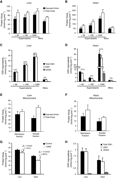
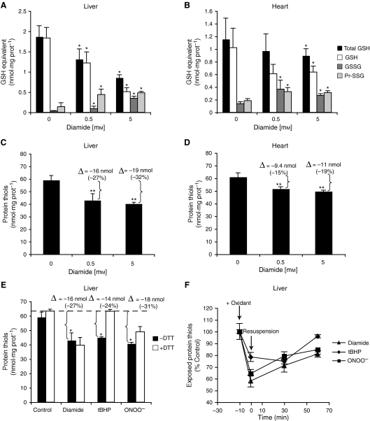
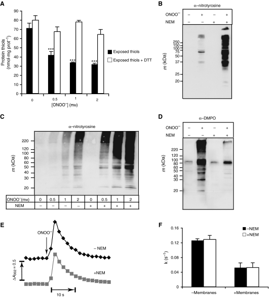
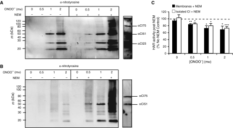
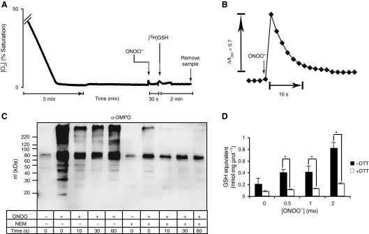
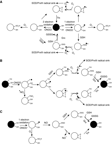
Cysteine plays a number of important roles in protecting the cell from oxidative damage through its thiol functional group. These defensive functions are generally considered to be carried out by the low molecular weight thiol glutathione and by cysteine residues in the active sites of proteins such as thioredoxin and peroxiredoxin. In addition, there are thiols exposed on protein surfaces that are not directly involved with protein function, although they can interact with the intracellular environment. In the present study, in subcellular fractions prepared from rat liver or heart, we show that the quantitatively dominant free thiols are those of cysteine residues exposed on protein surfaces and not those carried by glutathione. Within the mitochondrial matrix, the concentration of exposed protein thiols is 60–90 m m, which is approximately 26‐fold higher than the glutathione concentration in that compartment. This suggests that exposed protein thiols are of greater importance than glutathione for nonenzyme catalysed reactions of thiols with reactive oxygen and nitrogen species and with electrophiles within the cell. One such antioxidant role for exposed protein thiols may be to prevent protein oxidative damage. In the present study, in mitochondrial membranes and in complex I, we show that exposed protein thiols protect against tyrosine nitration and protein dysfunction caused by peroxynitrite. Therefore, exposed protein thiols are the dominant free thiol within the cell and may play a critical role in intracellular antioxidant defences against oxidative damage.
Overview
The claims and statistics of this paper haven’t been checked yet.
Run the check — free, no account needed. It reads the open-access text and checks the sources it cites.
Integrity signals
Registry
Cites
Statistics
Figures
Verification
Claims the paper makes
The claims and statistics of this paper haven’t been checked yet.
Run the check — free, no account needed. It reads the open-access text and checks the sources it cites.
Claims other papers make about this one
Each is checked against this paper’s own text: does it say what the citing paper says it does?
In the citing paper
“thiol residues are as capable as deprotonated amines of mediating nucleophilic attack, if not more so, and are found in large numbers in the mitochondrial matrix ((Requejo et al. 2010)).”
From The Mitochondrial Acylome Emerges: Proteomics, Regulation by Sirtuins, and Metabolic and Disease Implications (2018) · cites this paper as Requejo et al. 2010
Self-check
Percentages, sums and reported differences re-computed from the paper’s own numbers.
The self-check needs the paper’s full text.
Citations
Cites
Measurement of protein using bicinchoninic acid
Smith, P. K., Krohn, R. I., Hermanson, G. T., Mallia, A. K., Gartner, F. H., Provenzano, M. D., Fujimoto, E. K., Goeke, N. M., Olson, B. J., & Klenk, D. C. (1985) · Analytical Biochemistry · doi:10.1016/0003-2697(85)90442-7 · Cited by 18,482
How mitochondria produce reactive oxygen species
Murphy, M. P. (2009) · Biochemical Journal · doi:10.1042/bj20081386 · Cited by 8,207
Self-citationBeckman, J. S., Beckman, T. W., Chen, J., Marshall, P. A., & Freeman, B. A. (1990) · Proceedings of the National Academy of Sciences · doi:10.1073/pnas.87.4.1620 · Cited by 7,029
Schafer, F. Q., & Buettner, G. R. (2001) · Free Radical Biology and Medicine · doi:10.1016/s0891-5849(01)00480-4 · Cited by 4,587
Reconciling the chemistry and biology of reactive oxygen species
Winterbourn, C. C. (2008) · Nature Chemical Biology · doi:10.1038/nchembio.85 · Cited by 2,504
Peroxynitrite oxidation of sulfhydryls.
Radí, R., Beckman, J. S., Bush, K., & Freeman, B. Α. (1991) · Journal of Biological Chemistry · doi:10.1016/s0021-9258(20)64313-7 · Cited by 2,492
Peroxynitrite: biochemistry, pathophysiology and development of therapeutics
Szabó, C., Ischiropoulos, H., & Radi, R. (2007) · Nature Reviews Drug Discovery · doi:10.1038/nrd2222 · Cited by 2,134
A new collection method for the evaluation of nasal mucus proteins
Ruocco, L. A., Fattori, Romanelli, Martelloni, L., Casani, Samolewska, & Rezzonico (1998) · Clinical & Experimental Allergy · doi:10.1046/j.1365-2222.1998.00312.x · Cited by 1,296
Extensive Nitration of Protein Tyrosines in Human Atherosclerosis Detected by Immunohistochemistry
Beckman, J. S., Ye, Y. Z., Anderson, P. G., Chen, J., Accavitti, M. A., Tarpey, M. M., & White, C. R. (1994) · Biological Chemistry Hoppe-Seyler · doi:10.1515/bchm3.1994.375.2.81 · Cited by 1,122
Reactivity of biologically important thiol compounds with superoxide and hydrogen peroxide
Winterbourn, C. C., & Metodiewa, D. (1999) · Free Radical Biology and Medicine · doi:10.1016/s0891-5849(99)00051-9 · Cited by 799
Cited by
Sorted by citations
The thiol pool in human plasma: The central contribution of albumin to redox processes
Turell, L., Radí, R., & Álvarez, B. (2013) · Free Radical Biology and Medicine · doi:10.1016/j.freeradbiomed.2013.05.050 · Cited by 712
Lavie, L. (2014) · Sleep Medicine Reviews · doi:10.1016/j.smrv.2014.07.003 · Cited by 732
Biochemistry of Peroxynitrite and Protein Tyrosine Nitration
Ferrer‐Sueta, G., Campolo, N., Trujillo, M., Bartesaghi, S., Carballal, S., Romero, N., Álvarez, B., & Radí, R. (2018) · Chemical Reviews · doi:10.1021/acs.chemrev.7b00568 · Cited by 653
Heger, M., van Golen, R. F., Broekgaarden, M., & Michel, M. C. (2013) · Pharmacological Reviews · doi:10.1124/pr.110.004044 · Cited by 594
A Quantitative Tissue-Specific Landscape of Protein Redox Regulation during Aging
Xiao, H., Jedrychowski, M. P., Schweppe, D. K., Huttlin, E. L., Yu, Q., Heppner, D. E., Li, J., Long, J., Mills, E. L., Szpyt, J., et al. (2020) · Cell · doi:10.1016/j.cell.2020.02.012 · Cited by 412
Murphy, M. P. (2011) · Antioxidants and Redox Signaling · doi:10.1089/ars.2011.4289 · Cited by 351
Self-citationCarrico, C., Meyer, J. G., He, W., Gibson, B. W., & Verdin, E. (2018) · Cell Metabolism · doi:10.1016/j.cmet.2018.01.016 · Cited by 331
Cell signalling by reactive lipid species: new concepts and molecular mechanisms
Higdon, A. N., Diers, A. R., Oh, J. Y., Landar, A. L., & Darley-Usmar, V. M. (2012) · Biochemical Journal · doi:10.1042/bj20111752 · Cited by 327
Causes and consequences of cysteine S-glutathionylation
Grek, C. L., Zhang, J., Manevich, Y., Townsend, D. M., & Tew, K. D. (2013) · Journal of Biological Chemistry · doi:10.1074/jbc.r113.461368 · Cited by 312
Redox regulation of mitochondrial function with emphasis on cysteine oxidation reactions
Mailloux, R. J., Jin, X., & Willmore, W. G. (2013) · Redox Biology · doi:10.1016/j.redox.2013.12.011 · Cited by 321
Marzia Perluigi, A., Domenico, F. D., & Butterfield, D. A. (2023) · Physiological Reviews · doi:10.1152/physrev.00030.2022 · Cited by 322
Factors Affecting Protein Thiol Reactivity and Specificity in Peroxide Reduction
Ferrer‐Sueta, G., Manta, B., Botti, H., Radí, R., Trujillo, M., & Denicola, A. (2011) · Chemical Research in Toxicology · doi:10.1021/tx100413v · Cited by 295
Proteome-wide analysis of cysteine oxidation reveals metabolic sensitivity to redox stress
van der Reest, J., Lilla, S., Zheng, L., Zanivan, S. R., & Gottlieb, E. (2018) · Nature Communications · doi:10.1038/s41467-018-04003-3 · Cited by 273
Mitochondrial Respiratory Chain Complexes
Sousa, J. S., D’Imprima, E., & Vonck, J. (2018) · Sub-cellular biochemistry/Subcellular biochemistry · doi:10.1007/978-981-10-7757-9_7 · Cited by 263
Role of thiols in oxidative stress
Baba, S. P., & Bhatnagar, A. (2018) · Current Opinion in Toxicology · doi:10.1016/j.cotox.2018.03.005 · Cited by 240
Protein Oxidative Modifications: Beneficial Roles in Disease and Health.
Cai, Z., & Yan, L. (2013) · PubMed · Cited by 227
Mitochondrial Energy and Redox Signaling in Plants
Schwarzländer, M., & Finkemeier, I. (2012) · Antioxidants and Redox Signaling · doi:10.1089/ars.2012.5104 · Cited by 199
Generator-specific targets of mitochondrial reactive oxygen species
Bleier, L., Wittig, I., Heide, H., Steger, M., Brandt, U., & Dröse, S. (2014) · Free Radical Biology and Medicine · doi:10.1016/j.freeradbiomed.2014.10.511 · Cited by 188
Musatov, A., & Robinson, N. C. (2012) · Free Radical Research · doi:10.3109/10715762.2012.717273 · Cited by 187
Storkey, C., Davies, M. J., & Pattison, D. I. (2014) · Free Radical Biology and Medicine · doi:10.1016/j.freeradbiomed.2014.04.024 · Cited by 165
Glutathione and Glutaredoxin—Key Players in Cellular Redox Homeostasis and Signaling
Chai, Y.-C., & Mieyal, J. J. (2023) · Antioxidants · doi:10.3390/antiox12081553 · Cited by 162
Biotin conjugated organic molecules and proteins for cancer therapy: A review
Maiti, S., & Paira, P. (2018) · European Journal of Medicinal Chemistry · doi:10.1016/j.ejmech.2018.01.001 · Cited by 157
Mitochondrial respiratory chain complexes as sources and targets of thiol-based redox-regulation
Dröse, S., Brandt, U., & Wittig, I. (2014) · Biochimica et Biophysica Acta (BBA) - Proteins and Proteomics · doi:10.1016/j.bbapap.2014.02.006 · Cited by 155
Bazylewski, P. F., Divigalpitiya, R., & Fanchini, G. (2017) · RSC Advances · doi:10.1039/c6ra25879d · Cited by 153
Targeting Stimulator of Interferon Genes (STING): A Medicinal Chemistry Perspective
Zhang, H., You, Q., & Xu, X. (2019) · Journal of Medicinal Chemistry · doi:10.1021/acs.jmedchem.9b01039 · Cited by 153
Global redox proteome and phosphoproteome analysis reveals redox switch in Akt
Su, Z., Burchfield, J. G., Yang, P., Humphrey, S. J., Yang, G., Francis, D., Yasmin, S., Shin, S., Norris, D. M., Kearney, A. L., et al. (2019) · Nature Communications · doi:10.1038/s41467-019-13114-4 · Cited by 146
Activity-Based Sensing for Site-Specific Proteomic Analysis of Cysteine Oxidation
Shi, Y., & Carroll, K. S. (2019) · Accounts of Chemical Research · doi:10.1021/acs.accounts.9b00562 · Cited by 146
Regulation of mitochondrial processes by protein S-nitrosylation
Piantadosi, C. A. (2011) · Biochimica et Biophysica Acta (BBA) - General Subjects · doi:10.1016/j.bbagen.2011.03.008 · Cited by 147
Chouchani, E. T., Hurd, T. R., Nadtochiy, S. M., Brookes, P. S., Fearnley, I. M., Lilley, K. S., Smith, R. A. J., & Murphy, M. P. (2010) · Biochemical Journal · doi:10.1042/bj20100633 · Cited by 137
Self-citationRedox regulation of the actin cytoskeleton and its role in the vascular system
Xu, Q., Huff, L. P., Fujii, M., & Griendling, K. K. (2017) · Free Radical Biology and Medicine · doi:10.1016/j.freeradbiomed.2017.03.004 · Cited by 132
Chen, H., Qi, H., & Xiong, P. (2022) · Marine Drugs · doi:10.3390/md20070450 · Cited by 129
Musaogullari, A., & Chai, Y.-C. (2020) · International Journal of Molecular Sciences · doi:10.3390/ijms21218113 · Cited by 122
McDonagh, B., Sakellariou, G. K., Smith, N. T., Brownridge, P., & Jackson, M. J. (2014) · Journal of Proteome Research · doi:10.1021/pr5006394 · Cited by 111
Proteomic approaches to the characterization of protein thiol modification
Chouchani, E. T., James, A. M., Fearnley, I. M., Lilley, K. S., & Murphy, M. P. (2010) · Current Opinion in Chemical Biology · doi:10.1016/j.cbpa.2010.11.003 · Cited by 109
Self-citationThe relationship between oxidative stress and autoimmunity in Hashimoto's thyroiditis
Ateş, İ., Yılmaz, F. M., Altay, M., Yılmaz, N., Berker, D. S., & Güler, S. (2015) · European Journal of Endocrinology · doi:10.1530/eje-15-0617 · Cited by 106
Held, J. M., & Gibson, B. W. (2011) · Molecular & Cellular Proteomics · doi:10.1074/mcp.r111.013037 · Cited by 103
Cysteine-mediated redox signalling in the mitochondria
Bak, D. W., & Weerapana, E. (2014) · Molecular BioSystems · doi:10.1039/c4mb00571f · Cited by 102
Mitochondrial protein tyrosine nitration
Castro, L., Demicheli, V., Tórtora, V., & Radí, R. (2010) · Free Radical Research · doi:10.3109/10715762.2010.516254 · Cited by 101
STING palmitoylation as a therapeutic target
Hansen, A. L., Mukai, K., Schopfer, F. J., Taguchi, T., & Holm, C. K. (2019) · Cellular and Molecular Immunology · doi:10.1038/s41423-019-0205-5 · Cited by 102
Glutaredoxin 2 prevents aggregation of mutant SOD1 in mitochondria and abolishes its toxicity
Ferri, A., Fiorenzo, P., Nencini, M., Cozzolino, M., Pesaresi, M. G., Valle, C., Sepe, S., Moreno, S., & Carrı̀, M. T. (2010) · Human Molecular Genetics · doi:10.1093/hmg/ddq383 · Cited by 98
3-Mercaptopyruvate sulfur transferase is a protein persulfidase
Pedre, B., Talwar, D., Barayeu, U., Schilling, D., Luzarowski, M., Sokołowski, M., Glatt, S., & Dick, T. P. (2023) · Nature Chemical Biology · doi:10.1038/s41589-022-01244-8 · Cited by 96
Mitochondrial Redox Dysfunction and Environmental Exposures
Caito, S. W., & Aschner, M. (2015) · Antioxidants and Redox Signaling · doi:10.1089/ars.2015.6289 · Cited by 96
Pleonsil, P., Soogarun, S., & Suwanwong, Y. (2013) · International Journal of Biological Macromolecules · doi:10.1016/j.ijbiomac.2013.06.016 · Cited by 93
Batchelor‐McAuley, C., Tschulik, K., Neumann, C., Laborda, E., & Compton, R. G. (2014) · International Journal of Electrochemical Science · doi:10.1016/s1452-3981(23)07783-0 · Cited by 91
Giardia secretome highlights secreted tenascins as a key component of pathogenesis
Dubourg, A., Xia, D., Winpenny, J. P., Naimi, S. A., Bouzid, M., Sexton, D. W., Wastling, J. M., Hunter, P. R., & Tyler, K. M. (2018) · GigaScience · doi:10.1093/gigascience/giy003 · Cited by 91
Møller, I. M., Igamberdiev, A. U., Bykova, N. V., Finkemeier, I., Rasmusson, A. G., & Schwarzländer, M. (2020) · The Plant Cell · doi:10.1105/tpc.19.00535 · Cited by 89
Zhu, Z., Umehara, T., Okazaki, T., Goto, M., Fujita, Y., Hoque, S. A. M., Kawai, T., Zeng, W., & Shimada, M. (2019) · Frontiers in Physiology · doi:10.3389/fphys.2019.00252 · Cited by 88
Redox signaling pathways in unilateral ureteral obstruction (UUO)-induced renal fibrosis
Aranda‐Rivera, A. K., Cruz‐Gregorio, A., Aparicio‐Trejo, O. E., Ortega-Lozano, A. J., & Pedraza‐Chaverrí, J. (2021) · Free Radical Biology and Medicine · doi:10.1016/j.freeradbiomed.2021.05.034 · Cited by 87
Castro, H., Teixeira, F., Romão, S., Santos, M., Cruz, T., Flórido, M., Appelberg, R., Oliveira, P., Ferreira‐da‐Silva, F., & Tomás, A. M. (2011) · PLoS Pathogens · doi:10.1371/journal.ppat.1002325 · Cited by 83
Respiratory chain complex II as general sensor for apoptosis
Grimm, S. (2013) · Biochimica et Biophysica Acta (BBA) - Bioenergetics · doi:10.1016/j.bbabio.2012.09.009 · Cited by 82
Selective Inhibition of Deactivated Mitochondrial Complex I by Biguanides
Matsuzaki, S., & Humphries, K. M. (2015) · Biochemistry · doi:10.1021/bi501473h · Cited by 81
Studying the Effects of Cysteine Residues on Protein Interactions with Silver Nanoparticles
Siriwardana, K., Wang, A., Gadogbe, M., Collier, W. E., Fitzkee, N. C., & Zhang, D. (2015) · The Journal of Physical Chemistry C · doi:10.1021/jp512440z · Cited by 80
Watanabe, H., Imafuku, T., Otagiri, M., & Maruyama, T. (2017) · Journal of Pharmaceutical Sciences · doi:10.1016/j.xphs.2017.03.002 · Cited by 79
Selective and reversible photochemical derivatization of cysteine residues in peptides and proteins
Arumugam, S., Guo, J., Mbua, N. E., Friscourt, F., Lin, N., Nekongo, E. E., Boons, G., & Popik, V. V. (2014) · Chemical Science · doi:10.1039/c3sc51691a · Cited by 76
Horowitz, M. P., Milanese, C., Maio, R. D., Hu, X., Montero, L. M., Sanders, L. H., Tapias, V., Sepe, S., van Cappellen, W. A., Burton, E. A., et al. (2011) · Antioxidants and Redox Signaling · doi:10.1089/ars.2010.3629 · Cited by 76
Chemical Forms of Mercury in Human Hair Reveal Sources of Exposure
Manceau, A., Enescu, M., Simionovici, A., Lanson, M., González-Rey, M., Rovezzi, M., Tucoulou, R., Glatzel, P., Nagy, K. L., & Bourdineaud, J. (2016) · Environmental Science & Technology · doi:10.1021/acs.est.6b03468 · Cited by 75
Redox regulation of mitochondrial proteins and proteomes by cysteine thiol switches
Nietzel, T., Mostertz, J., Hochgräfe, F., & Schwarzländer, M. (2016) · Mitochondrion · doi:10.1016/j.mito.2016.07.010 · Cited by 75
de Groot, A., Roche, D., Fernandez, B., Ludanyi, M., Cruveiller, S., Pignol, D., Vallenet, D., Armengaud, J., & Blanchard, L. (2014) · Genome Biology and Evolution · doi:10.1093/gbe/evu069 · Cited by 72
Souza, C. F., Baldissera, M. D., Verdi, C. M., Santos, R. C. V., da Rocha, M. I. D. U. M., da Veiga, M. L., Silva, A. S. D., & Baldisserotto, B. (2019) · Microbial Pathogenesis · doi:10.1016/j.micpath.2019.04.007 · Cited by 72
Menger, K. E., James, A. M., Cochemé, H. M., Harbour, M. E., Chouchani, E. T., Ding, S., Fearnley, I. M., Partridge, L., & Murphy, M. P. (2015) · Cell Reports · doi:10.1016/j.celrep.2015.05.033 · Cited by 74
Self-citationTargeting Non-Catalytic Cysteine Residues Through Structure-Guided Drug Discovery
Hallenbeck, K. K., Turner, D., Renslo, A. R., & Arkin, M. R. (2016) · Current Topics in Medicinal Chemistry · doi:10.2174/1568026616666160719163839 · Cited by 73
Donida, B., Marchetti, D. P., Biancini, G. B., Deon, M., Manini, P. R., da Rosa, H. T., Moura, D. J., Saffi, J., Bender, F., Burin, M. G., et al. (2015) · Biochimica et Biophysica Acta (BBA) - Molecular Basis of Disease · doi:10.1016/j.bbadis.2015.02.004 · Cited by 68
Fernandes, C. G., Borges, C. G., Seminotti, B., Amaral, A. U., Knebel, L. A., Eichler, P., de Oliveira, A. B., Leipnitz, G., & Wajner, M. (2011) · Cellular and Molecular Neurobiology · doi:10.1007/s10571-011-9675-4 · Cited by 67
Nitro-Fatty Acid Logistics: Formation, Biodistribution, Signaling, and Pharmacology
Schopfer, F. J., & Khoo, N. K. H. (2019) · Trends in Endocrinology and Metabolism · doi:10.1016/j.tem.2019.04.009 · Cited by 67
Shu, N., Lorentzen, L. G., & Davies, M. J. (2019) · Free Radical Biology and Medicine · doi:10.1016/j.freeradbiomed.2019.04.026 · Cited by 66
Siriwardana, K., Wang, A., Vangala, K., Fitzkee, N. C., & Zhang, D. (2013) · Langmuir · doi:10.1021/la402239h · Cited by 63
Oyagbemi, A. A., Omóbòwálé, T. O., Saba, A. B., Olowu, E. R., Dada, R. O., & Akinrinde, A. S. (2015) · Journal of Dietary Supplements · doi:10.3109/19390211.2015.1103827 · Cited by 60
Turell, L., Vitturi, D. A., Coitiño, E. L., Lebrato, L., Möller, M. N., Sagasti, C., Salvatore, S. R., Woodcock, S. R., Álvarez, B., & Schopfer, F. J. (2016) · Journal of Biological Chemistry · doi:10.1074/jbc.m116.756288 · Cited by 61
Redox Metabolism in Mitochondria of Trypanosomatids
Tomás, A. M., & Castro, H. (2012) · Antioxidants and Redox Signaling · doi:10.1089/ars.2012.4948 · Cited by 59
Huang, Z., Wang, H., & Yang, W. (2015) · ACS Applied Materials & Interfaces · doi:10.1021/acsami.5b01552 · Cited by 58
New indole-based chalconoids as tubulin-targeting antiproliferative agents
Mirzaei, H., Shokrzadeh, M., Modanloo, M., Ziar, A., Riazi, G. H., & Emami, S. (2017) · Bioorganic Chemistry · doi:10.1016/j.bioorg.2017.09.005 · Cited by 57
Serien, D., & Sugioka, K. (2018) · Opto-Electronic Advances · doi:10.29026/oea.2018.180008 · Cited by 57
An injectable PEG hydrogel controlling neurotrophin-3 release by affinity peptides
Wang, J., Youngblood, R. L., Cassinotti, L. R., Skoumal, M., Corfas, G., & Shea, L. D. (2020) · Journal of Controlled Release · doi:10.1016/j.jconrel.2020.12.045 · Cited by 57
Cobley, J. N. (2020) · Antioxidants · doi:10.3390/antiox9100933 · Cited by 57
Dual regulation of SLC25A39 by AFG3L2 and iron controls mitochondrial glutathione homeostasis
Shi, X., DeCiucis, M., Grabinska, K. A., Kanyo, J., Liu, A., Lam, T. T., & Shen, H. (2024) · Molecular Cell · doi:10.1016/j.molcel.2023.12.008 · Cited by 54
Benavides, G. A., Liang, Q., Dodson, M. B., Darley-Usmar, V. M., & Zhang, J. (2013) · Free Radical Biology and Medicine · doi:10.1016/j.freeradbiomed.2013.09.006 · Cited by 53
Go, Y. (2011) · Journal of Proteomics & Bioinformatics · doi:10.4172/jpb.1000190 · Cited by 54
Photoactivatable protein labeling by singlet oxygen mediated reactions
To, T., Medzihradszky, K. F., Burlingame, A. L., DeGrado, W. F., Jo, H., & Shu, X. (2016) · Bioorganic & Medicinal Chemistry Letters · doi:10.1016/j.bmcl.2016.05.034 · Cited by 51
Taurine, Glutathione and Bioenergetics
Hansen, S. H., & Grunnet, N. (2013) · Advances in experimental medicine and biology · doi:10.1007/978-1-4614-6093-0_1 · Cited by 48
Mitochondrial Redox Metabolism in Trypanosomatids Is Independent of Tryparedoxin Activity
Castro, H., Romão, S., Carvalho, S., Teixeira, F., Sousa, C. F., & Tomás, A. M. (2010) · PLoS ONE · doi:10.1371/journal.pone.0012607 · Cited by 48
Why does mitochondrial complex I have so many subunits?
Hirst, J. (2011) · Biochemical Journal · doi:10.1042/bj20110918 · Cited by 48
Sundqvist, M., Christenson, K., Björnsdóttir, H., Osla, V., Karlsson, A., Dahlgren, C., Speert, D. P., Fasth, A. L., Brown, K. L., & Bylund, J. (2017) · Frontiers in Immunology · doi:10.3389/fimmu.2017.01828 · Cited by 49
Vanzin, C. S., Biancini, G. B., Sitta, Â., Wayhs, C. A. Y., Pereira, I. N., Rockenbach, F. J., García, S. C., Wyse, Â. T. D. S., Schwartz, I. V. D., Wajner, M., et al. (2011) · Molecular Genetics and Metabolism · doi:10.1016/j.ymgme.2011.06.013 · Cited by 46
Lumpuy‐Castillo, J., Amador‐Martínez, I., Díaz-Rojas, M., Lorenzo, Ó., Pedraza-Chaverrí, J., Sánchez‐Lozada, L. G., & Aparicio‐Trejo, O. E. (2024) · Redox Biology · doi:10.1016/j.redox.2024.103340 · Cited by 45
The properties of human disease mutations at protein interfaces
Livesey, B., & Marsh, J. A. (2022) · PLoS Computational Biology · doi:10.1371/journal.pcbi.1009858 · Cited by 46
Sun, Q., Sun, D., Song, L., Chen, Z., Chen, Z., Zhang, W., & Qian, J. (2016) · Analytical Chemistry · doi:10.1021/acs.analchem.6b00178 · Cited by 40
Storkey, C., Pattison, D. I., Ignasiak, M. T., Schiesser, C. H., & Davies, M. J. (2015) · Free Radical Biology and Medicine · doi:10.1016/j.freeradbiomed.2015.10.424 · Cited by 40
Davis, D. A., Bulut, H., Shrestha, P., Yaparla, A., Jaeger, H. K., Hattori, S.-I., Wingfield, P. T., Mieyal, J. J., Mitsuya, H., & Yarchoan, R. (2021) · mBio · doi:10.1128/mbio.02094-21 · Cited by 39
Powerhouse down: Complex II dissociation in the respiratory chain
Hwang, M., Rohlena, J., Dong, L., Neužil, J., & Grimm, S. (2014) · Mitochondrion · doi:10.1016/j.mito.2014.06.001 · Cited by 39
The Causes and Consequences of Nonenzymatic Protein Acylation
James, A. M., Smith, C. L., Smith, A. C., Robinson, A. J., Hoogewijs, K., & Murphy, M. P. (2018) · Trends in Biochemical Sciences · doi:10.1016/j.tibs.2018.07.002 · Cited by 40
Self-citationMiljković, J. L., Burger, N., Gawel, J. M., Mulvey, J. F., Norman, A. A. I., Nishimura, T., Tsujihata, Y., Logan, A., Sauchanka, O., Caldwell, S. T., et al. (2022) · Redox Biology · doi:10.1016/j.redox.2022.102429 · Cited by 39
Self-citationND3 Cys39 in complex I is exposed during mitochondrial respiration
Burger, N., James, A. M., Mulvey, J. F., Hoogewijs, K., Ding, S., Fearnley, I. M., Loureiro, M., Norman, A. A. I., Arndt, S., Mottahedin, A., et al. (2021) · Cell chemical biology · doi:10.1016/j.chembiol.2021.10.010 · Cited by 38
Self-citationImproved prediction of accessible surface area results in efficient energy function application
Iqbal, S., Mishra, A., & Hoque, M. T. (2015) · Journal of Theoretical Biology · doi:10.1016/j.jtbi.2015.06.012 · Cited by 38
Cherdkiatikul, T., & Suwanwong, Y. (2014) · SLAS DISCOVERY · doi:10.1177/1087057113520565 · Cited by 37
Quantification and identification of mitochondrial proteins containing vicinal dithiols
Requejo-Aguilar, R., Chouchani, E. T., James, A. M., Prime, T. A., Lilley, K. S., Fearnley, I. M., & Murphy, M. P. (2010) · Archives of Biochemistry and Biophysics · doi:10.1016/j.abb.2010.09.003 · Cited by 36
Self-citationN-Acetyl-l-Cysteine Protects Astrocytes against Proteotoxicity without Recourse to Glutathione
Gleixner, A. M., Hutchison, D. F., Sannino, S., Bhatia, T. N., Leak, L. C., Flaherty, P. T., Wipf, P., Brodsky, J. L., & Leak, R. K. (2017) · Molecular Pharmacology · doi:10.1124/mol.117.109926 · Cited by 36
James, A. M., Smith, A. C., Smith, C. L., Robinson, A. J., & Murphy, M. P. (2018) · Cell Reports · doi:10.1016/j.celrep.2018.07.007 · Cited by 36
Self-citationNedoboy, P. E., Morgan, P. E., Mocatta, T. J., Richards, M., Winterbourn, C. C., & Davies, M. J. (2014) · Free Radical Research · doi:10.3109/10715762.2014.947286 · Cited by 35
Carroll, L., Davies, M. J., & Pattison, D. I. (2015) · Free Radical Research · doi:10.3109/10715762.2015.1018247 · Cited by 36
Convenient detection of the thiol functional group using H/D isotope sensitive Raman spectroscopy
Bandyopadhyay, S., & Dey, A. (2014) · The Analyst · doi:10.1039/c3an02166a · Cited by 34
Metadata
- Authors
- Requejo, R., Hurd, T. R., Costa, N. J., & Murphy, M. P.
- Affiliations
- MRC Mitochondrial Biology Unit (GB)
- Venue
- The FEBS Journal
- Publisher
- Wiley
- Published
- 2010-02-24
- Volume
- 277
- Issue
- 6
- Pages
- 1465-1480
- ISSN
- 1742-464X (ISSN-L)
- 1742-4658
- PMC
- PMC2847196
- OpenAlex
- W1524076404
- Cited by
- 231
- References
- 52
- Citations by year
- 2026: 7 · 2025: 7 · 2024: 14 · 2023: 12 · 2022: 15 · 2021: 13 · 2020: 11 · 2019: 13 · 2018: 20 · 2017: 11 · 2016: 14 · 2015: 18 · 2014: 24 · 2013: 16 · 2012: 14
- FWCI
- 6.77
- Type
- journal article
- Language
- en
- Open access
- green
- www.ncbi.nlm.nih.gov
- Locations
- FEBS Journal (publishedVersion)
- PubMed Central (publishedVersion, cc-by)
- PubMed (publishedVersion)
- Funders
- Ministerio de Ciencia y Tecnología
- Medical Research Council
- Topics
- Life Sciences › Biochemistry, Genetics and Molecular Biology › Molecular Biology › Redox biology and oxidative stress
- Life Sciences › Biochemistry, Genetics and Molecular Biology › Biochemistry › Sulfur Compounds in Biology
- Health Sciences › Medicine › Biochemistry (medical) › Antioxidant Activity and Oxidative Stress
- Keywords
- cysteine residues, mitochondrial matrix, glutathione, oxidative damage, antioxidant defense, peroxynitrite, tyrosine nitration, protein oxidation, mitochondrial complex I, reactive oxygen species, reactive nitrogen species, electrophiles, rat liver, rat heart
- MeSH terms
- Animals, Cells, Cultured, Cysteine, Glutathione, Membrane Proteins, Mitochondria, Heart, Mitochondria, Liver, Sulfhydryl Compounds, Oxidative Stress, Rats
About these checks
Registry, retraction, Cites and statistics results are computed, not judged, with one exception: a printed reference that no index lists is read once by a model before it counts as a citation error. Claims are extracted by a model and kept only when their quote appears verbatim in the text; each cited source is then checked, with a model’s help, for whether it says what the claim relies on. Those verdicts can be wrong — read the claim’s quote and the cited source before relying on one.
Statistics and arithmetic are recomputed, not judged.
Confidence bands are shown only for fields calibrated on our annotated test set. None is calibrated yet, so no band is shown.
What counts as a claim ›How we measure confidence ›How we check this ›
Are you an author, or do you see a problem with this page? Request a review
