

The DOI matches its Crossref record; 61 of the 61 references it cites were found in a registry or an index.
Role of PGC-1α Signaling in Skeletal Muscle Health and Disease
Open accessAbstract
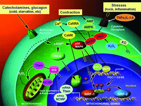
This paper reviews the current understanding of the molecular basis of the peroxisome proliferator-activated receptor-γ coactivator-1α (PGC-1α)-mediated pathway and discusses the role of PGC-1α in skeletal muscle atrophy caused by immobilization. PGC-1α is the master transcription regulator that stimulates mitochondrial biogenesis, by upregulating nuclear respiratory factors (NRF-1, 2) and mitochondrial transcription factor A (Tfam), which leads to increased mitochondrial DNA replication and gene transcription. PGC-1α also regulates cellular oxidant-antioxidant homeostasis by stimulating the gene expression of superoxide dismutase-2 (SOD2), catalase, glutathione peroxidase 1 (GPx1), and uncoupling protein (UCP). Recent reports from muscle-specific PGC-1α overexpression underline the importance of PGC-1α in atrophied skeletal muscle, demonstrate enhancement of the PGC-1α mitochondrial biogenic pathway, and reduced oxidative damage. Thus, PGC-1α appears to play a protective role against atrophy-linked skeletal muscle deterioration.
Overview
The claims and statistics of this paper haven’t been checked yet.
Run the check — free, no account needed. It reads the open-access text and checks the sources it cites.
Integrity signals
Registry
Cites
Statistics
Figures
Verification
Claims the paper makes
The claims and statistics of this paper haven’t been checked yet.
Run the check — free, no account needed. It reads the open-access text and checks the sources it cites.
Claims other papers make about this one
Each is checked against this paper’s own text: does it say what the citing paper says it does?
The source backs part of the claim, not all of it — or backs it for a different population, condition or comparison.
In the citing paper
“Through deacetylation, AMPK upregulates the expression of PGC-1α via the SIRT1 signaling pathway which in turn modulates FoxO transcriptional activity by repressing the expression of key atrogenes such as atrogin-1 and muscle RING-finger protein-1 (MuRF1), thereby preventing proteasome-mediated muscle protein degradation while simultaneously enhancing mitochondrial biogenesis, antioxidant defense,”
From Exercise-Induced Muscle–Fat Crosstalk: Molecular Mediators and Their Pharmacological Modulation for the Maintenance of Metabolic Flexibility in Aging (2025) · cites this paper as [103]
Experimental — accuracy not yet measuredThe cited source says what the claim attributes to it.
In the citing paper
“In addition, PPARGC1A inhibits the expression of multiple pro‐inflammatory mediators, including TNF‐α and IL‐6, and regulates antioxidant defense mechanisms (Kang & Li Ji, 2012), both of which in turn can activate autophagy.”
From Autophagy gene expression in skeletal muscle of older individuals is associated with physical performance, muscle volume and mitochondrial function in the study of muscle, mobility and aging ( SOMMA ) (2024) · cites this paper as Kang & Li Ji, 2012
In the citing paper
“ApN may also induce Ca2+ influx, thereby activating CaMKKβ, which leads to increased expression of peroxisome proliferator-activated receptor γ coactivator-1α (PGC-1α), a key player in mitochondrial function and biogenesis [42,43].”
From Adiponectin and Its Mimics on Skeletal Muscle: Insulin Sensitizers, Fat Burners, Exercise Mimickers, Muscling Pills … or Everything Together? (2020) · cites this paper as [43]
In the citing paper
“Second, activation of PGC-1α enhances the expression of several oxidative-stress detoxification enzymes and molecules involved in fatty-acid oxidation [43,51].”
From Adiponectin and Its Mimics on Skeletal Muscle: Insulin Sensitizers, Fat Burners, Exercise Mimickers, Muscling Pills … or Everything Together? (2020) · cites this paper as [43]
In the citing paper
“Eventually, as mentioned earlier, PGC-1α is also a regulator of cellular oxidant–antioxidant homeostasis by stimulating gene expression of several antioxidant enzymes such as superoxide dismutase-2, catalase, and glutathione peroxidase [43].”
From Adiponectin and Its Mimics on Skeletal Muscle: Insulin Sensitizers, Fat Burners, Exercise Mimickers, Muscling Pills … or Everything Together? (2020) · cites this paper as [43]
Self-check
Percentages, sums and reported differences re-computed from the paper’s own numbers.
The self-check needs the paper’s full text.
Citations
Cites
1 retracted
Wenz, T., Rossi, S. G., Rotundo, R. L., Spiegelman, B. M., & Moraes, C. T. (2009) · Proceedings of the National Academy of Sciences · doi:10.1073/pnas.0911570106 · Cited by 600
RetractedMootha, V. K., Lindgren, C. M., Eriksson, K.-F., Subramanian, A., Sihag, S., Lehar, J., Puigserver, P., Carlsson, E., Ridderstråle, M., Laurila, E., et al. (2003) · Nature Genetics · doi:10.1038/ng1180 · Cited by 10,197
THE NF-κB AND IκB PROTEINS: New Discoveries and Insights
Baldwin, A. S. (1996) · Annual Review of Immunology · doi:10.1146/annurev.immunol.14.1.649 · Cited by 5,882
Inflammation, stress, and diabetes
Wellen, K. E., & Hotamisligil, G. S. (2005) · Journal of Clinical Investigation · doi:10.1172/jci25102 · Cited by 4,143
Wu, Z., Puigserver, P., Andersson, U., Zhang, C., Adelmant, G., Mootha, V., Troy, A., Cinti, S., Lowell, B., Scarpulla, R. C., et al. (1999) · Cell · doi:10.1016/s0092-8674(00)80611-x · Cited by 4,139
A Cold-Inducible Coactivator of Nuclear Receptors Linked to Adaptive Thermogenesis
Puigserver, P., Wu, Z., Park, C. W., Graves, R., Wright, M., & Spiegelman, B. M. (1998) · Cell · doi:10.1016/s0092-8674(00)81410-5 · Cited by 3,864
Transcriptional co-activator PGC-1α drives the formation of slow-twitch muscle fibres
Lin, J., Wu, H., Tarr, P. T., Zhang, C.-Y., Wu, Z., Boss, O., Michael, L. F., Puigserver, P., Isotani, E., Olson, E. N., et al. (2002) · Nature · doi:10.1038/nature00904 · Cited by 2,546
St-Pierre, J., Drori, S., Uldry, M., Silvaggi, J. M., Rhee, J., Jäger, S., Handschin, C., Zheng, K., Lin, J., Yang, W., et al. (2006) · Cell · doi:10.1016/j.cell.2006.09.024 · Cited by 2,233
Metabolic control through the PGC-1 family of transcription coactivators
Lin, J., Handschin, C., & Spiegelman, B. M. (2005) · Cell Metabolism · doi:10.1016/j.cmet.2005.05.004 · Cited by 2,130
Puigserver, P., & Spiegelman, B. M. (2003) · Endocrine Reviews · doi:10.1210/er.2002-0012 · Cited by 2,076
Research works printed but not linked: 3
- 10.1016/S0092-8674(00)80611-X · DOI · Paper page
- 10.1038/nature00904 · DOI · Paper page
- Kang C., 2012, PGC‐1α enhances skeletal muscle recovery from disuse atrophy, Med. Sci. Sports Exer., 44, S501
Cited by
Sorted by citations
Items 101–200 of 295
Frye, R. E., Rose, S., Wynne, R. A., Bennuri, S. C., Blossom, S. J., Gilbert, K. M., Heilbrun, L. P., & Palmer, R. F. (2017) · Scientific Reports · doi:10.1038/s41598-017-04821-3 · Cited by 25
den Kamp, C. M. O., Gosker, H. R., Lagarde, S., Tan, D. Y. H., Snepvangers, F. J. M., Dingemans, A. C., LANGEN, R. C. J., & Schols, A. M. W. J. (2015) · Journal of Cachexia Sarcopenia and Muscle · doi:10.1002/jcsm.12007 · Cited by 25
Effect of maternal exercise on biochemical parameters in rats submitted to neonatal hypoxia-ischemia
Marcelino, T. B., Rodrigues, P. I. D. L., Miguel, P. M., Netto, C. A., Pereira, L. O., & Matté, C. (2015) · Brain Research · doi:10.1016/j.brainres.2015.06.024 · Cited by 25
Mitochondrial transplantation as a possible therapeutic option for sarcopenia
Türkel, İ., Özerkliğ, B., Yılmaz, M., Ülger, Ö., Kubat, G. B., & Tuncer, M. (2023) · Journal of Molecular Medicine · doi:10.1007/s00109-023-02326-3 · Cited by 25
Mechanisms of the NAD+ salvage pathway in enhancing skeletal muscle function
Su, M., Qiu, F., Li, Y., Che, T., Li, N., & Zhang, S. (2024) · Frontiers in Cell and Developmental Biology · doi:10.3389/fcell.2024.1464815 · Cited by 27
Beneficial Role of Exercise in the Modulation of mdx Muscle Plastic Remodeling and Oxidative Stress
Frinchi, M., Morici, G., Mudò, G., Bonsignore, M. R., & Liberto, V. D. (2021) · Antioxidants · doi:10.3390/antiox10040558 · Cited by 23
PGC-1α is associated with C2C12 Myoblast differentiation
Lin, Y., Zhao, Y., Li, R., Gong, J., Zheng, Y., & Wang, Y. (2014) · Open Life Sciences · doi:10.2478/s11535-014-0341-y · Cited by 24
Transglutaminase-2 regulates Wnt and FoxO3a signaling to determine the severity of osteoarthritis
Han, M.-S., Jung, Y., Kim, G.-W., & Han, S. W. (2020) · Scientific Reports · doi:10.1038/s41598-020-70115-w · Cited by 22
Wang, T., Sun, X., Zhang, Y., Wang, Q., Cheng, W., Gao, Y., Shi, X., & Jin, J. (2025) · Journal of Agricultural and Food Chemistry · doi:10.1021/acs.jafc.5c00300 · Cited by 21
Wang, Y., Sun, J., Xue, L., Sun, Y., Zhang, K., Fan, M., Qian, H., Li, Y., & Wang, L. (2025) · Journal of Agricultural and Food Chemistry · doi:10.1021/acs.jafc.5c03967 · Cited by 23
Chandran, S., Suggs, J. A., Wang, B. J., Han, A., Bhide, S., Cryderman, D. E., Moore, S. A., Bernstein, S. I., Wallrath, L. L., & Melkani, G. C. (2018) · Human Molecular Genetics · doi:10.1093/hmg/ddy332 · Cited by 20
Xu, Y., Tan, H., Liu, K., Wen, C., Pang, C., Liu, H., Xu, R. X., Li, Q., He, C., Nandakumar, K. S., et al. (2021) · Pharmacological Research · doi:10.1016/j.phrs.2021.105458 · Cited by 20
Corr, L. D., Field, A., Pufal, D., Clifford, T., Harper, L. D., & Naughton, R. J. (2021) · BMC Sports Science Medicine and Rehabilitation · doi:10.1186/s13102-021-00319-8 · Cited by 19
Yalçın, T., Kaya, S., Kuloğlu, T., & Yiğin, A. (2023) · Biological Trace Element Research · doi:10.1007/s12011-023-03656-6 · Cited by 19
Stress can affect mitochondrial energy metabolism and AMPK/SIRT1 signaling pathway in rats
Zhao, A.-R., Li, J., Wang, S., Bian, L.-H., Li, W., & Guo, J. (2023) · Brain Research Bulletin · doi:10.1016/j.brainresbull.2023.110770 · Cited by 19
Kim, Y. H., Jung, J. I., Jeon, Y. E., Kim, S. M., Oh, T. K., Lee, J., Moon, J. M., Kim, T. Y., & Kim, E. J. (2022) · Nutrition Research and Practice · doi:10.4162/nrp.2022.16.1.14 · Cited by 18
Hao, C., Ma, B., Gao, N., Jin, T., & Liu, X. (2022) · Journal of Pain Research · doi:10.2147/jpr.s359397 · Cited by 18
Ma, Y., Cai, G., Chen, J., Yang, X., Hua, G., Han, D., Li, X., Feng, D., & Deng, X. (2024) · BMC Genomics · doi:10.1186/s12864-023-09870-9 · Cited by 18
Zhang, T., Li, Z., Qin, Z., Cao, Y. L., Shan, T., Fang, Y., Tang, L., Jia, N., Jia, J., Jin, Z., et al. (2021) · Phytomedicine · doi:10.1016/j.phymed.2021.153516 · Cited by 18
Defective dystrophic thymus determines degenerative changes in skeletal muscle
Farini, A., Sitzia, C., Villa, C., Cassani, B., Tripodi, L., Legato, M., Belicchi, M., Bella, P., Lonati, C., Gatti, S., et al. (2021) · Nature Communications · doi:10.1038/s41467-021-22305-x · Cited by 18
Takanche, J. S., Kim, J.-S., Kim, J.-E., Han, S. H., & Yi, H. (2018) · Archives of Oral Biology · doi:10.1016/j.archoralbio.2018.01.018 · Cited by 18
Mohammadi, I., Mahdavi, A. H., Rabiee, F., Nasr‐Esfahani, M. H., & Ghaedi, K. (2020) · Gene · doi:10.1016/j.gene.2020.144394 · Cited by 17
Remels, A. H. V., Pansters, N. A., Gosker, H. R., Schols, A. M. W. J., & Langen, R. C. J. (2014) · American Journal of Physiology-Endocrinology and Metabolism · doi:10.1152/ajpendo.00452.2013 · Cited by 16
Rizk, F. H., Soliman, N. A., Kashef, S. M., & Saadany, A. A. E. (2022) · Journal of Physiology and Biochemistry · doi:10.1007/s13105-022-00925-1 · Cited by 19
Molecular Mechanisms Underlying TNFα-Induced Mitochondrial Biogenesis in Human Airway Smooth Muscle
Dasgupta, D., Bhat, S. M., Price, A. L., Delmotte, P. F., & Sieck, G. C. (2023) · International Journal of Molecular Sciences · doi:10.3390/ijms24065788 · Cited by 17
AMPK signaling pathway as a potential therapeutic target for Parkinson's disease
Athari, S. Z., Farajdokht, F., Keyhanmanesh, ., & Mohaddes, G. (2023) · Advanced Pharmaceutical Bulletin · doi:10.34172/apb.2024.013 · Cited by 16
Han, J.-W., Shin, S., Bae, H. R., Lee, H., Moon, S. Y., Seo, W. D., & Kwon, E. (2024) · Phytomedicine · doi:10.1016/j.phymed.2024.155747 · Cited by 17
Petrie, M. A., Taylor, E. B., Suneja, M., & Shields, R. K. (2021) · Physical Therapy · doi:10.1093/ptj/pzab243 · Cited by 16
Pereira, C., Severo, M., Neves, D., Ascensão, A. A. M. R., Magalhães, J., Guimarães, J. T., Monteiro, R., & Martins, M. J. (2015) · Hormone Molecular Biology and Clinical Investigation · doi:10.1515/hmbci-2014-0032 · Cited by 16
Kargl, C. K., Sullivan, B. P., Middleton, D., York, A. P. E., Burton, L. C., Brault, J. J., Kuang, S., & Gavin, T. P. (2022) · Experimental Physiology · doi:10.1113/ep090874 · Cited by 14
Lee, H., Kim, S. Y., & Lim, Y. (2022) · Nutrition Research · doi:10.1016/j.nutres.2022.12.007 · Cited by 17
Ndlovu, S. S., Chuturgoon, A. A., & Ghazi, T. (2023) · Plants · doi:10.3390/plants12071541 · Cited by 15
Aging‐associated decrease of PGC ‐1α promotes pain chronification
Wu, X., Yang, L., Li, Z., Gu, C., Jin, K., Luo, A., Rasheed, N. F., Fiutak, I., Chao, K., Chen, A., et al. (2024) · Aging Cell · doi:10.1111/acel.14177 · Cited by 15
Díaz‐Castro, J., Mira-Rufino, P. J., Moreno‐Fernández, J., Ríos, I. J. C., Ríos, L. J. C., Guisado, R., & Ochoa, J. J. (2020) · Food & Function · doi:10.1039/d0fo01147a · Cited by 15
Bai, Y., Zuo, J., Fang, X., Ma, R., Tian, T., Mo, F., Mu, Q., Zhang, Y., Yu, N., Bao, X., et al. (2021) · Oxidative Medicine and Cellular Longevity · doi:10.1155/2021/5566053 · Cited by 15
Coen, P. M., Huo, Z., Tranah, G. J., Barnes, H. N., Zhang, X., Wolff, C. A., Wu, K., Cawthon, P. M., Hepple, R. T., Toledo, F. G. S., et al. (2024) · Aging Cell · doi:10.1111/acel.14118 · Cited by 17
“In addition, PPARGC1A inhibits the expression of multiple pro-inflammatory mediators, including TNF-α and IL-6, and regu - lates antioxidant defense mechanisms (Kang & Li Ji, 2012 ), both of which in turn can activate autophagy.”
BackgroundExercise-Induced Molecular Adaptations in Chronic Non-Communicable Diseases—Narrative Review
Fuentes-Barría, H., Aguilera-Eguía, R. A., Alarcón-Rivera, M., López-Soto, O., Aristizábal-Hoyos, J. A., Roco-Videla, Á., Caviedes-Olmos, M., & Rojas-Gómez, D. M. (2025) · International Journal of Molecular Sciences · doi:10.3390/ijms262412096 · Cited by 15
Okamoto, M., Shimogishi, M., Nakamura, A., Suga, Y., Sugawara, K., Sato, M., Nishi, R., Fujisawa, A., Yamamoto, Y., & Kashiba, M. (2021) · Archives of Biochemistry and Biophysics · doi:10.1016/j.abb.2021.108988 · Cited by 14
Tung, Y., Liao, Y.-C., Yeh, T., Tsao, S., Chang, C., Shih, W.-T., & Huang, H.-Y. (2024) · Life Sciences · doi:10.1016/j.lfs.2024.122733 · Cited by 14
Androgens as the “old age stick” in skeletal muscle
Gentile, G., Stefano, F. D., Sorrentino, C., D’Angiolo, R., Lauretta, C., Giovannelli, P., Migliaccio, A., Castoria, G., & Donato, M. D. (2025) · Cell Communication and Signaling · doi:10.1186/s12964-025-02163-6 · Cited by 18
Wright, T. J., Davis, R. W., Holser, R. R., Hückstädt, L. A., Danesi, C. P., Porter, C. C., Widen, S. G., Williams, T. M., da Costa, D. P., & Sheffield‐Moore, M. (2020) · Frontiers in Physiology · doi:10.3389/fphys.2020.564555 · Cited by 15
Pioglitazone improves skeletal muscle functions in reserpine-induced fibromyalgia rat model
Hassan, F. E., Sakr, H. I., Mohie, P. M., Suliman, H. S., Mohamed, A. S., Attia, M. H., & Eid, D. (2021) · Annals of Medicine · doi:10.1080/07853890.2021.1916069 · Cited by 15
PGC-1α is responsible for survival of multiple myeloma cells under hyperglycemia and chemotherapy
Wen, Y., Cao, D., Zhou, H., Hu, Y., & Guo, T. (2015) · Oncology Reports · doi:10.3892/or.2015.3809 · Cited by 15
Kubat, G. B., Ülger, Ö., Atalay, Ö., Fatsa, T., Türkel, İ., Özerkliğ, B., Çelik, E., Ozenc, E., Şımşek, G., & Tuncer, M. (2024) · Journal of Muscle Research and Cell Motility · doi:10.1007/s10974-024-09676-6 · Cited by 13
Zhao, C., Hu, B., Zeng, X., Zhang, Z., Luo, W., Li, H., & Zhang, X. (2024) · Journal of Cellular Physiology · doi:10.1002/jcp.31351 · Cited by 13
Decreased PGC-1α Post-Cardiopulmonary Bypass Leads to Impaired Oxidative Stress in Diabetic Patients
Mahmood, E., Jeganathan, J., Feng, R., Saraf, M., Khabbaz, K. R., Mahmood, F., Senthilnathan, V., Liu, D., Chu, L. M., Parikh, S. M., et al. (2018) · The Annals of Thoracic Surgery · doi:10.1016/j.athoracsur.2018.08.009 · Cited by 13
Han, J. H., Kim, M.-J., Choi, H.-J., Jin, J. S., Lee, S., Bae, S., Ryu, D., & Ha, K. (2021) · Molecules · doi:10.3390/molecules26061579 · Cited by 13
Kim, Y. H., Jung, J. I., Jeon, Y. E., Kim, S. M., Hong, S. H., Kim, T. Y., & Kim, E. J. (2022) · Nutrition Research and Practice · doi:10.4162/nrp.2022.16.3.298 · Cited by 14
Estrogen-related Receptor Signaling in Skeletal Muscle Fitness
Sopariwala, D. H., Nguyen, H., & Narkar, V. A. (2023) · International Journal of Sports Medicine · doi:10.1055/a-2035-8192 · Cited by 13
Mthembu, S. X. H., Mazibuko-Mbeje, S. E., Ziqubu, K., Muvhulawa, N., Marcheggiani, F., Cirilli, I., Nkambule, B. B., Muller, C. J. F., Basson, A. K., Tiano, L., et al. (2023) · Pflügers Archiv - European Journal of Physiology · doi:10.1007/s00424-023-02890-0 · Cited by 13
Lee, S.-J., Im, M., Park, S. K., Kim, J. Y., So, E. Y., Liang, O. D., Kang, J., & Bae, G. (2020) · The American Journal of Chinese Medicine · doi:10.1142/s0192415x20500329 · Cited by 13
Alfaro, G. F., Novak, T. E., Rodning, S. P. P., & Moisá, S. J. (2020) · PLoS ONE · doi:10.1371/journal.pone.0235481 · Cited by 14
Liu, J., Li, Y., Gao, N., Ji, J., & He, Q. (2021) · Journal of Physiology and Biochemistry · doi:10.1007/s13105-021-00853-6 · Cited by 12
Halle, J. L., Counts, B. R., Zhang, Q., & Carson, J. A. (2022) · The FASEB Journal · doi:10.1096/fj.202200460r · Cited by 12
VMHdm/cSF-1 neuronal circuits regulate skeletal muscle PGC1-α via the sympathoadrenal drive
Yoshida, T., Fujitani, M., Farmer, S., Harada, A., Shi, Z. J., Lee, J. J., Tinajero, A., Singha, A. K., & Fujikawa, T. (2023) · Molecular Metabolism · doi:10.1016/j.molmet.2023.101792 · Cited by 11
Fowler, A., Knaus, K. R., Khuu, S., Khalilimeybodi, A., Schenk, S., Ward, S. R., Fry, A. C., Rangamani, P., & McCulloch, A. D. (2024) · Experimental Physiology · doi:10.1113/ep091712 · Cited by 10
Ogino, S., Ogino, N., Tomizuka, K., Eitoku, M., Okada, Y., Tanaka, Y., Suganuma, N., & Ogino, K. (2021) · Journal of Clinical Biochemistry and Nutrition · doi:10.3164/jcbn.21-24 · Cited by 10
Wang, S., Tang, C., Li, J., Wang, Z., Meng, F., Luo, G., Xin, H., Zhong, J., Wang, Y., Li, B., et al. (2022) · Animals · doi:10.3390/ani12202743 · Cited by 12
Membrane peroxidation in vertebrates: Potential role in metabolism and growth
Else, P. L. (2017) · European Journal of Lipid Science and Technology · doi:10.1002/ejlt.201600319 · Cited by 11
Theeuwes, W. F., Pansters, N. A. M., Gosker, H. R., Schols, A. M. W. J., Verhees, K. J. P., de Theije, C. C., van Gorp, R. H., KELDERS, M. C. J. M., Ronda, O. A. H. O., Haegens, A., et al. (2020) · Biochimica et Biophysica Acta (BBA) - Molecular Basis of Disease · doi:10.1016/j.bbadis.2020.165740 · Cited by 12
Effects of PGC-1α overexpression on the myogenic response during skeletal muscle regeneration
Washington, T. A., Haynie, W. S., Schrems, E. R., Perry, R. A., Brown, L. A., Williams, B. M., Rosa‐Caldwell, M. E., Lee, D. E., & Brown, J. L. (2022) · Sports Medicine and Health Science · doi:10.1016/j.smhs.2022.06.005 · Cited by 12
Denervation induces mitochondrial decline and exacerbates lysosome dysfunction in middle-aged mice
Triolo, M., Bhattacharya, D., & Hood, D. A. (2022) · Aging · doi:10.18632/aging.204365 · Cited by 12
Lee, S.-Y., Wu, S.-T., Du, C.-X., & Ku, H.-C. (2024) · Cardiovascular Toxicology · doi:10.1007/s12012-024-09884-z · Cited by 10
Luo, F., Ma, Z., Chen, H., Li, Z., Feng, J., Su, C., & Zhu, J. (2025) · Diabetology & Metabolic Syndrome · doi:10.1186/s13098-025-01634-7 · Cited by 10
Ding, Y., Wan, Q., & Liu, W. (2025) · Frontiers in Public Health · doi:10.3389/fpubh.2025.1615363 · Cited by 12
Immunoproteasome Inhibition Ameliorates Aged Dystrophic Mouse Muscle Environment
Tripodi, L., Molinaro, D., Fortunato, F., Mella, C., Cassani, B., Torrente, Y., & Farini, A. (2022) · International Journal of Molecular Sciences · doi:10.3390/ijms232314657 · Cited by 10
Hamarneh, S. R., Murphy, C. A., Shih, C. W., Frontera, W. R., Torriani, M., Irazoqui, J. E., & Makimura, H. (2014) · The Journal of Clinical Endocrinology & Metabolism · doi:10.1210/jc.2014-2711 · Cited by 10
Touron, J., Perrault, H., Maisonnave, L., Patrac, V., Walrand, S., Malpuech‐Brugère, C., Pereira, B., Burelle, Y., Costes, F., & Richard, R. (2022) · Journal of Applied Physiology · doi:10.1152/japplphysiol.00719.2021 · Cited by 10
Downregulation of PGC-1α during cisplatin-induced muscle atrophy in murine skeletal muscle
Sato, K., Yoshida, S., Miyauchi, Y., Sato, F., Kon, R., Ikarashi, N., Chiba, Y., Hosoe, T., & Sakai, H. (2023) · Biochimica et Biophysica Acta (BBA) - Molecular Basis of Disease · doi:10.1016/j.bbadis.2023.166877 · Cited by 9
Şahin, E., Orhan, C., Erten, F., Er, B., Acharya, M., Morde, A. A., Padigaru, M., & Şahin, K. (2021) · Antioxidants · doi:10.3390/antiox10111692 · Cited by 9
Catteau, M., Passerieux, E., Blervaque, L., Gouzi, F., Ayoub, B., Hayot, M., & Pomiès, P. (2021) · Cells · doi:10.3390/cells10113002 · Cited by 9
Antioxidant peroxiredoxin 3 expression is regulated by 17beta-estradiol in rat white adipose tissue
Bauzá-Thorbrügge, M., M. Galmés-Pascual, B., Sbert-Roig, M., J. García-Palmer, F., Gianotti, M., M. Proenza, A., & Lladó, I. (2017) · The Journal of Steroid Biochemistry and Molecular Biology · doi:10.1016/j.jsbmb.2017.05.008 · Cited by 8
Tamoxifen induced cardiac damage via the IL-6/p-STAT3/PGC-1α pathway
Meng, T., Zhang, D., Zhang, Y., Tian, P., Chen, J., Liu, A., Li, Y., Song, C., Zheng, Y., & Su, G. (2023) · International Immunopharmacology · doi:10.1016/j.intimp.2023.110978 · Cited by 8
Wang, Y., Wang, F., Sun, J., Xue, L., Sun, Y., Zhang, K., Fan, M., Qian, H., Yang, B., Du, J. B., et al. (2025) · Molecular Nutrition & Food Research · doi:10.1002/mnfr.70106 · Cited by 8
Mechanisms of Exercise in Improving Sarcopenia
Su, M., Shao, Y., Fang, Q., Li, Y., Qiu, F., Che, T., & Zhang, S. (2025) · Comprehensive physiology · doi:10.1002/cph4.70030 · Cited by 9
Liu, Y., Wang, P., Wen, C., Zheng, H., Tang, X.-R., Ling, Q., Liu, X., Qin, J., Tang, W., Yang, Z., et al. (2019) · Cryobiology · doi:10.1016/j.cryobiol.2019.07.002 · Cited by 8
Antioxidant and anti-ageing effects of oleuropein aglycone in canine skeletal muscle cells
Polacchini, G., Venerando, A., & Colitti, M. (2024) · Tissue and Cell · doi:10.1016/j.tice.2024.102369 · Cited by 8
Kim, A., Kim, Y. R., Jayasinghe, J. N. C., Park, M., Cha, S., Park, S. M., & Kim, N. S. (2025) · Free Radical Biology and Medicine · doi:10.1016/j.freeradbiomed.2025.09.022 · Cited by 7
Pharmacologic Treatments for the Preservation of Lean Body Mass During Weight Loss
Arora, G., Conde, K. R., & Desouza, C. V. (2026) · Journal of Clinical Medicine · doi:10.3390/jcm15020541 · Cited by 7
Paliwal, S., Fiumera, H. L., & Mohanty, S. (2020) · Mitochondrion · doi:10.1016/j.mito.2020.10.002 · Cited by 7
Sylviana, N., Christina, N., Goenawan, H., Pratiwi, Y. S., Setiawan, I. B., Tarawan, V. M., Nurhayati, T., Rezano, A., Juliati, J., Purba, A., et al. (2019) · Biomedical & Pharmacology Journal · doi:10.13005/bpj/1759 · Cited by 8
Liu, X., Trakooljul, N., Muráni, E., Krischek, C., Schellander, K., Wicke, M., Wimmers, K., & Ponsuksili, S. (2016) · Journal of Bioenergetics and Biomembranes · doi:10.1007/s10863-015-9639-3 · Cited by 7
Miao, Y., Dong, Y., Huang, P., Zhao, X., Huang, Z., Yao, J., He, L. N., & Xu, Q. (2017) · Oncotarget · doi:10.18632/oncotarget.18908 · Cited by 8
The Effects of Meteorin‐Like Protein on Diabetic Neuropathy: In Vivo and In Vitro Model Studies
Yüksel, F., Taslidere, A., Özcan, M., Tekin, Ç. S., & Tekin, S. (2025) · Journal of Biochemical and Molecular Toxicology · doi:10.1002/jbt.70323 · Cited by 6
Schmich, S. K. P., Keck, J., Bonaterra, G. A., Bertoune, M., Adam, A., Wilhelm, B., Slater, E. P., Schwarzbach, H., Fendrich, V., Kinscherf, R., et al. (2023) · Biomedicines · doi:10.3390/biomedicines11030912 · Cited by 6
Role of PPARα in inflammatory response of C2C12 myotubes
Shimizu, Y., Hamada, K., Guo, T., Hasegawa, C., Kuga, Y., Takeda, K., Yagi, T., Koyama, H., Takagi, H., Aotani, D., et al. (2023) · Biochemical and Biophysical Research Communications · doi:10.1016/j.bbrc.2023.149413 · Cited by 6
Pan, S., Ren, W., Zhao, Y., Cai, M., & Tian, Z. (2024) · Journal of Physiology and Biochemistry · doi:10.1007/s13105-024-01049-4 · Cited by 6
Hong, S., Nguyen, B. N., Min, H., Youn, H., Choi, S., Hitayezu, E., Cha, K.-H., Park, Y. T., Lee, C., Yoo, G., et al. (2024) · Microbiome · doi:10.1186/s40168-024-01971-1 · Cited by 8
Peng, H., Zhong, W., Zhao, H., Chen, L., Zhou, X. Q., Li, F., Zhu, W., & Li, G. (2015) · PubMed Central · Cited by 6
Clinical trials in mitochondrial diseases
Karaa, A., & Klopstock, T. (2023) · Handbook of clinical neurology · doi:10.1016/b978-0-12-821751-1.00002-6 · Cited by 6
Eun, S. Y., Chung, C. H., Cheon, Y., Park, G., Lee, C., Kim, J., & Lee, M. S. (2024) · Molecular Medicine Reports · doi:10.3892/mmr.2024.13250 · Cited by 6
Taghibeikzadehbadr, P., Shirian, S., & Sabouri, M. (2020) · Molecular Biology Reports · doi:10.1007/s11033-020-06017-x · Cited by 6
THE EFFECT OF DIFFERENT EXERCISE REGIMENS ON MITOCHONDRIAL BIOGENESIS AND PERFORMANCE
Psilander, N. (2014) · KTH Publication Database DiVA (KTH Royal Institute of Technology) · Cited by 6
Vabishchevich, V., Smith, R. T., & Bittel, A. J. (2026) · PLoS ONE · doi:10.1371/journal.pone.0339902 · Cited by 5
Kim, J., Yoon, Y. E., Shin, J., Lee, S. J., & Lee, S. (2024) · Journal of Functional Foods · doi:10.1016/j.jff.2024.106174 · Cited by 5
Yang, S. H., Yang, H. S., Ahn, B. M., Lee, S., Lee, S. J., Kim, J. S., Koo, Y. T., Lee, C. H., Kim, J. H., Park, J. H. Y., et al. (2024) · Food & Function · doi:10.1039/d3fo04204a · Cited by 5
Genotype Score for Iron Status Is Associated with Muscle Fiber Composition in Women
Takaragawa, M., Tobina, T., Shiose, K., Kakigi, R., Tsuzuki, T., Ichinoseki‐Sekine, N., Kumagai, H., Zempo, H., Miyamoto‐Mikami, E., Kobayashi, H., et al. (2021) · Genes · doi:10.3390/genes13010005 · Cited by 5
Ogino, H., Murano, K., Okuno, T., & Ueno, H. (2024) · Biological and Pharmaceutical Bulletin · doi:10.1248/bpb.b23-00662 · Cited by 4
Rett Syndrome: Thinking Beyond Brain Borders
Liu, Y., Guo, R., & Jaenisch, R. (2025) · Advances in experimental medicine and biology · doi:10.1007/978-3-031-89525-8_9 · Cited by 5
Inhibition of Adhesion Molecule Gene Expression and Cell Adhesion by the Metabolic Regulator PGC-1α
Minsky, N., & Roeder, R. G. (2016) · PLoS ONE · doi:10.1371/journal.pone.0165598 · Cited by 6
Metadata
- Authors
- Kang, C., & Li Ji, L.
- Affiliations
- University of Minnesota (US)
- ORCID
- Kang, Chounghun
- Venue
- Annals of the New York Academy of Sciences
- Publisher
- Wiley
- Published
- 2012-10
- Volume
- 1271
- Issue
- 1
- Pages
- 110-117
- ISSN
- 0077-8923 (ISSN-L)
- 1749-6632
- PMC
- PMC3499658
- OpenAlex
- W1524273338
- Cited by
- 291
- References
- 60
- Citations by year
- 2026: 24 · 2025: 32 · 2024: 35 · 2023: 31 · 2022: 20 · 2021: 30 · 2020: 30 · 2019: 17 · 2018: 18 · 2017: 12 · 2016: 9 · 2015: 15 · 2014: 11 · 2013: 7
- FWCI
- 0.96
- Type
- journal article
- Language
- en
- Open access
- bronze
- doi.org
- Locations
- Annals of the New York Academy of Sciences (publishedVersion)
- Europe PMC (PubMed Central) (publishedVersion, cc-by)
- PubMed Central (publishedVersion, cc-by)
- PubMed (publishedVersion)
- Topics
- Life Sciences › Biochemistry, Genetics and Molecular Biology › Molecular Biology › Muscle Physiology and Disorders
- Health Sciences › Medicine › Physiology (medical) › Adipose Tissue and Metabolism
- Life Sciences › Biochemistry, Genetics and Molecular Biology › Molecular Biology › Mitochondrial Function and Pathology
- Keywords
- PGC-1α, skeletal muscle atrophy, immobilization, mitochondrial biogenesis, nuclear respiratory factors, TFAM, oxidative stress, antioxidant enzymes, manganese superoxide dismutase, uncoupling proteins, muscle health
- MeSH terms
- Peroxisome Proliferator-Activated Receptor Gamma Coactivator 1-alpha, Aging, Animals, Heat-Shock Proteins, Humans, Muscular Atrophy, Musculoskeletal Diseases, Oxidoreductases, Transcription Factors, Signal Transduction, Exercise, Gene Expression Regulation, Enzymologic, Muscle, Skeletal, Mitochondrial Turnover
About these checks
Registry, retraction, Cites and statistics results are computed, not judged, with one exception: a printed reference that no index lists is read once by a model before it counts as a citation error. Claims are extracted by a model and kept only when their quote appears verbatim in the text; each cited source is then checked, with a model’s help, for whether it says what the claim relies on. Those verdicts can be wrong — read the claim’s quote and the cited source before relying on one.
Statistics and arithmetic are recomputed, not judged.
Confidence bands are shown only for fields calibrated on our annotated test set. None is calibrated yet, so no band is shown.
What counts as a claim ›How we measure confidence ›How we check this ›
Are you an author, or do you see a problem with this page? Request a review
