

The DOI matches its Crossref record; 77 of the 77 references it cites were found in a registry or an index.
Programmable and contractile materials through cell encapsulation in fibrous hydrogel assemblies
Open accessPublished version of a preprint
Abstract
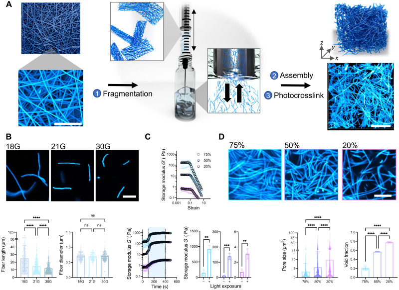
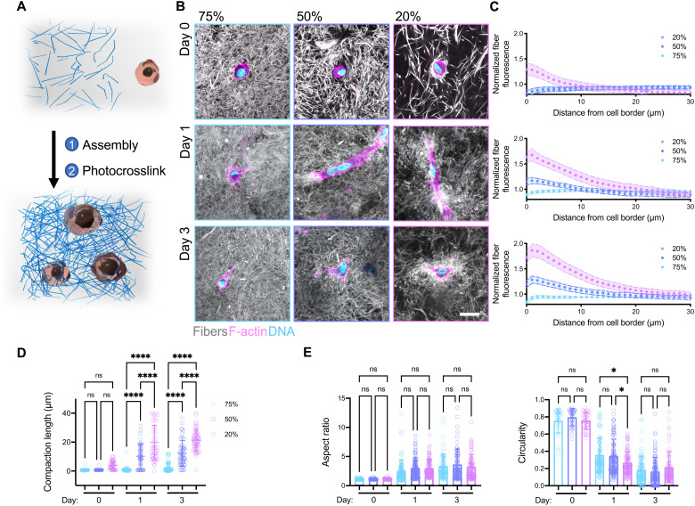
Synthetic fibrous materials were engineered to contract under cellular forces similar to natural extracellular matrix.
Overview
The claims and statistics of this paper haven’t been checked yet.
Run the check — free, no account needed. It reads the open-access text and checks the sources it cites.
Integrity signals
Registry
Cites
Statistics
Figures



Verification
Claims the paper makes
The claims and statistics of this paper haven’t been checked yet.
Run the check — free, no account needed. It reads the open-access text and checks the sources it cites.
Claims other papers make about this one
Each is checked against this paper’s own text: does it say what the citing paper says it does?
The cited source doesn’t support the claim as written.
In the citing paper
“Similar phenomena were observed in a study reported by Burdick et al., (2021) with higher fiber densities yielding higher strain [23].”
From Development of silk microfiber-reinforced bioink for muscle tissue engineering and in situ printing by a handheld 3D printer (2025) · cites this paper as [23]
Checked by a reasoning model — may be wrong.
Experimental — accuracy not yet measuredThe source backs part of the claim, not all of it — or backs it for a different population, condition or comparison.
In the citing paper
“Fiber-reinforced composites show enhanced mechanical strength and fiber-induced cell alignment in various cell studies[23, 26–30].”
From Development of silk microfiber-reinforced bioink for muscle tissue engineering and in situ printing by a handheld 3D printer (2025) · cites this paper as [23]
Experimental — accuracy not yet measuredThe source backs part of the claim, not all of it — or backs it for a different population, condition or comparison.
In the citing paper
“This finding is consistent with previous studies that have demonstrated similar outcomes, including fiber-oriented cell alignment [23, 27].”
From Development of silk microfiber-reinforced bioink for muscle tissue engineering and in situ printing by a handheld 3D printer (2025) · cites this paper as [23]
Experimental — accuracy not yet measuredThe source backs part of the claim, not all of it — or backs it for a different population, condition or comparison.
In the citing paper
“Literature suggests that higher fiber densities can impediment cell recruitment due to increased mechanical resistance, resulting in reduced cell spreading and increased cell circularity [23].”
From Development of silk microfiber-reinforced bioink for muscle tissue engineering and in situ printing by a handheld 3D printer (2025) · cites this paper as [23]
Experimental — accuracy not yet measuredThe cited source says what the claim attributes to it.
In the citing paper
“Fiber-reinforced hydrogel composites recently gained attention with their unique fibrous structure mimicking native tissue architecture [23–25].”
From Development of silk microfiber-reinforced bioink for muscle tissue engineering and in situ printing by a handheld 3D printer (2025) · cites this paper as [23]
Self-check
Percentages, sums and reported differences re-computed from the paper’s own numbers.
The self-check needs the paper’s full text.
Citations
Cites
Myofibroblasts and mechano-regulation of connective tissue remodelling
Tomasek, J. J., Gabbiani, G., Hinz, B., Chaponnier, C., & Brown, R. A. (2002) · Nature Reviews Molecular Cell Biology · doi:10.1038/nrm809 · Cited by 4,290 · cited 1×
“For example, cell contractility is responsible for many self-assembly or morphogenic processes in vivo, such as shaping tissues in embryonic development (8) and closing wounds during healing (9).”
BackgroundA practical guide to hydrogels for cell culture
Caliari, S. R., & Burdick, J. A. (2016) · Nature Methods · doi:10.1038/nmeth.3839 · Cited by 2,075
Self-citationNo evidence that carotenoid pigments boost either immune or antioxidant defenses in a songbird
Koch, R. E., Kavazis, A. N., Hasselquist, D., Hood, W. R., Zhang, Y., Toomey, M. B., & Hill, G. E. (2018) · Nature Communications · doi:10.1038/s41467-018-02974-x · Cited by 1,924
3D bioprinting of collagen to rebuild components of the human heart
Lee, A., Hudson, A. R., Shiwarski, D. J., Tashman, J. W., Hinton, T. J., Yerneni, S., Bliley, J. M., Campbell, P. G., & Feinberg, A. W. (2019) · Science · doi:10.1126/science.aav9051 · Cited by 1,782
Nonlinear elasticity in biological gels
Storm, C., Pastore, J. J., MacKintosh, F. C., Lubensky, T. C., & Janmey, P. A. (2005) · Nature · doi:10.1038/nature03521 · Cited by 1,703
Self-citationSLIM: Simultaneous Logic-in-Memory Computing Exploiting Bilayer Analog OxRAM Devices
Kingra, S. K., Parmar, V., Chang, C.-C., Hudec, B., Hou, T.-H., & Suri, M. (2020) · Scientific Reports · doi:10.1038/s41598-020-59121-0 · Cited by 1,439
Advanced maturation of human cardiac tissue grown from pluripotent stem cells
Ronaldson-Bouchard, K., Ma, S. P., Yeager, K., Chen, T., Song, L., Sirabella, D., Morikawa, K., Teles, D., Yazawa, M., & Vunjak-Novakovic, G. (2018) · Nature · doi:10.1038/s41586-018-0016-3 · Cited by 1,272
Yang, B.-J., Han, X.-X., Yin, L.-L., Xing, M.-Q., Xu, Z.-H., & Xue, H.-W. (2016) · Nature Communications · doi:10.1038/ncomms11388 · Cited by 1,202
Synthetic alternatives to Matrigel
Aisenbrey, E. A., & Murphy, W. L. (2020) · Nature Reviews Materials · doi:10.1038/s41578-020-0199-8 · Cited by 1,016 · cited 1×
“Furthermore, other ECM-derived matrices, such as Matrigel and decellularized ECM, are widely used in cell culture, in tissue engineering, and even as clinical therapies; however, their batch variability and animal sourcing are limiting (13).”
BackgroundBiomanufacturing of organ-specific tissues with high cellular density and embedded vascular channels
Skylar-Scott, M. A., Uzel, S. G. M., Nam, L. L., Ahrens, J. H., Truby, R. L., Damaraju, S., & Lewis, J. A. (2019) · Science Advances · doi:10.1126/sciadv.aaw2459 · Cited by 943
Cited by
Zhang, Y. S., Haghiashtiani, G., Hübscher, T., Kelly, D. J., Lee, J. M., Lutolf, M., McAlpine, M. C., Yeong, W. Y., Zenobi-Wong, M., & Malda, J. (2021) · Nature Reviews Methods Primers · doi:10.1038/s43586-021-00073-8 · Cited by 391
Engineered hydrogels for mechanobiology
Blache, U., Ford, E. M., Ha, B., Rijns, L., Chaudhuri, O., Dankers, P. Y. W., Kloxin, A. M., Snedeker, J. G., & Gentleman, E. (2022) · Nature Reviews Methods Primers · doi:10.1038/s43586-022-00179-7 · Cited by 227
Light-based vat-polymerization bioprinting
Levato, R., Dudaryeva, O., Garciamendez-Mijares, C. E., Kirkpatrick, B. E., Rizzo, R., Schimelman, J., Anseth, K. S., Chen, S., Zenobi-Wong, M., & Zhang, Y. S. (2023) · Nature Reviews Methods Primers · doi:10.1038/s43586-023-00231-0 · Cited by 176
Ding, A., Tang, F., & Alsberg, E. (2025) · Chemical Reviews · doi:10.1021/acs.chemrev.4c00070 · Cited by 165
Granular Hydrogels in Biofabrication: Recent Advances and Future Perspectives
Daly, A. C. (2023) · Advanced Healthcare Materials · doi:10.1002/adhm.202301388 · Cited by 145
Metadata
- Authors
- Davidson, M. D., Prendergast, M. E., Ban, E., Xu, K. L., Mickel, G., Mensah, P., Dhand, A., Janmey, P. A., Shenoy, V. B., & Burdick, J. A.
- Affiliations
- University of Pennsylvania (US)
- Corresponding author
- Burdick, Jason A.
- ORCID
- Venue
- Science Advances
- Published
- 2021-11-12
- Volume
- 7
- Issue
- 46
- Pages
- eabi8157-eabi8157
- ISSN
- 2375-2548 (ISSN-L)
- PMC
- PMC8580309
- OpenAlex
- W3212440101
- Cited by
- 101
- References
- 76
- Citations by year
- 2026: 15 · 2025: 23 · 2024: 25 · 2023: 22 · 2022: 11 · 2021: 4
- FWCI
- 4.92
- Type
- journal article
- Language
- en
- Open access
- gold
- doi.org
- Licence
- cc-by-nc
- Locations
- Science Advances (publishedVersion, cc-by-nc)
- bioRxiv (Cold Spring Harbor Laboratory) (acceptedVersion)
- PubMed Central (publishedVersion, cc-by-nc)
- PubMed (publishedVersion)
- Funders
- National Science Foundation (1720530, CMMI: 15‐48571, 1548571)
- University of Pennsylvania (1720530, DMR- 1720530)
- National Institutes of Health
- Materials Research Science and Engineering Center, Harvard University (DMR‐1720530, 1720530)
- National Institute of Diabetes and Digestive and Kidney Diseases (F32DK117568)
- National Institute of Arthritis and Musculoskeletal and Skin Diseases (R01AR056624)
- Division of Materials Research (DMR17-20530, 17-20530)
- Division of Civil, Mechanical and Manufacturing Innovation (CMMI 1548571, 15–48571, DMR-1720530)
- Topics
- Physical Sciences › Materials Science › Biomaterials › Hydrogels: synthesis, properties, applications
- Physical Sciences › Engineering › Biomedical Engineering › 3D Printing in Biomedical Research
- Life Sciences › Biochemistry, Genetics and Molecular Biology › Cell Biology › Cellular Mechanics and Interactions
- Keywords
- fibrous hydrogels, cell encapsulation, cell contraction, extracellular matrix mimicry, strain stiffening, fiber alignment, mesenchymal stem cells, photocrosslinking, self-healing hydrogels, shear thinning, extrusion-based bioprinting, photopatterning, microtissues, biofabrication, tissue engineering
About these checks
Registry, retraction, Cites and statistics results are computed, not judged, with one exception: a printed reference that no index lists is read once by a model before it counts as a citation error. Claims are extracted by a model and kept only when their quote appears verbatim in the text; each cited source is then checked, with a model’s help, for whether it says what the claim relies on. Those verdicts can be wrong — read the claim’s quote and the cited source before relying on one.
Statistics and arithmetic are recomputed, not judged.
Confidence bands are shown only for fields calibrated on our annotated test set. None is calibrated yet, so no band is shown.
What counts as a claim ›How we measure confidence ›How we check this ›
Are you an author, or do you see a problem with this page? Request a review
How to cite
Cite CoChat for the checks on this page, and the paper for its own findings.
- This page
- CoChat. (2026). Citation check: Programmable and contractile materials through cell encapsulation in fibrous hydrogel assemblies. https://app.cochat.ai/papers/10.1126/sciadv.abi8157
- The paper
- Davidson, M. D., Prendergast, M. E., Ban, E., Xu, K. L., Mickel, G., Mensah, P., Dhand, A., Janmey, P. A., Shenoy, V. B., & Burdick, J. A. (2021). Programmable and contractile materials through cell encapsulation in fibrous hydrogel assemblies. Science Advances. https://doi.org/10.1126/sciadv.abi8157
Data sources
Bibliographic data: Semantic Scholar, OpenAlex, Crossref.
Contains information from the Semantic Scholar Academic Graph Datasets, made available under the ODC Attribution License (ODC-BY 1.0).
Semantic Scholar’s data is described in: Kinney, R., et al. (2023). The Semantic Scholar Open Data Platform. arXiv:2301.10140.
