

The DOI matches its Crossref record; 104 of the 106 references it cites were found in a registry or an index, and 2 are not checked yet; 21 of the 39 sources its claims cite were checked for support.
Scaffolding protein functional sites using deep learning
Open accessPublished version of a preprint
Abstract
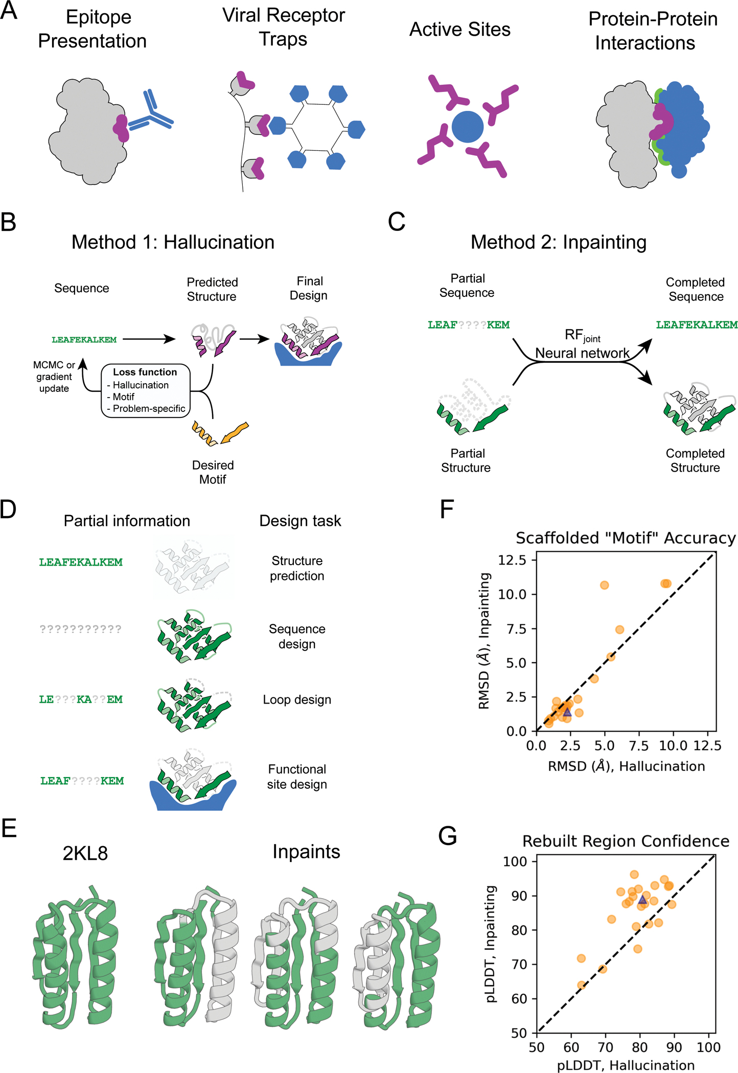
The binding and catalytic functions of proteins are generally mediated by a small number of functional residues held in place by the overall protein structure. Here, we describe deep learning approaches for scaffolding such functional sites without needing to prespecify the fold or secondary structure of the scaffold. The first approach, “constrained hallucination,” optimizes sequences such that their predicted structures contain the desired functional site. The second approach, “inpainting,” starts from the functional site and fills in additional sequence and structure to create a viable protein scaffold in a single forward pass through a specifically trained RoseTTAFold network. We use these two methods to design candidate immunogens, receptor traps, metalloproteins, enzymes, and protein-binding proteins and validate the designs using a combination of in silico and experimental tests.
Verified 2026-10-05 · 2 references not checked yet
Overview
CoChat Summary
TL;DR
The paper introduces two deep learning methods, constrained hallucination and inpainting, for scaffolding functional sites into de novo protein structures without pre-specifying scaffold fold or secondary structure. It validates designs in silico and experimentally across immunogens, receptor traps, metalloproteins, enzymes, and protein binders.
Key findings
- AlphaFold recapitulated designed structures with overall and motif RMSD typically <2 Å and <1 Å, respectively, and pLDDT >80; inpainting was much faster (1–10 s/design on an NVIDIA RTX2080 GPU) than hallucination (5–20 min/design).
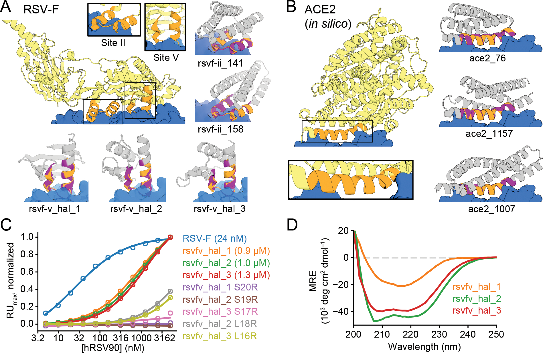
- For RSV-F site V, 37 hallucinated scaffolds were expressed in E. coli and three bound hRSV90 with Kd’s of 0.9–1.3 μM; four inpainted designs bound by yeast display but were poorly expressed in E. coli.
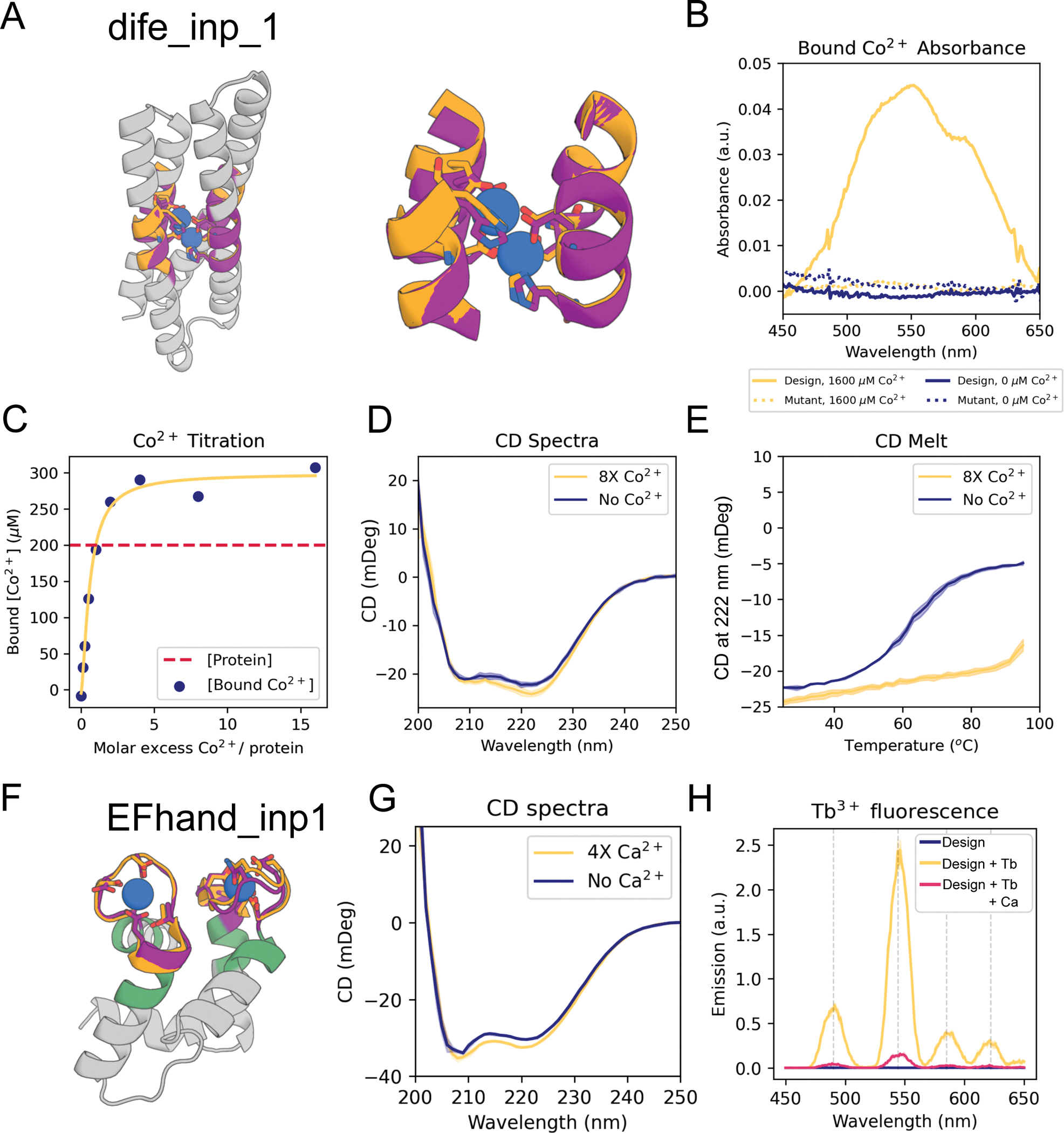
- For the E. coli bacterioferritin di-iron site, 96 inpainted designs were tested: 76 had soluble expression, at least 8 showed a Co2+-binding spectroscopic shift, and 3 had CD spectra consistent with the designed fold and were stabilized by metal binding; mutation of metal-binding residues abolished binding.
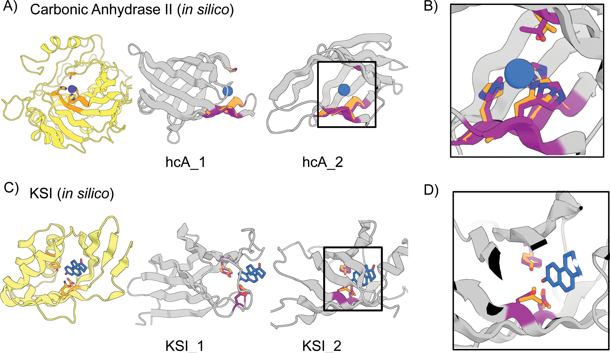
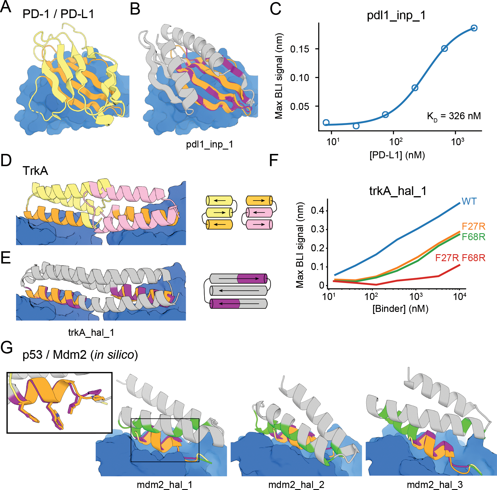
- For PD-L1, of 31 designs tested, pdl1_inp_1 bound PD-L1 with KD = 326 nM, compared with HAC PD-1 KD = 110 pM and WT PD-1 KD = 3.9 μM; carbonic anhydrase II active-site designs were <100 residues versus the 261-residue native protein.
Methods
- Constrained hallucination optimized sequences with a composite loss combining a hallucination loss and a motif reconstruction loss using trRosetta and later RosettaFold; inpainting used RFjoint, trained on masked/inpainting sequence and structure recovery, to complete missing sequence and structure in a single forward pass.
- In silico validation used AlphaFold; experimental validation for selected designs included E. coli expression, yeast display, binding assays (Kd, BLI), circular dichroism, metal/terbium fluorescence, and mutagenesis.
- Functional motifs were derived from native proteins including RSV-F sites II and V, hACE2, bacterioferritin di-iron, EF-hand, carbonic anhydrase II, Δ5-3-ketosteroid isomerase, PD-1/PD-L1, p53/Mdm2, and TrkA-NGF.
Limitations
- Stated in the paper: AlphaFold has limited ability to predict protein stability or mutational effects; hallucination is compute-intensive; inpainting usually requires larger input motifs and underperforms hallucination when the missing region is large; some inpainted RSV-F site V designs were poorly expressed in E. coli; di-iron hallucination designs were not tested due to buried polar residues; and side-chain placement for ketosteroid isomerase was difficult, requiring a two-stage approach.
- Evident from the text: many designs were validated only in silico (e.g., receptor traps, enzyme active sites, some Mdm2 binders), and experimental hit rates were often low (e.g., 3/37 RSV-F site V binders, 3/96 di-iron designs with CD/stabilization, 1/31 PD-L1 binders); inpainting also generated less structural diversity than hallucination.
Model-written summary. Check it against the paper before relying on it.
Integrity signals
Registry
Cites
Statistics
Figures
Verification
Claims the paper makes
9 Valid · 0 Not valid · 12 Not checked · 71 Own results and interpretations
Every source the paper cites for this claim was checked and supports it.
In the paper
“trRosetta can also be used to design sequences that fold into a target backbone structure by carrying out sequence optimization using a structure recapitulation loss function that rewards similarity of the predicted structure to the target structure (13).”
§ Scaffolding protein functional sites using deep learning > Abstract
What the sources say
- Supportedcites [13] Norn C, Wicky BIM, Juergens D, Liu S, Kim D, Tischer D, Koepnick B, Anishchenko I, Players F, Baker D, Ovchinnikov S, Protein sequence design by conformational landscape optimization. Proc. Natl. Acad. Sci. 118 (2021), doi: 10.1073/pnas.2017228118. PMC7980421 33712545 source
The Results and Discussions section, “Sequence Design by Gradient Backpropagation” (page not provided), describes optimizing amino acid sequences with a loss function based on the probabilities of the target structure’s observed interresidue distances and orientations. This supports the claim’s description of a structure-recapitulation loss that rewards agreement with the target backbone. (LLM, conf=0.91)
plainstates a cause- Asserted: passes — The authors put it forward as true.
- Checkable: passes — It can be checked against evidence.
- Load-bearing: passes — The paper’s conclusions depend on it.
- Based: passes — The paper gives a reason why it holds.
- Supported
The cited sources support only part of this claim — read each source’s verdict below.
In the paper
“We previously demonstrated that the trRosetta structure-prediction neural network (11) can be used to generate new proteins by maximizing the trRosetta output probability that a sequence folds to some (unspecified) three dimensional structure during Monte Carlo sampling in sequence space (12).”
§ Scaffolding protein functional sites using deep learning > Abstract
What the sources say
- Not checked yetcites [11] Yang J, Anishchenko I, Park H, Peng Z, Ovchinnikov S, Baker D, Improved protein structure prediction using predicted interresidue orientations. Proc. Natl. Acad. Sci. (2020), doi: 10.1073/pnas.1914677117. PMC6983395 31896580 source
- Partly supportedcites [12] Anishchenko I, Pellock SJ, Chidyausiku TM, Ramelot TA, Ovchinnikov S, Hao J, Bafna K, Norn C, Kang A, Bera AK, DiMaio F, Carter L, Chow CM, Montelione GT, Baker D, De novo protein design by deep network hallucination. Nature. 600, 547– 552 (2021). 34853475 PMC9293396 sourceExperimental — accuracy not yet measured
Split into 2 atoms — 1 confirmed, 1 partially supported, 0 disputed, 0 misattributed, 0 unverifiable. Expand the claim to see per-atom verdicts.
boostedstates a cause- Asserted: passes — The authors put it forward as true.
- Checkable: passes — It can be checked against evidence.
- Load-bearing: passes — The paper’s conclusions depend on it.
- Based: passes — The paper gives a reason why it holds.
The cited sources support only part of this claim — read each source’s verdict below.
In the paper
“Previous methods can scaffold functional sites made up of one or two contiguous chain segments (6–10), but with the exception of helical bundles (8) these do not extend readily to more complex sites composed of three or more chain segments, and the generated backbones are not guaranteed to be designable (encodable by some amino acid sequence).”
§ Scaffolding protein functional sites using deep learning > Abstract
What the sources say
- Supportedcites [6] Procko E, Berguig GY, Shen BW, Song Y, Frayo S, Convertine AJ, Margineantu D, Booth G, Correia BE, Cheng Y, Schief WR, Hockenbery DM, Press OW, Stoddard BL, Stayton PS, Baker D, A Computationally Designed Inhibitor of an Epstein-Barr Viral Bcl-2 Protein Induces Apoptosis in Infected Cells. Cell. 157, 1644– 1656 (2014). 24949974 PMC4079535 source
The citation to this paper is attached to the helical-bundle exception, rather than to the general claim about the limits of previous methods. In the Results sections on grafting and de novo design (page not provided), the paper describes using 3- and 4-helix bundles to build a binding interface with contacts beyond the grafted BH3 helix. (LLM, conf=0.86)
- Could not verifycites [7] Correia BE, Bates JT, Loomis RJ, Baneyx G, Carrico C, Jardine JG, Rupert P, Correnti C, Kalyuzhniy O, Vittal V, Connell MJ, Stevens E, Schroeter A, Chen M, MacPherson S, Serra AM, Adachi Y, Holmes MA, Li Y, Klevit RE, Graham BS, Wyatt RT, Baker D, Strong RK, Crowe JE, Johnson PR, Schief WR, Proof of principle for epitope-focused vaccine design. Nature. 507, 201– 206 (2014). 24499818 PMC4260937 sourceExperimental — accuracy not yet measured
The Design of Epitope-Scaffolds section describes a helix-turn-helix epitope and a three-helix-bundle template, while Computational Method describes the paper’s Fold from Loops approach. Neither passage assesses whether previous methods extend to functional sites made of three or more chain segments; searches for that limit and the helical-bundle exception found no supporting passage in our copy. (LLM, conf=0.72)
- Supportedcites [8] Silva D-A, Yu S, Ulge UY, Spangler JB, Jude KM, Labão-Almeida C, Ali LR, Quijano-Rubio A, Ruterbusch M, Leung I, Biary T, Crowley SJ, Marcos E, Walkey CD, Weitzner BD, Pardo-Avila F, Castellanos J, Carter L, Stewart L, Riddell SR, Pepper M, Bernardes GJL, Dougan M, Garcia KC, Baker D, De novo design of potent and selective mimics of IL-2 and IL-15. Nature. 565, 186– 191 (2019). 30626941 PMC6521699 source
The citation is attached to the helical-bundle exception. In “Computational design of IL-2/IL-15 mimics that bind and activate IL-2Rβγc” (page unavailable), the paper describes building a connected protein around the three helices H1, H3 and H4 that form the receptor-binding interface, supporting that exception. (LLM, conf=0.91)
- Partly supportedcites [9] Sesterhenn F, Yang C, Bonet J, Cramer JT, Wen X, Wang Y, Chiang C-I, Abriata LA, Kucharska I, Castoro G, Vollers SS, Galloux M, Dheilly E, Rosset S, Corthésy P, Georgeon S, Villard M, Richard C-A, Descamps D, Delgado T, Oricchio E, Rameix-Welti M-A, Más V, Ervin S, Eléouët J-F, Riffault S, Bates JT, Julien J-P, Li Y, Jardetzky T, Krey T, Correia BE, De novo protein design enables the precise induction of RSV-neutralizing antibodies. Science. 368 (2020), doi: 10.1126/science.aay5051. PMC7391827 32409444 sourceExperimental — accuracy not yet measured
The Introduction (page not provided) says earlier grafting work largely used simple motifs such as linear helical segments and identifies irregular, multi-segment functional sites as a challenge. The Discussion & Conclusions (page not provided) also describes limitations of template-based design. Neither passage establishes the specific three-or-more-segment threshold or identifies helical bundles as an exception. (LLM, conf=0.82)
- Not checked yetcites [10] Yang C, Sesterhenn F, Bonet J, van Aalen EA, Scheller L, Abriata LA, Cramer JT, Wen X, Rosset S, Georgeon S, Jardetzky T, Krey T, Fussenegger M, Merkx M, Correia BE, Bottom-up de novo design of functional proteins with complex structural features. Nat. Chem. Biol, 1– 9 (2021). 33398169 source
plain- Asserted: passes — The authors put it forward as true.
- Checkable: passes — It can be checked against evidence.
- Load-bearing: passes — The paper’s conclusions depend on it.
- Based: passes — The paper gives a reason why it holds.
- Supported
The cited sources support only part of this claim — read each source’s verdict below.
In the paper
“Previous methods can scaffold functional sites made up of one or two contiguous chain segments (6–10), but with the exception of helical bundles (8) these do not extend readily to more complex sites composed of three or more chain segments, and the generated backbones are not guaranteed to be designable (encodable by some amino acid sequence).”
§ Scaffolding protein functional sites using deep learning > Abstract
What the sources say
- Partly supportedcites [6] Procko E, Berguig GY, Shen BW, Song Y, Frayo S, Convertine AJ, Margineantu D, Booth G, Correia BE, Cheng Y, Schief WR, Hockenbery DM, Press OW, Stoddard BL, Stayton PS, Baker D, A Computationally Designed Inhibitor of an Epstein-Barr Viral Bcl-2 Protein Induces Apoptosis in Infected Cells. Cell. 157, 1644– 1656 (2014). 24949974 PMC4079535 sourceExperimental — accuracy not yet measured
The Results section, under “BHRF1-binding proteins created by grafting Bim-BH3 side chains to a helical scaffold” (page unavailable), describes grafting the interacting residues of a single BH3 helix onto protein scaffolds. This supports the one-contiguous-segment example, but our copy does not establish that previous methods scaffold functional sites made up of two contiguous chain segments. (LLM, conf=0.82)
- Supportedcites [7] Correia BE, Bates JT, Loomis RJ, Baneyx G, Carrico C, Jardine JG, Rupert P, Correnti C, Kalyuzhniy O, Vittal V, Connell MJ, Stevens E, Schroeter A, Chen M, MacPherson S, Serra AM, Adachi Y, Holmes MA, Li Y, Klevit RE, Graham BS, Wyatt RT, Baker D, Strong RK, Crowe JE, Johnson PR, Schief WR, Proof of principle for epitope-focused vaccine design. Nature. 507, 201– 206 (2014). 24499818 PMC4260937 source
The Abstract (page not available) says that previously developed side-chain and backbone grafting methods transplanted continuous or discontinuous epitopes onto scaffold proteins. This supports the attributed point that previous methods could scaffold functional sites comprising contiguous chain segments; the claim does not attribute a limitation to exactly two segments to this paper. (LLM, conf=0.86)
- Could not verifycites [8] Silva D-A, Yu S, Ulge UY, Spangler JB, Jude KM, Labão-Almeida C, Ali LR, Quijano-Rubio A, Ruterbusch M, Leung I, Biary T, Crowley SJ, Marcos E, Walkey CD, Weitzner BD, Pardo-Avila F, Castellanos J, Carter L, Stewart L, Riddell SR, Pepper M, Bernardes GJL, Dougan M, Garcia KC, Baker D, De novo design of potent and selective mimics of IL-2 and IL-15. Nature. 565, 186– 191 (2019). 30626941 PMC6521699 sourceExperimental — accuracy not yet measured
The computational-design section describes this paper’s method for scaffolding an IL-2 binding interface formed by three helices (H1, H3 and H4). It does not address whether previous methods could scaffold functional sites made up of one or two contiguous chain segments; searches for prior scaffolding methods found no passage establishing that point. The paper does, however, fit the helical-bundle exception separately identified in the full citing sentence. (LLM, conf=0.76)
- Supportedcites [9] Sesterhenn F, Yang C, Bonet J, Cramer JT, Wen X, Wang Y, Chiang C-I, Abriata LA, Kucharska I, Castoro G, Vollers SS, Galloux M, Dheilly E, Rosset S, Corthésy P, Georgeon S, Villard M, Richard C-A, Descamps D, Delgado T, Oricchio E, Rameix-Welti M-A, Más V, Ervin S, Eléouët J-F, Riffault S, Bates JT, Julien J-P, Li Y, Jardetzky T, Krey T, Correia BE, De novo protein design enables the precise induction of RSV-neutralizing antibodies. Science. 368 (2020), doi: 10.1126/science.aay5051. PMC7391827 32409444 source
The Introduction (page unavailable) describes earlier grafting methods using linear helical segments. The Results section, ‘De novo design of immunogens presenting structurally complex epitopes’ (page unavailable), adds that one earlier study grafted a discontinuous HIV epitope into a scaffold. Together, these support the claim that previous methods could scaffold sites comprising a single segment or more than one segment. (LLM, conf=0.85)
- Not checked yetcites [10] Yang C, Sesterhenn F, Bonet J, van Aalen EA, Scheller L, Abriata LA, Cramer JT, Wen X, Rosset S, Georgeon S, Jardetzky T, Krey T, Fussenegger M, Merkx M, Correia BE, Bottom-up de novo design of functional proteins with complex structural features. Nat. Chem. Biol, 1– 9 (2021). 33398169 source
plain- Asserted: passes — The authors put it forward as true.
- Checkable: passes — It can be checked against evidence.
- Load-bearing: passes — The paper’s conclusions depend on it.
- Based: passes — The paper gives a reason why it holds.
- Partly supported
The cited sources support only part of this claim — read each source’s verdict below.
In the paper
“Previous methods can scaffold functional sites made up of one or two contiguous chain segments (6–10), but with the exception of helical bundles (8) these do not extend readily to more complex sites composed of three or more chain segments, and the generated backbones are not guaranteed to be designable (encodable by some amino acid sequence).”
§ Scaffolding protein functional sites using deep learning > Abstract
What the sources say
- Partly supportedcites [6] Procko E, Berguig GY, Shen BW, Song Y, Frayo S, Convertine AJ, Margineantu D, Booth G, Correia BE, Cheng Y, Schief WR, Hockenbery DM, Press OW, Stoddard BL, Stayton PS, Baker D, A Computationally Designed Inhibitor of an Epstein-Barr Viral Bcl-2 Protein Induces Apoptosis in Infected Cells. Cell. 157, 1644– 1656 (2014). 24949974 PMC4079535 sourceExperimental — accuracy not yet measured
The Results section, under “In silico folding probability correlates with binding activity” (page unavailable), says that functional protein design has a low success rate and that inactive designs may fold improperly or have unstable designed states. This supports the general point that a generated design is not guaranteed to work, but it does not establish the more specific claim that a generated backbone cannot be encoded by any amino acid sequence. (LLM, conf=0.79)
- Could not verifycites [7] Correia BE, Bates JT, Loomis RJ, Baneyx G, Carrico C, Jardine JG, Rupert P, Correnti C, Kalyuzhniy O, Vittal V, Connell MJ, Stevens E, Schroeter A, Chen M, MacPherson S, Serra AM, Adachi Y, Holmes MA, Li Y, Klevit RE, Graham BS, Wyatt RT, Baker D, Strong RK, Crowe JE, Johnson PR, Schief WR, Proof of principle for epitope-focused vaccine design. Nature. 507, 201– 206 (2014). 24499818 PMC4260937 sourceExperimental — accuracy not yet measured
The Abstract (page unavailable) says the earlier grafting methods used scaffold proteins of known structure, but does not say whether their generated backbones were guaranteed to be designable. The Computational Method and Methods > Fold From Loops sections describe sequence design and filtering for the new method; a search for designability of backbones from previous methods found no passage addressing that point. (LLM, conf=0.72)
- Could not verifycites [8] Silva D-A, Yu S, Ulge UY, Spangler JB, Jude KM, Labão-Almeida C, Ali LR, Quijano-Rubio A, Ruterbusch M, Leung I, Biary T, Crowley SJ, Marcos E, Walkey CD, Weitzner BD, Pardo-Avila F, Castellanos J, Carter L, Stewart L, Riddell SR, Pepper M, Bernardes GJL, Dougan M, Garcia KC, Baker D, De novo design of potent and selective mimics of IL-2 and IL-15. Nature. 565, 186– 191 (2019). 30626941 PMC6521699 sourceExperimental — accuracy not yet measured
The Methods section (page unavailable) describes how this paper generated backbones and then performed sequence design and filtering. Searches for prior scaffolding methods and backbone designability found no passage saying that backbones generated by previous methods are not guaranteed to be designable. (LLM, conf=0.72)
- Could not verifycites [9] Sesterhenn F, Yang C, Bonet J, Cramer JT, Wen X, Wang Y, Chiang C-I, Abriata LA, Kucharska I, Castoro G, Vollers SS, Galloux M, Dheilly E, Rosset S, Corthésy P, Georgeon S, Villard M, Richard C-A, Descamps D, Delgado T, Oricchio E, Rameix-Welti M-A, Más V, Ervin S, Eléouët J-F, Riffault S, Bates JT, Julien J-P, Li Y, Jardetzky T, Krey T, Correia BE, De novo protein design enables the precise induction of RSV-neutralizing antibodies. Science. 368 (2020), doi: 10.1126/science.aay5051. PMC7391827 32409444 sourceExperimental — accuracy not yet measured
The Introduction and Discussion & Conclusions describe limitations of earlier scaffolding approaches, including destabilization and the need for extensive optimization. Neither passage says whether the generated backbones are guaranteed to be encodable by an amino acid sequence; a search for backbone designability found no such statement in our copy. (LLM, conf=0.72)
- Not checked yetcites [10] Yang C, Sesterhenn F, Bonet J, van Aalen EA, Scheller L, Abriata LA, Cramer JT, Wen X, Rosset S, Georgeon S, Jardetzky T, Krey T, Fussenegger M, Merkx M, Correia BE, Bottom-up de novo design of functional proteins with complex structural features. Nat. Chem. Biol, 1– 9 (2021). 33398169 source
plain- Asserted: passes — The authors put it forward as true.
- Checkable: passes — It can be checked against evidence.
- Load-bearing: passes — The paper’s conclusions depend on it.
- Based: passes — The paper gives a reason why it holds.
- Partly supported
59 more claims · 3 with their cited sources checked
Create a free account to see all 92 claims and to verify the 16 references still open.
- Probably not a substantial claim. 5
- It may state two separate claims as one. 23
Claims other papers make about this one
Each is checked against this paper’s own text: does it say what the citing paper says it does?
The source backs part of the claim, not all of it — or backs it for a different population, condition or comparison.
In the citing paper
“Experimental characterization showed that the method can scaffold a wide range of protein functional motifs with atomic accuracy19”
From De novo design of protein structure and function with RFdiffusion (2023) · cites this paper as [4]
Experimental — accuracy not yet measuredThe source backs part of the claim, not all of it — or backs it for a different population, condition or comparison.
In the citing paper
“but the approach fails on minimalist site descriptions that do not sufficiently constrain the overall fold and, because it is deterministic, can produce only a limited diversity of designs for a given problem.”
From De novo design of protein structure and function with RFdiffusion (2023) · cites this paper as [4]
Experimental — accuracy not yet measuredThe source backs part of the claim, not all of it — or backs it for a different population, condition or comparison.
In the citing paper
“Experimental characterization showed that the method can scaffold a wide range of protein functional motifs with atomic accuracy19”
From De novo design of protein structure and function with RFdiffusion (2023) · cites this paper as [19]
Experimental — accuracy not yet measuredThe source backs part of the claim, not all of it — or backs it for a different population, condition or comparison.
In the citing paper
“This measure of in silico success has been found to correlate with experimental success4,7,26 and is significantly more stringent than template modelling (TM)-score-based metrics used elsewhere5,16,27–29”
From De novo design of protein structure and function with RFdiffusion (2023) · cites this paper as [4]
Experimental — accuracy not yet measuredThe check wasn’t confident either way, so it gives no verdict — never disputed.
In the citing paper
“we established an in silico benchmark test (Supplementary Table 9) comprising 25 motif-scaffolding design problems addressed in six recent publications encompassing several design methodologies4,5,29,38–40”
From De novo design of protein structure and function with RFdiffusion (2023) · cites this paper as [4]
Experimental — accuracy not yet measured
Citations
Cites
Adam: A Method for Stochastic Optimization
Kingma, D. P., & Ba, J. (2014) · arXiv · doi:10.48550/arxiv.1412.6980 · Cited by 98,946
Basic local alignment search tool
Altschul, S. F., Gish, W., Miller, W., Myers, E. W., & Lipman, D. J. (1990) · Journal of Molecular Biology · doi:10.1006/jmbi.1990.9999 · Cited by 98,669
BERT: Pre-training of Deep Bidirectional Transformers for Language Understanding
Devlin, J., Chang, M.-W., Lee, K., & Toutanova, K. (2018) · arXiv · doi:10.48550/arxiv.1810.04805 · Cited by 73,173
BERT: Pre-training of Deep Bidirectional Transformers for Language Understanding
Devlin, J., Chang, M.-W., Lee, K., & Toutanova, K. (2019) · Proceedings of the 2019 Conference of the North American Chapter of the Association for Computational Linguistics: Human Language Technologies, Volume 1 (Long and Short Papers) · doi:10.18653/v1/n19-1423 · Cited by 54,325
Highly accurate protein structure prediction with AlphaFold
Jumper, J., Evans, R., Pritzel, A., Green, T., Figurnov, M., Ronneberger, O., Tunyasuvunakool, K., Bates, R., Žídek, A., Potapenko, A., et al. (2021) · Nature · doi:10.1038/s41586-021-03819-2 · Cited by 43,764
Berman, H. M. (2000) · Nucleic Acids Research · doi:10.1093/nar/28.1.235 · Cited by 40,196
Comparison of multiple Amber force fields and development of improved protein backbone parameters
Hornak, V., Abel, R., Okur, A., Strockbine, B., Roitberg, A., & Simmerling, C. (2006) · Proteins: Structure, Function, and Bioinformatics · doi:10.1002/prot.21123 · Cited by 6,914
Protein production by auto-induction in high-density shaking cultures
Studier, F. W. (2005) · Protein Expression and Purification · doi:10.1016/j.pep.2005.01.016 · Cited by 5,931
Accurate prediction of protein structures and interactions using a 3-track network
Baek, M., DiMaio, F., Anishchenko, I., Dauparas, J., Ovchinnikov, S., Lee, G. R., Wang, J., Cong, Q., Kinch, L. N., Schaeffer, R. D., et al. (2021) · doi:10.1101/2021.06.14.448402 · Cited by 5,549
Self-citationMMseqs2 enables sensitive protein sequence searching for the analysis of massive data sets
Steinegger, M., & Söding, J. (2017) · Nature Biotechnology · doi:10.1038/nbt.3988 · Cited by 4,840
Research works printed but not linked: 1
- Z. Lin T. Sercu Y. LeCun A. Rives “Deep generative models create new and diverse protein structures ” 35th Conference on Neural Information Processing Systems (NeurIPS 2021) 6 to 14 December 2021.
Cited by
Evolutionary-scale prediction of atomic level protein structure with a language model
Lin, Z., Akin, H., Rao, R., Hie, B., Zhu, Z., Lu, W., Smetanin, N., Verkuil, R., Kabeli, O., Shmueli, Y., et al. (2022) · doi:10.1101/2022.07.20.500902 · Cited by 5,990
Watson, J. L., Juergens, D., Bennett, N. R., Trippe, B. L., Yim, J., Eisenach, H. E., Ahern, W., Borst, A. J., Ragotte, R. J., Milles, L. F., et al. (2022) · doi:10.1101/2022.12.09.519842 · Cited by 2,369
“generative modeling of images when predicting the noise added in the forward process (rather than ), we reasoned that by predicting we could better leverage the inductive biases of x 0 x 0 RoseTTAFold pre-trained for structure prediction to produce realistic structures (as in [4]).”
Self-citationDe novo design of protein structure and function with RFdiffusion
Watson, J. L., Juergens, D., Bennett, N. R., Trippe, B. L., Yim, J., Eisenach, H. E., Ahern, W., Borst, A. J., Ragotte, R. J., Milles, L. F., et al. (2023) · Nature · doi:10.1038/s41586-023-06415-8 · Cited by 2,325
“From left to right: (1) symmetric design model, (2) AF2 prediction of design following sequence design with ProteinMPNN, (3) 2D class averages showing both top and side views (scale bar, 60 Å for all class averages) and (4) 3D reconstructions from class averages with the design model fit into the density map.”
Self-citationGeneralized biomolecular modeling and design with RoseTTAFold All-Atom
Krishna, R., Wang, J., Ahern, W., Sturmfels, P., Venkatesh, P., Kalvet, I., Lee, G. R., Morey-Burrows, F. S., Anishchenko, I., Humphreys, I. R., et al. (2024) · Science · doi:10.1126/science.adl2528 · Cited by 978
Self-citationFrom nature to industry: Harnessing enzymes for biocatalysis
Buller, R., Lutz, S., Kazlauskas, R. J., Snajdrova, R., Moore, J. C., & Bornscheuer, U. T. (2023) · Science · doi:10.1126/science.adh8615 · Cited by 706
Metadata
- Authors
- Wang, J., Lisanza, S., Juergens, D., Tischer, D., Watson, J. L., Castro, K. M., Ragotte, R., Saragovi, A., Milles, L. F., Baek, M., Anishchenko, I., Yang, W., Hicks, D. R., Expòsit, M., Schlichthaerle, T., Chun, J.-H., Dauparas, J., Bennett, N., Wicky, B. I. M., Muenks, A., DiMaio, F., Correia, B., Ovchinnikov, S., & Baker, D.
- Affiliations
- University of Washington (US)
- École Polytechnique Fédérale de Lausanne (CH)
- Harvard University (US)
- Howard Hughes Medical Institute (US)
- Corresponding author
- Wang, Jue
- Lisanza, Sidney
- Juergens, David
- Tischer, Doug
- Watson, Joseph L.
- Ovchinnikov, Sergey
- Baker, David
- ORCID
- Wang, Jue
- Lisanza, Sidney
- Juergens, David
- Tischer, Doug
- Watson, Joseph L.
- Castro, Karla M.
- Ragotte, Robert
- Saragovi, Amijai
- Milles, Lukas F.
- Baek, Minkyung
- Anishchenko, Ivan
- Yang, Wei
- Hicks, Derrick R.
- Expòsit, Marc
- Schlichthaerle, Thomas
- Chun, Jung-Ho
- Dauparas, Justas
- Bennett, Nathaniel
- Wicky, Basile I. M.
- Muenks, Andrew
- DiMaio, Frank
- Correia, Bruno
- Ovchinnikov, Sergey
- Baker, David
- Venue
- Science
- Published
- 2022-07-22
- Volume
- 377
- Issue
- 6604
- Pages
- 387-394
- ISSN
- 0036-8075 (ISSN-L)
- 1095-9203
- PMC
- PMC9621694
- OpenAlex
- W4286488966
- Cited by
- 487
- References
- 94
- Citations by year
- 2026: 84 · 2025: 107 · 2024: 124 · 2023: 121 · 2022: 49
- FWCI
- 32.74
- Type
- journal article
- Language
- en
- Open access
- green
- doi.org
- Licence
- cc-by-nc-nd
- Locations
- Science (publishedVersion)
- bioRxiv (Cold Spring Harbor Laboratory) (acceptedVersion, cc-by-nc-nd)
- PubMed (publishedVersion)
- PubMed Central (acceptedVersion, cc-by)
- Infoscience (Ecole Polytechnique Fédérale de Lausanne) (submittedVersion, cc-by-nc-nd)
- MPG.PuRe (Max Planck Society) (submittedVersion)
- Seoul National University Open Repository (Seoul National University) (submittedVersion)
- Funders
- Howard Hughes Medical Institute
- Washington Research Foundation
- Amgen
- Open Philanthropy Project
- Fundación Bancaria Caixa d'Estalvis i Pensions de Barcelona
- National Institutes of Health (DP5OD026389, HHSN272201700059C)
- National Institute on Aging (U19AG065156)
- National Cancer Institute (R01CA240339)
- National Institute of Allergy and Infectious Diseases (HHSN272201700059C)
- Topics
- Life Sciences › Biochemistry, Genetics and Molecular Biology › Molecular Biology › Protein Structure and Dynamics
- Life Sciences › Biochemistry, Genetics and Molecular Biology › Molecular Biology › Machine Learning in Bioinformatics
- Life Sciences › Pharmacology, Toxicology and Pharmaceutics › Drug Discovery › Computational Drug Discovery Methods
- Keywords
- protein scaffolding, protein design, deep learning, inpainting, RoseTTAFold, immunogen, metalloproteins, enzyme design, protein structure prediction
- MeSH terms
- Deep Learning, Binding Sites, Catalysis, Protein Binding, Proteins, Protein Engineering, Protein Structure, Secondary, Protein Folding
About these checks
Registry, retraction, Cites and statistics results are computed, not judged, with one exception: a printed reference that no index lists is read once by a model before it counts as a citation error. Claims are extracted by a model and kept only when their quote appears verbatim in the text; each cited source is then checked, with a model’s help, for whether it says what the claim relies on. Those verdicts can be wrong — read the claim’s quote and the cited source before relying on one.
The claim verdicts this report counts are experimental: their accuracy is not yet measured.
A language model drew the claims from the paper on 2026-10-05, under version 3 of the claim definition (model deepseek-v4.1-flash, prompt extract-v3).
A second model labelled each claim on 2026-10-05 (model jev-1.13-20260917, definition revision gold-guideline-v0.2).
Cited sources are checked with a language model’s help (gpt-6-sol).
Statistics and arithmetic are recomputed, not judged.
Confidence bands are shown only for fields calibrated on our annotated test set. None is calibrated yet, so no band is shown.
What counts as a claim ›How we measure confidence ›How we check this ›
Are you an author, or do you see a problem with this page? Request a review
How to cite
Cite CoChat for the checks on this page, and the paper for its own findings.
- This page
- CoChat. (2026). Citation check: Scaffolding protein functional sites using deep learning. https://app.cochat.ai/papers/10.1126/science.abn2100
- The paper
- Wang, J., Lisanza, S., Juergens, D., Tischer, D., Watson, J. L., Castro, K. M., Ragotte, R., Saragovi, A., Milles, L. F., Baek, M., Anishchenko, I., Yang, W., Hicks, D. R., Expòsit, M., Schlichthaerle, T., Chun, J.-H., Dauparas, J., Bennett, N., Wicky, B. I. M., . . . Baker, D. (2022). Scaffolding protein functional sites using deep learning. Science. https://doi.org/10.1126/science.abn2100
Data sources
Bibliographic data: Semantic Scholar, OpenAlex, Crossref.
Contains information from the Semantic Scholar Academic Graph Datasets, made available under the ODC Attribution License (ODC-BY 1.0).
Semantic Scholar’s data is described in: Kinney, R., et al. (2023). The Semantic Scholar Open Data Platform. arXiv:2301.10140.
