

The DOI matches its Crossref record; 53 of the 53 references it cites were found in a registry or an index.
The Emerging Pathogen Candida auris: Growth Phenotype, Virulence Factors, Activity of Antifungals, and Effect of SCY-078, a Novel Glucan Synthesis Inhibitor, on Growth Morphology and Biofilm Formation
Open accessAbstract
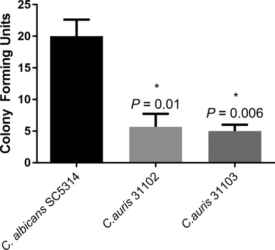
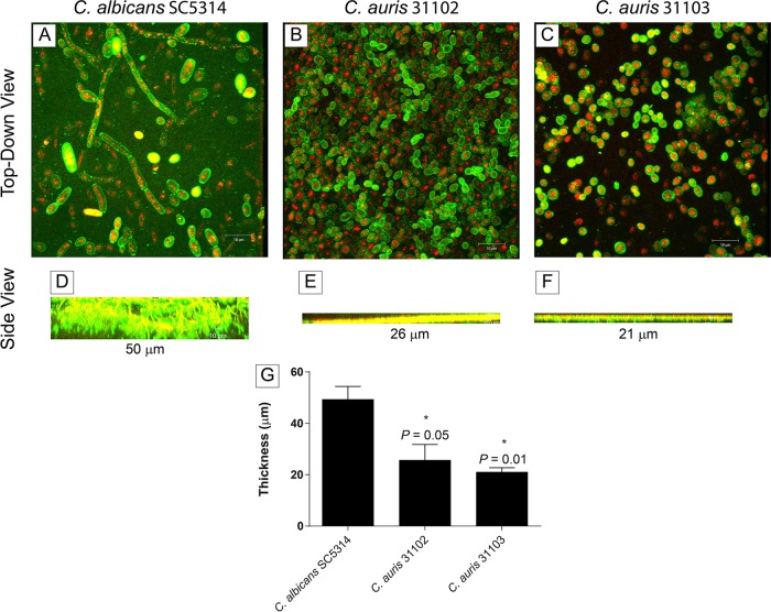
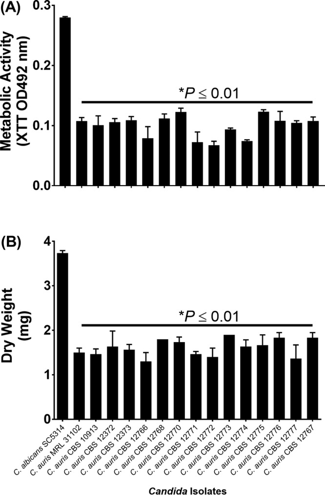
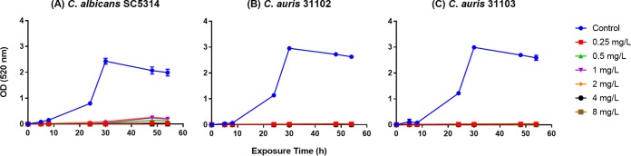
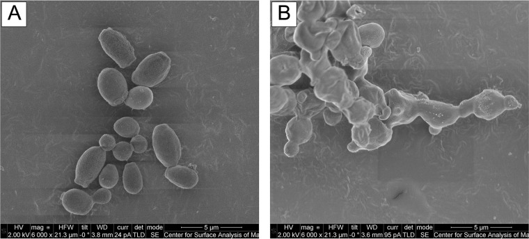
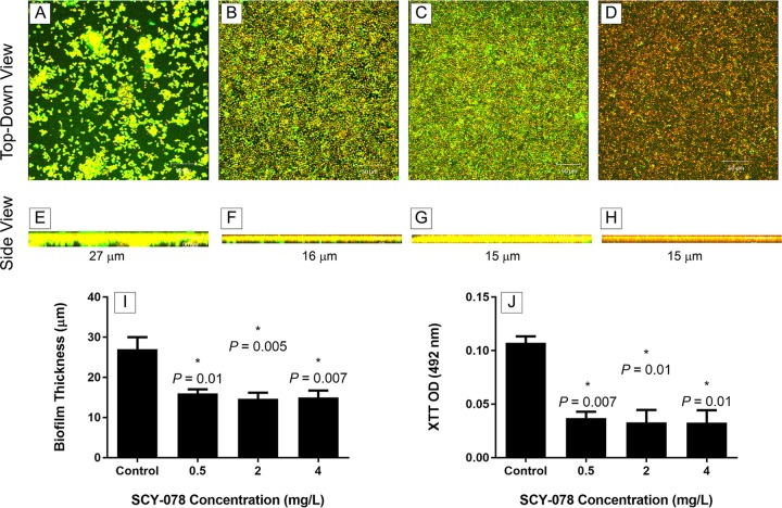
Candida auris , a new multidrug-resistant Candida spp. which is associated with invasive infection and high rates of mortality, has recently emerged. Here, we determined the virulence factors (germination, adherence, biofilm formation, phospholipase and proteinase production) of 16 C. auris isolates and their susceptibilities to 11 drugs belonging to different antifungal classes, including a novel orally bioavailable 1,3-β- d -glucan synthesis inhibitor (SCY-078). We also examined the effect of SCY-078 on the growth, ultrastructure, and biofilm-forming abilities of C. auris . Our data showed that while the tested strains did not germinate, they did produce phospholipase and proteinase in a strain-dependent manner and had a significantly reduced ability to adhere and form biofilms compared to that of Candida albicans ( P = 0.01). C. auris isolates demonstrated reduced susceptibility to fluconazole and amphotericin B, while, in general, they were susceptible to the remaining drugs tested. SCY-078 had an MIC 90 of 1 mg/liter against C. auris and caused complete inhibition of the growth of C. auris and C. albicans . Scanning electron microscopy analysis showed that SCY-078 interrupted C. auris cell division, with the organism forming abnormal fused fungal cells. Additionally, SCY-078 possessed potent antibiofilm activity, wherein treated biofilms demonstrated significantly reduced metabolic activity and a significantly reduced thickness compared to the untreated control ( P < 0.05 for both comparisons). Our study shows that C. auris expresses several virulence determinants (albeit to a lesser extent than C. albicans ) and is resistant to fluconazole and amphotericin B. SCY-078, the new orally bioavailable antifungal, had potent antifungal/antibiofilm activity against C. auris , indicating that further evaluation of this antifungal is warranted.
Overview
The claims and statistics of this paper haven’t been checked yet.
Run the check — free, no account needed. It reads the open-access text and checks the sources it cites.
Integrity signals
Registry
Cites
Statistics
Figures
Verification
Claims the paper makes
The claims and statistics of this paper haven’t been checked yet.
Run the check — free, no account needed. It reads the open-access text and checks the sources it cites.
Claims other papers make about this one
Each is checked against this paper’s own text: does it say what the citing paper says it does?
In the citing paper
“Regarding its anti-biofilm activity, somewhat unsurprisingly since it shares the same target with echinocandins, ibrexafungerp has been shown to display activity against biofilms formed by different Candida species, including C. albicans, C. parapsilosis, C. tropicalis, C. glabrata [44], and most recently C. auris [45].”
From Antifungal therapy of Candida biofilms: Past, present and future (2023) · cites this paper as [45]
In the citing paper
“Importantly, SCY-078 appears to be active against echinocandin-resistant and azole-resistant Candida strains, including C. auris [111,112,113].”
From Treatment of Invasive Candidiasis: A Narrative Review (2018) · cites this paper as [112]
In the citing paper
“However, exposure to various cues that induce C. albicans filamentation does not induce morphogenesis in C. auris, suggesting divergence in the circuitry governing the yeast-to-filament transition between the two Candida species (23).”
From Genetic Analysis of Candida auris Implicates Hsp90 in Morphogenesis and Azole Tolerance and Cdr1 in Azole Resistance (2019) · cites this paper as [23]
Self-check
Percentages, sums and reported differences re-computed from the paper’s own numbers.
The self-check needs the paper’s full text.
Citations
Cites
The UNITE database for molecular identification of fungi – recent updates and future perspectives
Abarenkov, K., Nilsson, R. H., Larsson, K., Alexander, I. J., Eberhardt, U., Erland, S., Høiland, K., Kjøller, R., Larsson, E., Pennanen, T., et al. (2010) · New Phytologist · doi:10.1111/j.1469-8137.2009.03160.x · Cited by 1,911
Satoh, K., Makimura, K., Hasumi, Y., Nishiyama, Y., Uchida, K., & Yamaguchi, H. (2009) · Microbiology and Immunology · doi:10.1111/j.1348-0421.2008.00083.x · Cited by 1,381
Potential Role of Phospholipases in Virulence and Fungal Pathogenesis
Ghannoum, M. A. (2000) · Clinical Microbiology Reviews · doi:10.1128/cmr.13.1.122-143.2000 · Cited by 823
Self-citationMedically Important Fungi: A Guide to Identification
Larone, D. H. (1976) · Cited by 809
Kuhn, D. M., George, T., Chandra, J., Mukherjee, P. K., & Ghannoum, M. A. (2002) · Antimicrobial Agents and Chemotherapy · doi:10.1128/aac.46.6.1773-1780.2002 · Cited by 775
Self-citationFirst hospital outbreak of the globally emerging Candida auris in a European hospital
Schelenz, S., Hagen, F., Rhodes, J. L., Abdolrasouli, A., Chowdhary, A., Hall, A., Ryan, L., Shackleton, J., Trimlett, R., Meis, J. F., et al. (2016) · Antimicrobial Resistance & Infection Control · doi:10.1186/s13756-016-0132-5 · Cited by 768
Medically important fungi: A guide to identification
Ajello, L. (1989) · Diagnostic Microbiology and Infectious Disease · doi:10.1016/0732-8893(89)90040-0 · Cited by 667
Saville, S. P., Lazzell, A. L., Monteagudo, C., & Lopez-Ribot, J. L. (2003) · Eukaryotic Cell · doi:10.1128/ec.2.5.1053-1060.2003 · Cited by 655
Plate method for detection of phospholipase activity inCandida albicans
Price, M. F., Wilkinson, I. D., & Gentry, L. O. (1982) · Medical Mycology · doi:10.1080/00362178285380031 · Cited by 651
Biofilm formation by Candida species on the surface of catheter materials in vitro
Hawser, S. P., & Douglas, L. J. (1994) · Infection and Immunity · doi:10.1128/iai.62.3.915-921.1994 · Cited by 599
Research works printed but not linked: 3
- Odds FC . 1988. Candida and candidosis: a review and bibliography, 2nd ed, vol 1. Bailliere Tindall, London, England.
- Kwon-Chung KJ BennettJE. 1992. Medical mycology. Lea & Febiger, Philadelphia, PA.
- Clinical and Laboratory Standards Institute. 2008. Reference method for broth dilution antifungal susceptibility testing of yeasts, 3rd ed. Approved standard M27-A3. Clinical and Laboratory Standards Institute, Wayne, PA.
Cited by
Sorted by citations
Chowdhary, A., Sharma, C., & Meis, J. F. (2017) · PLOS Pathogens · doi:10.1371/journal.ppat.1006290 · Cited by 750
Candida auris: a Review of the Literature
Jeffery-Smith, A., Taori, S. K., Schelenz, S., Jeffery, K. J., Johnson, E. M., Borman, A. M., Candida auris Incident Management Team, Manuel, R. J., & Brown, C. S. (2017) · Clinical Microbiology Reviews · doi:10.1128/cmr.00029-17 · Cited by 624
Chowdhary, A., Prakash, A., Sharma, C., Kordalewska, M., Kumar, A., Sarma, S., Tarai, B., Singh, A., Upadhyaya, G., Upadhyay, S., et al. (2018) · Journal of Antimicrobial Chemotherapy · doi:10.1093/jac/dkx480 · Cited by 577
A Candida auris Outbreak and Its Control in an Intensive Care Setting
Eyre, D. W., Sheppard, A. E., Madder, H., Moir, I., Moroney, R., Quan, T. P., Griffiths, D., George, S., Butcher, L., Morgan, M., et al. (2018) · New England Journal of Medicine · doi:10.1056/nejmoa1714373 · Cited by 517
COVID-19-associated fungal infections
Hoenigl, M., Seidel, D., Sprute, R., Cunha, C., Oliverio, M., Goldman, G. H., Ibrahim, A. S., & Carvalho, A. (2022) · Nature Microbiology · doi:10.1038/s41564-022-01172-2 · Cited by 482
Metadata
- Authors
- Larkin, E., Hager, C., Chandra, J., Mukherjee, P. K., Retuerto, M., Salem, I., Long, L., Isham, N., Kovanda, L., Borroto-Esoda, K., Wring, S., Angulo, D., & Ghannoum, M.
- Affiliations
- University Hospitals of Cleveland (US)
- Astellas Pharma (United States) (US)
- Scynexis Inc., Jersey City, New Jersey, USA
- Scynexis (United States) (US)
- Corresponding author
- Ghannoum, Mahmoud
- ORCID
- Venue
- Antimicrobial Agents and Chemotherapy
- Publisher
- American Society for Microbiology
- Published
- 2017-05
- Volume
- 61
- Issue
- 5
- ISSN
- 0066-4804 (ISSN-L)
- 1098-6596
- PMC
- PMC5404565
- OpenAlex
- W2589939125
- Cited by
- 395
- References
- 53
- Citations by year
- 2026: 24 · 2025: 25 · 2024: 31 · 2023: 36 · 2022: 30 · 2021: 49 · 2020: 67 · 2019: 51 · 2018: 42 · 2017: 39
- FWCI
- 42.10
- Type
- journal article
- Language
- en
- Open access
- hybrid
- doi.org
- Licence
- Locations
- Antimicrobial Agents and Chemotherapy (publishedVersion, cc-by)
- Europe PMC (PubMed Central) (publishedVersion, cc-by)
- PubMed Central (publishedVersion, cc-by)
- PubMed (publishedVersion)
- Funders
- Case Western Reserve University
- National Institutes of Health
- National Institute of Arthritis and Musculoskeletal and Skin Diseases (P30AR039750)
- National Institute of Dental and Craniofacial Research (R01DE024228)
- Topics
- Health Sciences › Medicine › Infectious Diseases › Antifungal resistance and susceptibility
- Life Sciences › Biochemistry, Genetics and Molecular Biology › Molecular Biology › Fungal and yeast genetics research
- Health Sciences › Medicine › Infectious Diseases › Fungal Infections and Studies
- Keywords
- Candida auris, antifungal susceptibility, fluconazole resistance, amphotericin B resistance, biofilm formation, antibiofilm activity, fungal virulence factors, phospholipase, proteolytic enzymes, fungal adhesion, cell division inhibition, Candida albicans comparison, multidrug-resistant fungi
- MeSH terms
- Amphotericin B, Antifungal Agents, Candida, Candida albicans, Candidiasis, Cell Adhesion, Cell Division, Glucans, Glycosides, Humans, Microbial Sensitivity Tests, Peptide Hydrolases, Phospholipases, Triterpenes, Fluconazole, Biofilms, Drug Resistance, Multiple, Fungal, Virulence Factors
About these checks
Registry, retraction, Cites and statistics results are computed, not judged, with one exception: a printed reference that no index lists is read once by a model before it counts as a citation error. Claims are extracted by a model and kept only when their quote appears verbatim in the text; each cited source is then checked, with a model’s help, for whether it says what the claim relies on. Those verdicts can be wrong — read the claim’s quote and the cited source before relying on one.
Statistics and arithmetic are recomputed, not judged.
Confidence bands are shown only for fields calibrated on our annotated test set. None is calibrated yet, so no band is shown.
What counts as a claim ›How we measure confidence ›How we check this ›
Are you an author, or do you see a problem with this page? Request a review
