

The DOI matches its Crossref record; 57 of the 57 references it cites were found in a registry or an index.
Evaluating the risk of ovarian cancer before surgery using the ADNEX model to differentiate between benign, borderline, early and advanced stage invasive, and secondary metastatic tumours: prospective multicentre diagnostic study
Open accessAbstract
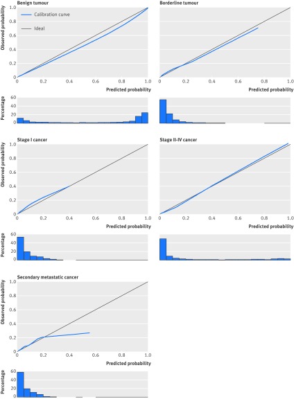
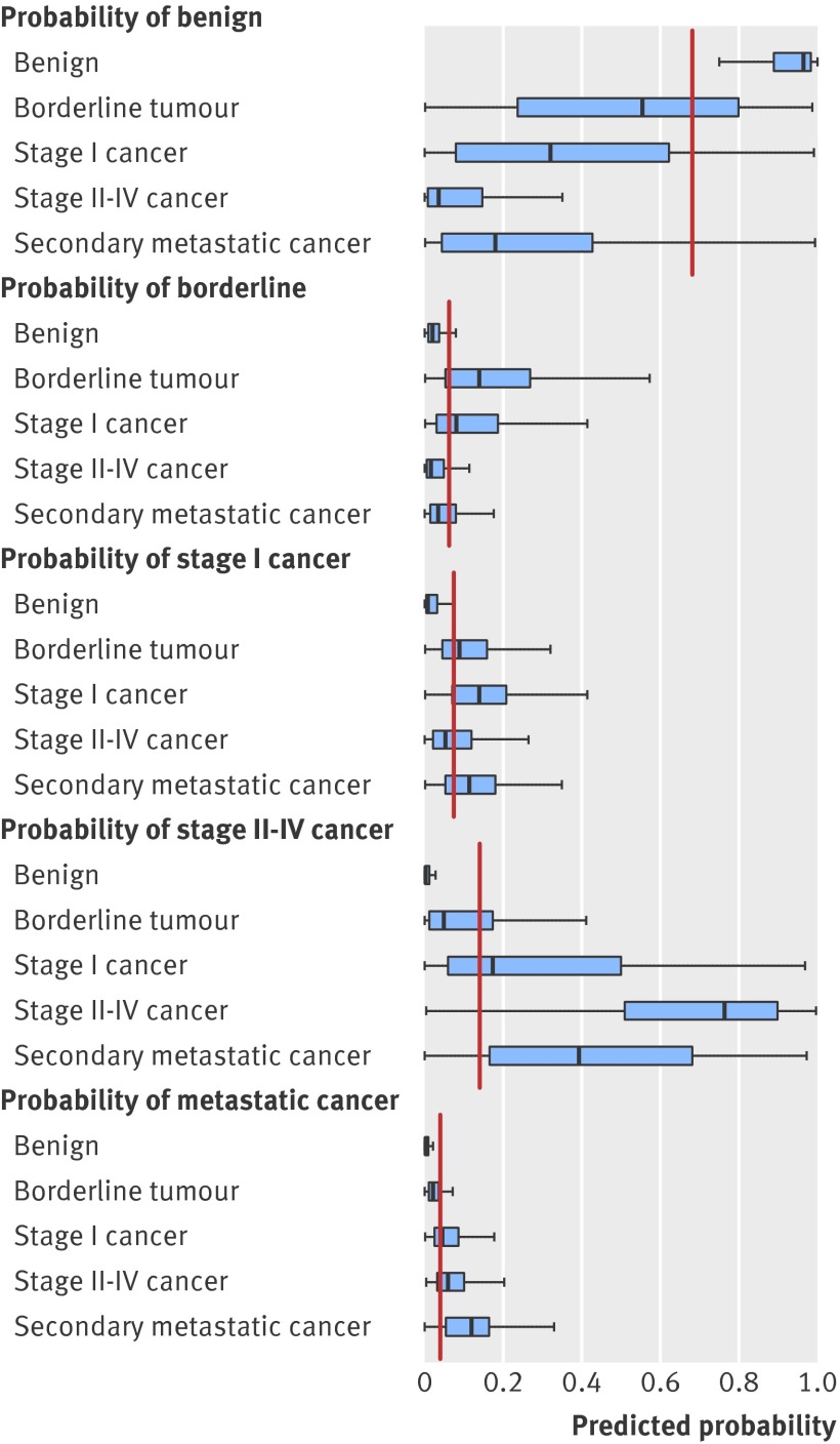
OBJECTIVES: To develop a risk prediction model to preoperatively discriminate between benign, borderline, stage I invasive, stage II-IV invasive, and secondary metastatic ovarian tumours. DESIGN: Observational diagnostic study using prospectively collected clinical and ultrasound data. SETTING: 24 ultrasound centres in 10 countries. PARTICIPANTS: Women with an ovarian (including para-ovarian and tubal) mass and who underwent a standardised ultrasound examination before surgery. The model was developed on 3506 patients recruited between 1999 and 2007, temporally validated on 2403 patients recruited between 2009 and 2012, and then updated on all 5909 patients. MAIN OUTCOME MEASURES: Histological classification and surgical staging of the mass. RESULTS: The Assessment of Different NEoplasias in the adneXa (ADNEX) model contains three clinical and six ultrasound predictors: age, serum CA-125 level, type of centre (oncology centres v other hospitals), maximum diameter of lesion, proportion of solid tissue, more than 10 cyst locules, number of papillary projections, acoustic shadows, and ascites. The area under the receiver operating characteristic curve (AUC) for the classic discrimination between benign and malignant tumours was 0.94 (0.93 to 0.95) on temporal validation. The AUC was 0.85 for benign versus borderline, 0.92 for benign versus stage I cancer, 0.99 for benign versus stage II-IV cancer, and 0.95 for benign versus secondary metastatic. AUCs between malignant subtypes varied between 0.71 and 0.95, with an AUC of 0.75 for borderline versus stage I cancer and 0.82 for stage II-IV versus secondary metastatic. Calibration curves showed that the estimated risks were accurate. CONCLUSIONS: The ADNEX model discriminates well between benign and malignant tumours and offers fair to excellent discrimination between four types of ovarian malignancy. The use of ADNEX has the potential to improve triage and management decisions and so reduce morbidity and mortality associated with adnexal pathology.
Overview
The claims and statistics of this paper haven’t been checked yet.
Run the check — free, no account needed. It reads the open-access text and checks the sources it cites.
Integrity signals
Registry
Cites
Statistics
Figures
Verification
Claims the paper makes
The claims and statistics of this paper haven’t been checked yet.
Run the check — free, no account needed. It reads the open-access text and checks the sources it cites.
Claims other papers make about this one
Each is checked against this paper’s own text: does it say what the citing paper says it does?
In the citing paper
“The ADNEX model, developed by the International Ovarian Tumor Analysis (IOTA) consortium (authors BVC and DT are part of this consortium), preoperatively estimates the probability of malignancy in women with an ovarian tumour who are scheduled for surgery. 31”
From Evaluation of performance measures in predictive artificial intelligence models to support medical decisions: overview and guidance (2025) · cites this paper as [31]
In the citing paper
“ADNEX was developed on the data from 5909 individuals recruited between 1999 and 2012 in 24 secondary and tertiary care centres from 10 countries (Italy, Belgium, Sweden, Czech Republic, Poland, France, United Kingdom, China, Spain, and Canada).”
From Evaluation of performance measures in predictive artificial intelligence models to support medical decisions: overview and guidance (2025) · cites this paper as [31]
Self-check
Percentages, sums and reported differences re-computed from the paper’s own numbers.
The self-check needs the paper’s full text.
Citations
Cites
(1990) · Choice Reviews Online · doi:10.5860/choice.27-3338 · Cited by 35,305
Hosmer, D. W., & Lemeshow, S. (2000) · doi:10.1002/0471722146 · Cited by 20,302
Siegel, R., Ma, J., Zou, Z., & Jemal, A. (2014) · CA: A Cancer Journal for Clinicians · doi:10.3322/caac.21208 · Cited by 11,499
SEER Cancer Statistics Review, 1975-2003
Ries, L. A. G., Harkins, D., Krapcho, M. F., Mariotto, A. B., Miller, B., Feuer, E. J., Clegg, L. X., Eisner, M. P., Horner, M., Howlader, N., et al. (2006) · Digital Archive @ GSU · Cited by 11,404
Regression Modeling Strategies
Harrell, F. E. (2001) · Springer series in statistics · doi:10.1007/978-1-4757-3462-1 · Cited by 9,270
Sterne, J. A. C., White, I. R., Carlin, J. B., Spratt, M., Royston, P., Kenward, M. G., Wood, A. M., & Carpenter, J. R. (2009) · BMJ · doi:10.1136/bmj.b2393 · Cited by 7,299
Signet-Ring Cell Carcinoma of the Colon: A Case Report and Review of the Literature
Park, P. Y., Goldin, T. A., Chang, J. C. S., Markman, M. M., & Kundranda, M. N. (2015) · Case Reports in Oncology · doi:10.1159/000441772 · Cited by 5,273
Clinical Prediction Models: A Practical Approach to Development, Validation, and Updating
Steyerberg, E. W. (2019) · Statistics for Biology and Health · doi:10.1007/978-3-030-16399-0 · Cited by 3,281
Rubin, M., & Stephen, C. D. (1993) · Cited by 3,149
Rao, S. J. (2003) · Journal of the American Statistical Association · doi:10.1198/jasa.2003.s263 · Cited by 1,967
Cited by
Sorted by citations
Items 101–200 of 510
Sundar, S. S., Rick, C., Dowling, F., Au, P., Snell, K. I. E., Rai, N., Champaneria, R., Stobart, H., Neal, R. D., Davenport, C. F., et al. (2016) · BMJ Open · doi:10.1136/bmjopen-2015-010333 · Cited by 27
Nucleosome footprinting in plasma cell-free DNA for the pre-surgical diagnosis of ovarian cancer
Vanderstichele, A., Busschaert, P., Landolfo, C., Olbrecht, S., Coosemans, A., Froyman, W., Loverix, L., Concin, N., Braicu, E. I., Wimberger, P., et al. (2022) · npj Genomic Medicine · doi:10.1038/s41525-022-00300-5 · Cited by 27
Qian, L., Du, Q., Jiang, M., Yuan, F., Chen, H., & Feng, W. (2021) · Frontiers in Oncology · doi:10.3389/fonc.2021.673722 · Cited by 24
Phillips, C. H., Guo, Y., Strachowski, L. M., Jha, P., Reinhold, C., & Andreotti, R. F. (2022) · Canadian Association of Radiologists Journal · doi:10.1177/08465371221108057 · Cited by 25
Basta, A., Bidziński, M., Bieńkiewicz, A., Blecharz, P., Bodnar, L., Jach, R., Knapp, P., Kojs, Z., Kotarski, J., Markowska, J., et al. (2017) · Ginekologia Onkologiczna/Current Gynecologic Oncology · doi:10.15557/cgo.2017.0001 · Cited by 25
Ovarian Strumal Carcinoid: Case Report, Systematic Literature Review and Pooled Analysis
Turla, A., Zamparini, M., Milione, M., Grisanti, S., Amoroso, V., Pedersini, R., Cosentini, D., & Berruti, A. (2022) · Frontiers in Endocrinology · doi:10.3389/fendo.2022.871210 · Cited by 24
Coosemans, A., Baert, T., Ceusters, J., Busschaert, P., Landolfo, C., Verschuere, T., Rompuy, A. V., Vanderstichele, A., Froyman, W., Neven, P., et al. (2019) · International Journal of Gynecological Cancer · doi:10.1136/ijgc-2019-000521 · Cited by 24
Guo, Y., Jiang, T., Ouyang, L., Li, X., He, W., Zhang, Z., Shen, H., You, Z., Yang, G., & Lai, H. (2020) · Gynecologic Oncology · doi:10.1016/j.ygyno.2020.12.006 · Cited by 23
Safety and efficiency of performing transvaginal ultrasound-guided tru-cut biopsy for pelvic masses
Verschuere, H., Froyman, W., den Bosch, T. V., Hoefs, M. V., Kaijser, J., Schoubroeck, D. V., Rompuy, A. V., Vergote, I. B., & Timmerman, D. (2021) · Gynecologic Oncology · doi:10.1016/j.ygyno.2021.03.026 · Cited by 23
Koch, A. H., Jeelof, L. S., Muntinga, C. L. P., Gootzen, T. A., van de Kruis, N. M. A., Nederend, J., Boers, T. G. W., van der Sommen, F., & Piek, J. M. J. (2023) · Insights into Imaging · doi:10.1186/s13244-022-01345-x · Cited by 25
“…have been developed by the International Ovarian tumor analysis (IOTA) group, such as the Assessment of Different NEo-plasias in the adneXa (ADNEX) model, which combines six ultrasound features together with patients age, serum CA125 and type of center (oncology referral center vs other) [3, 4].”
MethodMachine learning and radiomics for segmentation and classification of adnexal masses on ultrasound
Barcroft, J. F., Linton‐Reid, K., Landolfo, C., Al‐Memar, M., Parker, N., Kyriacou, C., Munaretto, M., Fantauzzi, M., Cooper, N., Yazbek, J., et al. (2024) · npj Precision Oncology · doi:10.1038/s41698-024-00527-8 · Cited by 26
“ADNEX (with CA-125) is the best-performing ultrasound-based model with an AUC of 0.94, sensitivity of 0.865 ( fi xed speci fi city 0.90) and speci fi city of 0.866 ( fi xed sensitivity 0.90) 9,11 .”
BackgroundMethodInfluentialMoro, F., Vagni, M., Tran, H. E., Bernardini, F., Mascilini, F., Ciccarone, F., Nero, C., Giannarelli, D., Boldrini, L., Fagotti, A., et al. (2024) · Ultrasound in Obstetrics and Gynecology · doi:10.1002/uog.27680 · Cited by 25
Gehringer, C. K., Martin, G. P., CALSTER, B. V., Hyrich, K. L., Verstappen, S. M. M., & Sergeant, J. C. (2024) · Journal of Clinical Epidemiology · doi:10.1016/j.jclinepi.2024.111481 · Cited by 22
“MPMs have been applied to several clinical settings(2,18–21), however, the adoption of this approach is still relatively scarce(13).”
MethodInfluentialNohuz, E., Simone, L. D., & Chene, G. (2018) · Journal of Gynecology Obstetrics and Human Reproduction · doi:10.1016/j.jogoh.2018.04.012 · Cited by 23
Factors associated with misdiagnosis of frozen section of mucinous borderline ovarian tumor
Zhang, W., Jia, S., Xiang, Y., Yang, J., Jia, C., & Leng, J. (2018) · Journal of International Medical Research · doi:10.1177/0300060518795582 · Cited by 22
Guideline No. 404: Initial Investigation and Management of Benign Ovarian Masses
Wolfman, W., Thurston, J., Yeung, G., & Glanc, P. (2020) · Journal of Obstetrics and Gynaecology Canada · doi:10.1016/j.jogc.2020.01.014 · Cited by 23
Djokovic, D., Pinto, P., van Herendael, B. J., Laganà, A. S., Thomas, V., & Keckstein, J. (2021) · European Journal of Obstetrics & Gynecology and Reproductive Biology · doi:10.1016/j.ejogrb.2021.06.035 · Cited by 22
He, P., Wang, J., Duan, W., Song, C., Yu, Y., & Wu, Q. (2021) · Journal of Ovarian Research · doi:10.1186/s13048-021-00922-w · Cited by 21
“The Assessment of Different NEoplasias in the adneXa (ADNEX) model is the first predictive multiclass model developed by the IOTA and is able to differentiate between benign tumours, borderline ovarian tumours (BOTs), stage I ovarian cancer (OC), stage II-IV OC and secondary metastatic ovarian cancers [12].”
BackgroundResultInfluentialNgu, S., Chai, Y. K., Choi, K. M., Leung, T. W., Li, J., Kwok, S. T., Chu, M. M. Y., Tse, K. Y., Cheung, V. Y. T., Ngan, H., et al. (2022) · Cancers · doi:10.3390/cancers14030810 · Cited by 22
Radiomics and Radiogenomics of Ovarian Cancer
Panico, C., Avesani, G., Zormpas‐Petridis, K., Rundo, L., Nero, C., & Sala, E. (2023) · Radiologic Clinics of North America · doi:10.1016/j.rcl.2023.02.006 · Cited by 24
Poonyakanok, V., Tanmahasamut, P., Jaishuen, A., Wongwananuruk, T., Asumpinwong, C., Panichyawat, N., & Chantrapanichkul, P. (2021) · Gynecologic and Obstetric Investigation · doi:10.1159/000513517 · Cited by 21
Davenport, C. F., Rai, N., Sharma, P., Deeks, J. J., Berhane, S., Mallett, S., Saha, P., Solanki, R., Bayliss, S. E., Snell, K. I. E., et al. (2022) · Cancers · doi:10.3390/cancers14153621 · Cited by 22
“Three studies were found, two from single centres [45,46] and one multicentre study [47].”
BackgroundA Deep Learning Model System for Diagnosis and Management of Adnexal Masses
Li, J., Chen, Y., Zhang, M., Zhang, P., He, K., Yan, F., Li, J., Xu, H. L., Burkhoff, D., Luo, Y., et al. (2022) · Cancers · doi:10.3390/cancers14215291 · Cited by 21
“If more than one mass was detected, then the mass with the most complex morphology or, in the case of a similar morphology, the largest diameter, was used [16,17].”
MethodCzekierdowski, A., Stachowicz, N., Smoleń, A., Kluz, T., Łoziński, T., Miturski, A., & Kraczkowski, J. J. (2021) · Diagnostics · doi:10.3390/diagnostics11030414 · Cited by 20
“According to the results of the IOTA group’s recent studies, a cut-off risk set at 20% is related to the best balance between ADNEX model sensitivity and specificity [17].”
MethodResultPeng, X., Ma, Y., Wang, L., Li, H., Zheng, X., & Liu, Y. (2021) · International Journal of General Medicine · doi:10.2147/ijgm.s328010 · Cited by 20
Wilson, M. P., Katlariwala, P., & Low, G. (2020) · Radiology · doi:10.1148/radiol.2020200437 · Cited by 20
Silvestre, L., Martins, W. P., & dos Reis, F. J. C. (2015) · Journal of Ovarian Research · doi:10.1186/s13048-015-0174-y · Cited by 19
“Among our cases, 43 % were malignant, this prevalence is similar to the reported prevalence of 42 % of malignancy in oncology centers and 17 % in other centers [27].”
ResultComparison of Ultrasound Scores in Differentiating between Benign and Malignant Adnexal Masses
Pelayo, M., Pelayo‐Delgado, I., Saúco, J. S., Sánchez-Zurdo, J., Abarca-Martínez, L., Corraliza-Galán, V., Martín-Gromaz, C., Pablos-Antona, M. J., Zurita-Calvo, J., & Alcázar, J. L. (2023) · Diagnostics · doi:10.3390/diagnostics13071307 · Cited by 20
Ultrasound evaluation of ovarian masses and assessment of the extension of ovarian malignancy
Moro, F., Esposito, R., Landolfo, C., Froyman, W., Timmerman, D., Bourne, T., Scambia, G., Valentin, L., & Testa, A. C. (2021) · British Journal of Radiology · doi:10.1259/bjr.20201375 · Cited by 20
Seebacher, V., Aust, S., D’Andrea, D., Grimm, C., Reiser, E., Tiringer, D., von Mersi, H., Polterauer, S., Reinthaller, A., & Helmy-Bader, S. (2017) · PLoS ONE · doi:10.1371/journal.pone.0182383 · Cited by 21
Landolfo, C., Achten, E., Ceusters, J., Baert, T., Froyman, W., Heremans, R., Vanderstichele, A., Thirion, G., Hoylandt, A. V., Claes, S., et al. (2020) · Gynecologic Oncology · doi:10.1016/j.ygyno.2020.09.025 · Cited by 18
Huang, X., Wang, Z., Zhang, M., & Luo, H. (2021) · Frontiers in Oncology · doi:10.3389/fonc.2021.684257 · Cited by 17
de Oca, M. K. M., Dotters-Katz, S. K., Kuller, J. A., & Previs, R. A. (2021) · Obstetrical & Gynecological Survey · doi:10.1097/ogx.0000000000000909 · Cited by 20
“Symptoms of ovarian cancer are vague and can mimic other conditions such as pregnancy or gastrointestinal-related disorders.(68,71) Several studies have investigated the ability of ultrasound to correctly diagnose an adnexal mass in pregnancy.”
BackgroundAas‐Eng, M. K., Montanari, E., Lieng, M., Keckstein, J., & Hudelist, G. (2020) · Seminars in Reproductive Medicine · doi:10.1055/s-0040-1718740 · Cited by 18
Dai, W., Wu, Y., Ling, Y.-T., Zhao, J., Zhang, S., Gu, Z.-W., Gong, L., Zhu, M.-N., Dong, S., Xu, S., et al. (2024) · EClinicalMedicine · doi:10.1016/j.eclinm.2024.102923 · Cited by 18
Liu, L., Cai, W., Tian, H., Wu, B., Zhang, J., Wang, T., Hao, Y., & Yue, G. (2024) · Frontiers in Oncology · doi:10.3389/fonc.2024.1377489 · Cited by 18
Giourga, M., Petropoulos, I., Stavros, S., Potiris, A., Gerede, A., Sapantzoglou, I., Fanaki, M., Papamattheou, E., Karasmani, C., Karampitsakos, T., et al. (2024) · Journal of Clinical Medicine · doi:10.3390/jcm13144123 · Cited by 19
Spagnol, G., Marchetti, M., Carollo, M., Bigardi, S., Tripepi, M., Facchetti, E., Tommasi, O. D., Vitagliano, A., Cavallin, F., Tozzi, R., et al. (2024) · Cancers · doi:10.3390/cancers16223790 · Cited by 19
Zheng, X., Lyu, G., Gan, Y., Hu, M., Liu, X., Chen, S., Wu, X., Yang, W., Ye, F., Yan, X.-B., et al. (2020) · European Radiology · doi:10.1007/s00330-020-07113-z · Cited by 17
Jiang, M., Le, Q., Yang, B., Yuan, F., & Chen, H. (2021) · Journal of Ovarian Research · doi:10.1186/s13048-021-00805-0 · Cited by 17
“The IOTA ADNEX model [10] contains nine predictors (three clinical and six ultrasound variables): age, serum CA125 level, type of centre (oncology referral centre vs non-oncology centre), maximum diameter of the lesion, maximal diameter of the largest solid part of the lesion, more than 10 cyst locules (yes or no), nu…”
MethodESGO/ISUOG/IOTA/ESGE Consensus Statement on preoperative diagnosis of ovarian tumours
Timmerman, D., Planchamp, F., Bourne, T., Landolfo, C., du Bois, A., Chiva, L. M., Cibula, D., Concin, N., Fischerová, D., Froyman, W., et al. (2021) · Facts Views and Vision in ObGyn · doi:10.52054/fvvo.13.2.016 · Cited by 17
Predictive Analytics: The Application of J48 Algorithm on Grocery Data to Predict Obesity
Daud, N., Noor, N. L. M., Aljunid, S. A., Noordin, N., & Teng, N. I. M. F. (2018) · IEEE Conference on Big Data and Analytics (ICBDA) · doi:10.1109/icbdaa.2018.8629623 · Cited by 17
“Calibration refers to the assessment of how well a model could predict, in comparison to the actual events [24].”
BackgroundStukan, M., Badocha, M., & Ratajczak, K. (2019) · BMC Cancer · doi:10.1186/s12885-019-5629-x · Cited by 17
Basic Gynecologic Ultrasound Examination (Level I): DEGUM, ÖGUM, and SGUM Recommendations
Grab, D., Merz, E., Eichhorn, K., Tutschek, B., Kagan, K. O., Heling, K., Faber, R., Klaritsch, P., Rempen, A., Kähler, C., et al. (2022) · Ultraschall in der Medizin - European Journal of Ultrasound · doi:10.1055/a-1851-5157 · Cited by 19
Spagnol, G., Marchetti, M., Tommasi, O. D., Vitagliano, A., Cavallin, F., Tozzi, R., Saccardi, C., & Noventa, M. (2023) · Gynecologic Oncology · doi:10.1016/j.ygyno.2023.08.012 · Cited by 16
Weichert, J., Welp, A., Scharf, J. L., Dracopoulos, C., Becker, W.-H., & Gembicki, M. (2021) · Geburtshilfe und Frauenheilkunde · doi:10.1055/a-1522-3029 · Cited by 19
Joyeux, E., Miras, T., Masquin, I., Duglet, P.-E., Astruc, K., & Douvier, S. (2016) · Gynécologie Obstétrique & Fertilité · doi:10.1016/j.gyobfe.2016.07.007 · Cited by 16
Cytokines in peritoneal fluid of ovarian neoplasms
de Lima, C. A., Rodrigues, I. S. S., Martins-Filho, A., Micheli, D. C., Tavares‐Murta, B. M., Murta, E. F. C., & Nomelini, R. S. (2019) · Journal of Obstetrics and Gynaecology · doi:10.1080/01443615.2019.1633516 · Cited by 16
“ADNEX discriminates between benign and malignant tumours, offering a more refined approach to treatment for women with some adnexal mass, and for the diagnosis and treatment of patients with ovarian neoplasia (Van Calster et al. 2014; Froyman et al. 2017).”
BackgroundVara, J., Pagliuca, M., Springer, S., Canales, J. G., Brotóns, I., Yakcich, J., AJOSSA, S., Pascual, M. A., Guerriero, S., & Alcázar, J. L. (2023) · Diagnostics · doi:10.3390/diagnostics13040673 · Cited by 17
Lombardi, A., Arezzo, F., Sciascio, E. D., Ardito, C. A., Mongelli, M., Lillo, N. D., Fascilla, F. D., Silvestris, E., Kardhashi, A., Putino, C., et al. (2023) · Artificial Intelligence in Medicine · doi:10.1016/j.artmed.2023.102697 · Cited by 15
Machine learning models in evaluating the malignancy risk of ovarian tumors: a comparative study
He, X., Bai, X.-H., Chen, H., & Feng, W. (2024) · Journal of Ovarian Research · doi:10.1186/s13048-024-01544-8 · Cited by 16
Otify, M., Laios, A., Shamy, T. E., D’Angelo, A., & Amso, N. N. (2019) · European Journal of Obstetrics & Gynecology and Reproductive Biology · doi:10.1016/j.ejogrb.2019.11.016 · Cited by 15
Ultrasound Assessment of Adnexal Pathology: Standardized Methods and Different Levels of Experience
Tavoraitė, I., Kronlachner, L., Opolskiene, G., & Bartkevičienė, D. (2021) · Medicina · doi:10.3390/medicina57070708 · Cited by 14
Barcroft, J. F., Pandrich, M., Forno, S. D., Cooper, N., Linton‐Reid, K., Landolfo, C., Timmerman, D., Saso, S., & Bourne, T. (2024) · Ultrasound in Obstetrics and Gynecology · doi:10.1002/uog.27707 · Cited by 14
Yue, X., Zhong, L., Wang, Y., Zhang, C., Chen, X., Wang, S., Hu, J., Hu, J., Wang, C., & Liu, X. (2022) · Ultrasound in Medicine & Biology · doi:10.1016/j.ultrasmedbio.2022.02.001 · Cited by 15
Pelayo, M., Saúco, J. S., Sánchez-Zurdo, J., Abarca-Martínez, L., Borrero, C., Sainz‐Bueno, J. A., Alcázar, J. L., & Pelayo‐Delgado, I. (2023) · Diagnostics · doi:10.3390/diagnostics13132152 · Cited by 17
“The ADNEX model (Assessment of Different Neoplasia in the Adnexa) was published by the IOTA group as another complementary score that calculate the probability of malignancy [7].”
MethodDevelopment of a fuzzy-driven system for ovarian tumor diagnosis
Żywica, P., Dyczkowski, K., Wójtowicz, A., Stachowiak, A., Szubert, S., & Moszyński, R. (2016) · Journal of Applied Biomedicine · doi:10.1016/j.bbe.2016.08.003 · Cited by 14
“Among these, the most important are the LR1 and LR2 models, based on logistic regression [6], and the most recent IOTA model – ADNEX [7].”
Role of ultrasound in advanced peritoneal malignancies
Blasis, I. D., Moruzzi, M. C., Moro, F., Mascilini, F., Cianci, S., Alletti, S. G., Turco, L. C., Garganese, G., Scambia, G., & Testa, A. C. (2019) · Minerva Medica · doi:10.23736/s0026-4806.19.06103-2 · Cited by 14
Pascual, M. A., Vancraeynest, L., Timmerman, S., Ceusters, J., Ledger, A., Graupera, B., Rodríguez, I., Valero, B., Landolfo, C., Testa, A. C., et al. (2024) · Ultrasound in Obstetrics and Gynecology · doi:10.1002/uog.27642 · Cited by 16
Past, Present and Future Ultrasonographic Techniques for Analyzing Ovarian Masses
Guerriero, S., Saba, L., Alcázar, J. L., Pascual, M. A., AJOSSA, S., Perniciano, M., Piras, A., Sedda, F., Peddes, C., Fabbri, P., et al. (2015) · Women s Health · doi:10.2217/whe.15.11 · Cited by 13
Performance of the IOTA ADNEX model combined with HE4 for identifying early-stage ovarian cancer
Yang, S., Tang, J. Y., Rong, Y., Wang, M., Long, J., Chen, C., & Wang, C. (2022) · Frontiers in Oncology · doi:10.3389/fonc.2022.949766 · Cited by 12
Moro, F., Scavello, I., Maseroli, E., Rastrelli, G., Poma, C. B., Bonin, C., Dassie, F., Federici, S., Fiengo, S., Guccione, L., et al. (2022) · Journal of Endocrinological Investigation · doi:10.1007/s40618-022-01939-8 · Cited by 13
Miyoshi, A., Miyatake, T., Hara, T., Tanaka, A., Komura, N., Komiya, S., Kanao, S., Takeda, M., Mimura, M., Nagamatsu, M., et al. (2015) · Case Reports in Obstetrics and Gynecology · doi:10.1155/2015/414019 · Cited by 13
“However, the size of borderline ovarian tumors is relatively large, so they are often suspected of being ovarian cancers, having a much direr prognosis [3].”
BackgroundPerformance of the IOTA ADNEX Model on Selected Group of Patients with Borderline Ovarian Tumours
Gaurilčikas, A., Gedgaudaitė, M., Čižauskas, A., Atstupėnaitė, V., Paškauskas, S., Gaurilcikiene, D., Biržietis, T., Vaitkienė, D., & Nadišauskienė, R. J. (2020) · Medicina · doi:10.3390/medicina56120690 · Cited by 13
“For the calculation of risk, the ADNEX model uses three clinical variables (patients’ age, Ca125 tumour marker, and whether the ultrasound scan was performed in an oncology center or not) and six ultrasound variables (maximal diameter of the lesion, maximal diameter of the largest solid part, presence of more than 10…”
MethodInfluentialUltrasonographic evaluation of ovarian mass for predicting malignancy in pregnant women
Lee, S. J., Kim, Y. H., Lee, M., Ko, H. S., Oh, S., Seol, H., Kim, J. W., Ahn, K. H., Na, S., Seong, W. J., et al. (2021) · Gynecologic Oncology · doi:10.1016/j.ygyno.2021.09.007 · Cited by 13
Landolfo, C., Ceusters, J., Valentin, L., Froyman, W., Van Gorp, T., Heremans, R., Baert, T., Wouters, R., Vankerckhoven, A., Van Rompuy, A.-S., et al. (2024) · British Journal of Cancer · doi:10.1038/s41416-024-02578-x · Cited by 14
“The Assessment of Different NEoplasias in the adneXa (ADNEX) is a multinomial logistic regression model developed by the International Ovarian Tumour Analysis (IOTA) group [12].”
Hu, Y., Chen, B., Dong, H., Sheng, B., Xiao, Z., Li, J., Tian, W., & Lv, F. (2023) · Frontiers in Oncology · doi:10.3389/fonc.2023.1101297 · Cited by 12
Bruno, M., Capanna, G., Stanislao, V., Ciuffreda, R., Tabacco, S., Fantasia, I., Florio, C. D., Stabile, G., D’Alfonso, A., Guido, M., et al. (2023) · Diagnostics · doi:10.3390/diagnostics13203247 · Cited by 12
“These methods include the evaluation of different NEOplasias in the adneXa (ADNEX) model [20] introduced in 2014 and the simple rule risk (SRR) model proposed in 2016 [21].”
MethodLedger, A., Ceusters, J., Valentin, L., Testa, A. C., Holsbeke, C. V., Franchi, D., Bourne, T., Froyman, W., Timmerman, D., & CALSTER, B. V. (2023) · BMC Medical Research Methodology · doi:10.1186/s12874-023-02103-3 · Cited by 12
Borges, A. L., Brito, M., Ambrósio, P., Condeço, R., Pinto, P., Ambrósio, B., Mahomed, F., Gama, J. M. R., Bernardo, M. J., Gouveia, A., et al. (2024) · Ultrasound in Obstetrics and Gynecology · doi:10.1002/uog.27641 · Cited by 12
Meys, E. M. J., Jeelof, L. S., Ramaekers, B. L. T., Dirksen, C. D., Kooreman, L. F. S., Slangen, B. F. M., Kruitwagen, R. F. P. M., & Gorp, T. V. (2018) · European Journal of Cancer · doi:10.1016/j.ejca.2018.05.003 · Cited by 12
Moro, F., Mascilini, F., Pasciuto, T., Leombroni, M., Destri, M. L., Blasis, I. D., Garofalo, S., Scambia, G., & Testa, A. C. (2019) · International Journal of Gynecological Cancer · doi:10.1136/ijgc-2019-000373 · Cited by 12
“Ultrasound examination is also an effective tool in distinguishing a primary ovarian cancer from metastasis.(8) 9 However, adnexal masses may be difficult to classify during pregnancy as hormonal changes associated with the pregnancy may cause alterations in the sonographic appearances of ovarian masses and, for insta…”
MethodColon carcinoma presenting as ovarian metastasis
Kemps, P. G., Bol, M., Steller, E. J. A., de Pont, L. M. H., Holterhues, C., van Gerven, L., & Kolkman, W. (2021) · Radiology Case Reports · doi:10.1016/j.radcr.2021.06.072 · Cited by 12
Horała, A., Plewa, S., Dereziński, P., Klupczyńska, A., Matysiak, J., Nowak‐Markwitz, E., & Kokot, Z. J. (2021) · International Journal of Environmental Research and Public Health · doi:10.3390/ijerph18042167 · Cited by 12
Esquivel, A. L., Rodríguez, N., Ayala, N., Buriticá, C., Gómez, A., Velandia, A., Rodríguez, N., & Alcázar, J. L. (2021) · Journal of Ultrasound in Medicine · doi:10.1002/jum.15728 · Cited by 12
“Within these models, the IOTA Simple Rules and the IOTA ADNEX model are the best predictive tests validated externally, because both predict ovarian cancer in patients with persistent adnexal masses.(2,7) The IOTA Simple Rules is based on five ultrasound features suggestive of benignity and five ultrasound features su…”
MethodWhitney, H. M., Yoeli‐Bik, R., Abramowicz, J. S., Li, L., Li, H., Longman, R. E., Lengyel, E. R., & Giger, M. L. (2024) · Journal of medical imaging · doi:10.1117/1.jmi.11.4.044505 · Cited by 11
“…such as the International Ovarian Tumor Analysis (IOTA) Simple Rules, 12 the IOTA Assessment of Different NEoplasias in the adneXa model, 13 and the American College of Radiology Ovarian-Adnexal Reporting and Data (O-RADS) 14 risk stratification system with the goal of reducing false…”
Yang, Y., Li, J., Chen, H., & Feng, W. (2022) · Diagnostics · doi:10.3390/diagnostics12102348 · Cited by 11
“Sonographic Parameters According to the judging principle [15], 258 left-sided and 232 right-sided ovary cysts were analyzed.”
MethodInfluentialCherukuri, S., Jajoo, S., & Dewani, D. (2022) · Cureus · doi:10.7759/cureus.31194 · Cited by 11
Borges, A. L., Reis‐de‐Carvalho, C., Chorão, M., Pereira, H., & Djokovic, D. (2021) · World Journal of Clinical Cases · doi:10.12998/wjcc.v9.i10.2334 · Cited by 11
CALSTER, B. V. (2016) · Ultrasound in Obstetrics and Gynecology · doi:10.1002/uog.17391 · Cited by 11
Prediction Models of Adnexal Masses: State-of-the-Art Review
Mina, M., Kosmas, I. P., Tsakiridis, I. G., Mamopoulos, A. M., Kalogiannidis, I. A., Athanasiadis, A. P., & Dagklis, T. (2021) · Obstetrical & Gynecological Survey · doi:10.1097/ogx.0000000000000873 · Cited by 10
Wang, T., Cui, W., Nie, F., Huang, X. J., Huang, L., Liu, L., Zhu, Y., & Zheng, R. (2023) · Ultrasound in Medicine & Biology · doi:10.1016/j.ultrasmedbio.2023.05.012 · Cited by 10
Application of O-RADS US combined with MV-Flow to diagnose ovarian-adnexal tumors
Ruan, L., Liu, H., Xiang, H., Ni, Y., Feng, Y., Zhou, H., & Qi, M. (2023) · ULTRASONOGRAPHY · doi:10.14366/usg.23061 · Cited by 10
“In 2014, Van Calster et al. [11] developed a risk prediction model, the Assessment of Different Neoplasia in the Adnexa (ADNEX), to distinguish between benign, borderline, invasive in stage I, invasive in stages II-IV, and secondary metastatic ovarian tumors preoperatively.”
BackgroundLems, E., Leemans, J. C., Lok, C. A. R., Bongers, M. Y., & Geomini, P. M. A. J. (2023) · European Journal of Obstetrics & Gynecology and Reproductive Biology · doi:10.1016/j.ejogrb.2023.09.018 · Cited by 10
Ovarian Cysts and Tumors in Adolescents
Stanković, Z. B. (2024) · Obstetrics and Gynecology Clinics of North America · doi:10.1016/j.ogc.2024.08.006 · Cited by 10
Moro, F., Momi, M., Ledger, A., Barreñada, L., Ceusters, J., Sturla, D., Mor, E., Fornari, L., Mascilini, F., Ciccarone, F., et al. (2025) · American Journal of Obstetrics and Gynecology · doi:10.1016/j.ajog.2025.07.017 · Cited by 10
CALSTER, B. V., van Smeden, M., van Amsterdam, W. A. C., Coemans, M., Wynants, L., & Steyerberg, E. W. (2025) · Annual Review of Statistics and Its Application · doi:10.1146/annurev-statistics-042324-123749 · Cited by 14
Grandi, G., Fiocchi, F., Cortesi, L., Toss, A., Boselli, F., Sammarini, M., Sighinolfi, G., & Facchinetti, F. (2021) · Menopause The Journal of The North American Menopause Society · doi:10.1097/gme.0000000000001883 · Cited by 10
“presence of more than ten cysts [yes/no], number of papillary projections [0, 1, 2, 3,>3], presence of acoustic shadows [yes/ no], and presence of ascites [yes/no]).(12)”
BackgroundMethodPoonyakanok, V., Tanmahasamut, P., & Jaishuen, A. (2023) · Journal of obstetrics and gynaecology research · doi:10.1111/jog.15624 · Cited by 10
Giourga, M., Pouliakis, A., Vlastarakos, P., Stavrou, S., Tsiriva, M., Gerede, A., Daskalakis, G., Voros, C., Drakakis, P., & Domali, E. (2023) · Ultrasound International Open · doi:10.1055/a-2044-2855 · Cited by 10
Bartl, T., Karacs, J., Kreuzinger, C., Pfaffinger, S., Kendler, J., Ciocsirescu, C., Wolf, A., Reinthaller, A., Meyer, E. L., Brandstetter, M., et al. (2021) · Cancers · doi:10.3390/cancers13051076 · Cited by 10
“Numerous mathematical models have been proposed to date as potential aids in clinical decision making such as nomograms to interpret established biomarkers more comparatively or more complex risk prediction models such as the broadly validated IOTA ADNEX [18,19].”
MethodShould We Manage Large Ovarian Cysts Laparoscopically?
Paul, P. G., Chopade, G., Patil, S. R., Das, T., Thomas, M., & Garg, R. A. (2016) · Journal of Gynecologic Surgery · doi:10.1089/gyn.2016.0009 · Cited by 10
Bayesian Decision Curve Analysis With Bayesdca
Cruz, G. N. F., & Korthauer, K. D. (2024) · Statistics in Medicine · doi:10.1002/sim.10277 · Cited by 13
Chankrachang, A., Lattiwongsakorn, W., Tantipalakorn, C., & Tongsong, T. (2025) · Journal of Clinical Medicine · doi:10.3390/jcm14082776 · Cited by 9
Almeida, G., Bort, M. C., & Alcázar, J. L. (2025) · Journal of Clinical Ultrasound · doi:10.1002/jcu.24048 · Cited by 9
“Subsequently, in 2014, the IOTA group developed the ADNEX (Assessment of Different NEoplasias in the adneXa) model (Van Calster et al. 2014).”
Hack, K., & Glanc, P. (2019) · Obstetrics and Gynecology Clinics of North America · doi:10.1016/j.ogc.2019.07.002 · Cited by 9
Accuracy of Magnetic Resonance Imaging for Identifying Ovarian Cancer in a Community-Based Setting
Lin, R., Hung, Y., Cheng, J. N., & Suh‐Burgmann, E. (2022) · Women s Health Reports · doi:10.1089/whr.2021.0106 · Cited by 9
Sensor detection in gynaecological medicine
Thompson, M. E., Ahmadi, S., Davoudian, K., Franier, B. D. L., Lotay, N., & Bernardini, M. Q. (2022) · Sensors & Diagnostics · doi:10.1039/d2sd00090c · Cited by 11
Current Update on PET/MRI in Gynecological Malignancies—A Review of the Literature
Virarkar, M., Vulasala, S. S., Calimano-Ramirez, L. F., Singh, A., Lall, C. G., & Bhosale, P. (2023) · Current Oncology · doi:10.3390/curroncol30010083 · Cited by 9
“(71%), which may lead to unnecessary surgical intervention [84,85].”
BackgroundMorcellation and risk of malignancy in presumed ovarian fibromas/fibrothecomas
Froyman, W., Landolfo, C., Amant, F. C., den Bosch, T. V., Vergote, I. B., Coosemans, A., Testa, A. C., Valentin, L., Bourne, T., CALSTER, B. V., et al. (2016) · The Lancet Oncology · doi:10.1016/s1470-2045(16)00022-x · Cited by 9
Metadata
- Authors
- Van Calster, B., Van Hoorde, K., Valentin, L., Testa, A. C., Fischerova, D., Van Holsbeke, C., Savelli, L., Franchi, D., Epstein, E., Kaijser, J., Van Belle, V., Czekierdowski, A., Guerriero, S., Fruscio, R., Lanzani, C., Scala, F., Bourne, T., Timmerman, D., & International Ovarian Tumour Analysis (IOTA) group
- Affiliations
- Lund University (SE)
- Università Cattolica del Sacro Cuore (IT)
- IRCCS Azienda Ospedliero-Universitaria di Bologna Policlinico di Sant'Orsola (IT)
- European Institute of Oncology (IT)
- Karolinska University Hospital (SE)
- Universitair Ziekenhuis Leuven (BE)
- Medical University of Lublin (PL)
- Azienda Ospedaliero-Universitaria Cagliari (IT)
- University of Milan (IT)
- Istituto Nazionale Tumori IRCCS "Fondazione G. Pascale" (IT)
- ORCID
- Venue
- BMJ
- Publisher
- BMJ
- Published
- 2014-10-15
- Volume
- 349
- Issue
- oct07 3
- Pages
- g5920-g5920
- ISSN
- 1756-1833 (ISSN-L)
- PMC
- PMC4198550
- OpenAlex
- W2099878111
- Cited by
- 510
- References
- 57
- Citations by year
- 2026: 52 · 2025: 60 · 2024: 70 · 2023: 54 · 2022: 55 · 2021: 59 · 2020: 36 · 2019: 24 · 2018: 30 · 2017: 19 · 2016: 27 · 2015: 19 · 2014: 4
- FWCI
- 27.87
- Type
- journal article
- Language
- en
- Open access
- hybrid
- doi.org
- Licence
- cc-by-nc
- Locations
- BMJ (publishedVersion, cc-by-nc)
- PubMed Central (publishedVersion, cc-by-nc)
- PubMed (publishedVersion)
- BOA (University of Milano-Bicocca) (submittedVersion, cc-by-nc)
- Lirias (KU Leuven) (submittedVersion, cc-by-nc)
- UNICA IRIS Institutional Research Information System (University of Cagliari) (submittedVersion, other-oa)
- Lund University Publications (Lund University) (submittedVersion, other-oa)
- HighWire Press Open Archive (submittedVersion)
- PubliCatt (Università Cattolica del Sacro Cuore) (submittedVersion, cc-by-nc)
- Spiral (Imperial College London) (submittedVersion, other-oa)
- Funders
- National Institute for Health and Care Research
- Imperial College London
- Imperial College Healthcare NHS Trust
- Fonds Wetenschappelijk Onderzoek (G049312N)
- iMinds
- Malmö Högskola
- Skånes universitetssjukhus
- Vlaamse regering (G049312N)
- Medical Research Council
- Topics
- Health Sciences › Medicine › Oncology › Ovarian cancer diagnosis and treatment
- Life Sciences › Biochemistry, Genetics and Molecular Biology › Genetics › BRCA gene mutations in cancer
- Health Sciences › Medicine › Pathology and Forensic Medicine › Breast Lesions and Carcinomas
- Keywords
- ovarian cancer, ADNEX model, preoperative risk prediction, ovarian ultrasonography, borderline ovarian tumors, CA125, surgical staging, adnexal mass, triage
- MeSH terms
- Adnexal Diseases, Adult, Female, Humans, Neoplasm Staging, Ovarian Neoplasms, Predictive Value of Tests, Prospective Studies, Ultrasonography, Models, Statistical, Risk Assessment
About these checks
Registry, retraction, Cites and statistics results are computed, not judged, with one exception: a printed reference that no index lists is read once by a model before it counts as a citation error. Claims are extracted by a model and kept only when their quote appears verbatim in the text; each cited source is then checked, with a model’s help, for whether it says what the claim relies on. Those verdicts can be wrong — read the claim’s quote and the cited source before relying on one.
Statistics and arithmetic are recomputed, not judged.
Confidence bands are shown only for fields calibrated on our annotated test set. None is calibrated yet, so no band is shown.
What counts as a claim ›How we measure confidence ›How we check this ›
Are you an author, or do you see a problem with this page? Request a review
