

The DOI matches its Crossref record; 57 of the 57 references it cites were found in a registry or an index.
Evaluating the risk of ovarian cancer before surgery using the ADNEX model to differentiate between benign, borderline, early and advanced stage invasive, and secondary metastatic tumours: prospective multicentre diagnostic study
Open accessAbstract
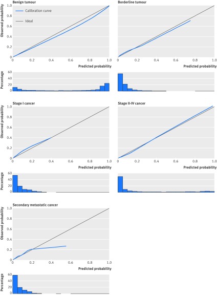
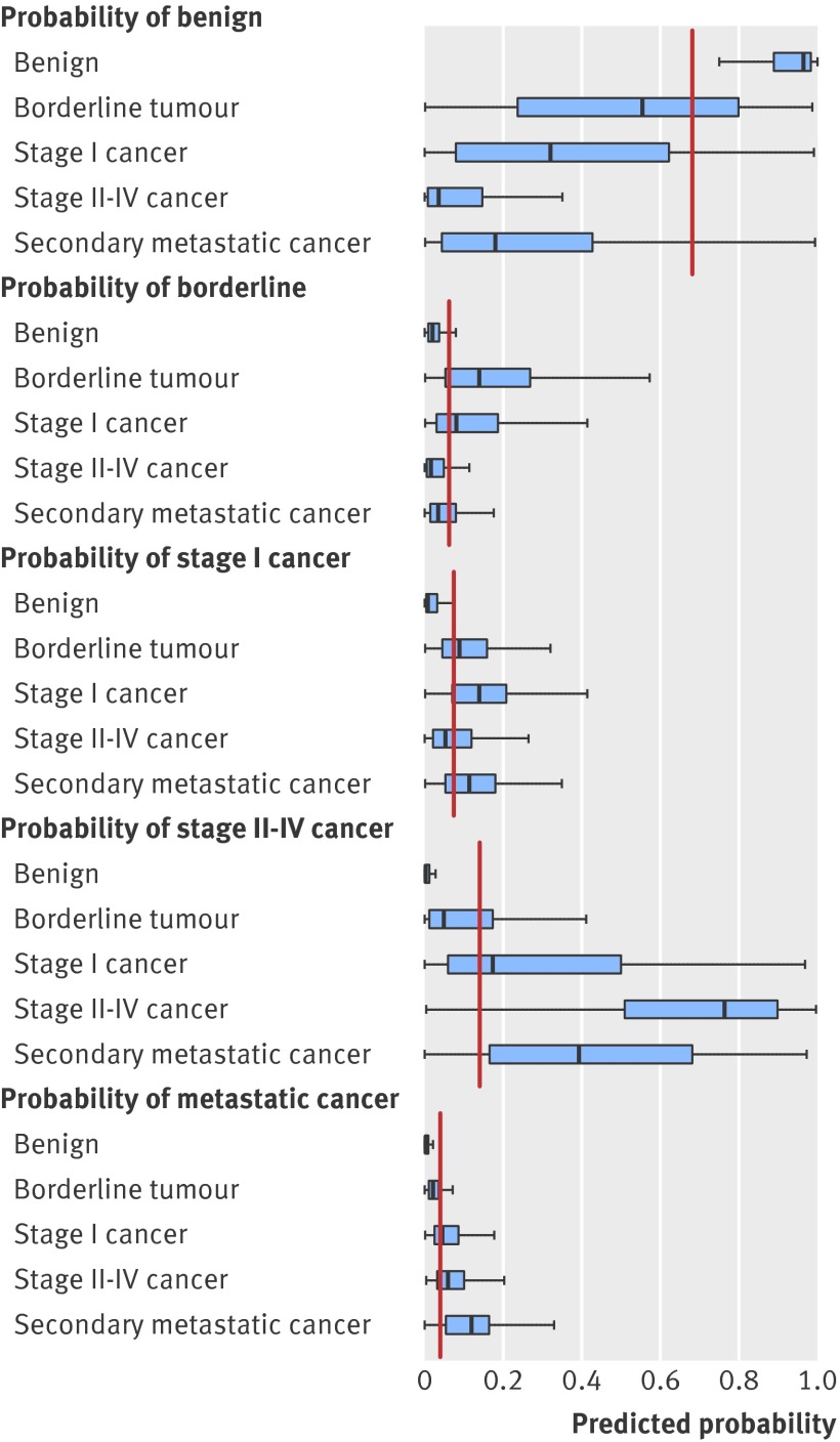
OBJECTIVES: To develop a risk prediction model to preoperatively discriminate between benign, borderline, stage I invasive, stage II-IV invasive, and secondary metastatic ovarian tumours. DESIGN: Observational diagnostic study using prospectively collected clinical and ultrasound data. SETTING: 24 ultrasound centres in 10 countries. PARTICIPANTS: Women with an ovarian (including para-ovarian and tubal) mass and who underwent a standardised ultrasound examination before surgery. The model was developed on 3506 patients recruited between 1999 and 2007, temporally validated on 2403 patients recruited between 2009 and 2012, and then updated on all 5909 patients. MAIN OUTCOME MEASURES: Histological classification and surgical staging of the mass. RESULTS: The Assessment of Different NEoplasias in the adneXa (ADNEX) model contains three clinical and six ultrasound predictors: age, serum CA-125 level, type of centre (oncology centres v other hospitals), maximum diameter of lesion, proportion of solid tissue, more than 10 cyst locules, number of papillary projections, acoustic shadows, and ascites. The area under the receiver operating characteristic curve (AUC) for the classic discrimination between benign and malignant tumours was 0.94 (0.93 to 0.95) on temporal validation. The AUC was 0.85 for benign versus borderline, 0.92 for benign versus stage I cancer, 0.99 for benign versus stage II-IV cancer, and 0.95 for benign versus secondary metastatic. AUCs between malignant subtypes varied between 0.71 and 0.95, with an AUC of 0.75 for borderline versus stage I cancer and 0.82 for stage II-IV versus secondary metastatic. Calibration curves showed that the estimated risks were accurate. CONCLUSIONS: The ADNEX model discriminates well between benign and malignant tumours and offers fair to excellent discrimination between four types of ovarian malignancy. The use of ADNEX has the potential to improve triage and management decisions and so reduce morbidity and mortality associated with adnexal pathology.
Overview
The claims and statistics of this paper haven’t been checked yet.
Run the check — free, no account needed. It reads the open-access text and checks the sources it cites.
Integrity signals
Registry
Cites
Statistics
Figures
Verification
Claims the paper makes
The claims and statistics of this paper haven’t been checked yet.
Run the check — free, no account needed. It reads the open-access text and checks the sources it cites.
Claims other papers make about this one
Each is checked against this paper’s own text: does it say what the citing paper says it does?
In the citing paper
“The ADNEX model, developed by the International Ovarian Tumor Analysis (IOTA) consortium (authors BVC and DT are part of this consortium), preoperatively estimates the probability of malignancy in women with an ovarian tumour who are scheduled for surgery. 31”
From Evaluation of performance measures in predictive artificial intelligence models to support medical decisions: overview and guidance (2025) · cites this paper as [31]
In the citing paper
“ADNEX was developed on the data from 5909 individuals recruited between 1999 and 2012 in 24 secondary and tertiary care centres from 10 countries (Italy, Belgium, Sweden, Czech Republic, Poland, France, United Kingdom, China, Spain, and Canada).”
From Evaluation of performance measures in predictive artificial intelligence models to support medical decisions: overview and guidance (2025) · cites this paper as [31]
Self-check
Percentages, sums and reported differences re-computed from the paper’s own numbers.
The self-check needs the paper’s full text.
Citations
Cites
(1990) · Choice Reviews Online · doi:10.5860/choice.27-3338 · Cited by 35,305
Hosmer, D. W., & Lemeshow, S. (2000) · doi:10.1002/0471722146 · Cited by 20,302
Siegel, R., Ma, J., Zou, Z., & Jemal, A. (2014) · CA: A Cancer Journal for Clinicians · doi:10.3322/caac.21208 · Cited by 11,499
SEER Cancer Statistics Review, 1975-2003
Ries, L. A. G., Harkins, D., Krapcho, M. F., Mariotto, A. B., Miller, B., Feuer, E. J., Clegg, L. X., Eisner, M. P., Horner, M., Howlader, N., et al. (2006) · Digital Archive @ GSU · Cited by 11,404
Regression Modeling Strategies
Harrell, F. E. (2001) · Springer series in statistics · doi:10.1007/978-1-4757-3462-1 · Cited by 9,270
Sterne, J. A. C., White, I. R., Carlin, J. B., Spratt, M., Royston, P., Kenward, M. G., Wood, A. M., & Carpenter, J. R. (2009) · BMJ · doi:10.1136/bmj.b2393 · Cited by 7,299
Signet-Ring Cell Carcinoma of the Colon: A Case Report and Review of the Literature
Park, P. Y., Goldin, T. A., Chang, J. C. S., Markman, M. M., & Kundranda, M. N. (2015) · Case Reports in Oncology · doi:10.1159/000441772 · Cited by 5,273
Clinical Prediction Models: A Practical Approach to Development, Validation, and Updating
Steyerberg, E. W. (2019) · Statistics for Biology and Health · doi:10.1007/978-3-030-16399-0 · Cited by 3,281
Rubin, M., & Stephen, C. D. (1993) · Cited by 3,149
Rao, S. J. (2003) · Journal of the American Statistical Association · doi:10.1198/jasa.2003.s263 · Cited by 1,967
Cited by
Sorted by citations
Andreotti, R. F., Timmerman, D., Strachowski, L. M., Froyman, W., Benacerraf, B. R., Bennett, G. L., Bourne, T., Brown, D. L., Coleman, B. G., Frates, M. C., et al. (2019) · Radiology · doi:10.1148/radiol.2019191150 · Cited by 530
van den Goorbergh, R., van Smeden, M., Timmerman, D., & CALSTER, B. V. (2022) · Journal of the American Medical Informatics Association · doi:10.1093/jamia/ocac093 · Cited by 427
Timmerman, D., CALSTER, B. V., Testa, A. C., Savelli, L., Fischerová, D., Froyman, W., Wynants, L., Holsbeke, C. V., Epstein, E., Franchi, D., et al. (2016) · American Journal of Obstetrics and Gynecology · doi:10.1016/j.ajog.2016.01.007 · Cited by 337
Meys, E. M. J., Kaijser, J., Kruitwagen, R. F. P. M., Slangen, B. F. M., CALSTER, B. V., Aertgeerts, B., Verbakel, J. Y. J., Timmerman, D., & Gorp, T. V. (2016) · European Journal of Cancer · doi:10.1016/j.ejca.2016.01.007 · Cited by 281
“Someof thesehavenotbeenvalidatedextensivelybut seem promising, such as the ADNEX-model [83].”
BackgroundClinical Use of Cancer Biomarkers in Epithelial Ovarian Cancer
Sölétormos, G., Duffy, M. J., Hassan, S. O. A., Verheijen, R. H. M., Tholander, B., Bast, R. C., Gaarenstroom, K. N., Sturgeon, C. M., Bonfrèr, J. M. G., Petersen, P. H., et al. (2015) · International Journal of Gynecological Cancer · doi:10.1097/igc.0000000000000586 · Cited by 255
“Assessment of Different NEoplasias in the adneXa.(33) The National Institute for Health and Care Excellence has introduced guidelines for early detection of ovarian cancer in symptomatic women for use by general practitioners.”
Three myths about risk thresholds for prediction models
on behalf of the Topic Group ‘Evaluating diagnostic tests and prediction models’ of the STRATOS initiative, Wynants, L., van Smeden, M., McLernon, D. J., Timmerman, D., Steyerberg, E. W., & Van Calster, B. (2019) · BMC Medicine · doi:10.1186/s12916-019-1425-3 · Cited by 213
“It was constructed using multinomial logistic regression and validated with more recent data and in other centers [14].”
MethodFroyman, W., Landolfo, C., Campo, B. D. C., Wynants, L., Sladkevicius, P., Testa, A. C., Holsbeke, C. V., Domali, E., Fruscio, R., Epstein, E., et al. (2019) · The Lancet Oncology · doi:10.1016/s1470-2045(18)30837-4 · Cited by 192
Gao, Y., Zeng, S., Xu, X., Li, H., Yao, S., Song, K., Li, X., Chen, L., Tang, J., Xing, H., et al. (2022) · The Lancet Digital Health · doi:10.1016/s2589-7500(21)00278-8 · Cited by 185
Andreotti, R. F., Timmerman, D., Benacerraf, B. R., Bennett, G. L., Bourne, T., Brown, D. L., Coleman, B. G., Frates, M. C., Froyman, W., Goldstein, S. R., et al. (2018) · Journal of the American College of Radiology · doi:10.1016/j.jacr.2018.07.004 · Cited by 178
“- Terms developed by the IOTA Group that are strongly evidence based were incorporated as a package modified by descriptors that are IOTA and non-IOTA based.”
BackgroundInfluentialPate, A., Riley, R. D., Collins, P. G. S., van Smeden, M., CALSTER, B. V., Ensor, J., & Martin, G. P. (2023) · Statistical Methods in Medical Research · doi:10.1177/09622802231151220 · Cited by 172
Westwood, M. E., Ramaekers, B. L. T., Lang, S., Grimm, S. E., Deshpande, S., de Kock, S., Armstrong, N., Joore, M. A., & Kleijnen, J. (2018) · Health Technology Assessment · doi:10.3310/hta22440 · Cited by 162
ESGO/ISUOG/IOTA/ESGE Consensus Statement on pre-operative diagnosis of ovarian tumors
Timmerman, D., Planchamp, F., Bourne, T., Landolfo, C., du Bois, A., Chiva, L. M., Cibula, D., Concin, N., Fischerová, D., Froyman, W., et al. (2021) · International Journal of Gynecological Cancer · doi:10.1136/ijgc-2021-002565 · Cited by 158
“36 –61 A study aiming to validate the ADNEX model when applied by Level II examiners has confirmed that it can be used successfully by less-experienced examiners.”
MethodRutten, M. J., van Meurs, H. S., van de Vrie, R., Gaarenstroom, K. N., Naaktgeboren, C. A., Gorp, T. V., ter Brugge, H. G., Hofhuis, W., Schreuder, H. W. R., Arts, H. J. G., et al. (2017) · Journal of Clinical Oncology · doi:10.1200/jco.2016.69.2962 · Cited by 145
Basha, M. A. A., Metwally, M. I., Gamil, S. A., Khater, H. M., Aly, S. A., Sammak, A. A. E., Zaitoun, M., Khattab, E. M., Azmy, T. M., Alayouty, N. A., et al. (2020) · European Radiology · doi:10.1007/s00330-020-07143-7 · Cited by 127
CALSTER, B. V., Valentin, L., Froyman, W., Landolfo, C., Ceusters, J., Testa, A. C., Wynants, L., Sladkevicius, P., Holsbeke, C. V., Domali, E., et al. (2020) · BMJ · doi:10.1136/bmj.m2614 · Cited by 133
Meys, E. M. J., Jeelof, L. S., Achten, N. M. J., Slangen, B. F. M., Lambrechts, S., Kruitwagen, R. F. P. M., & Gorp, T. V. (2016) · Ultrasound in Obstetrics and Gynecology · doi:10.1002/uog.17225 · Cited by 111
ESGO/ISUOG/IOTA/ESGE Consensus Statement on preoperative diagnosis of ovarian tumors
Timmerman, D., Planchamp, F., Bourne, T., Landolfo, C., du Bois, A., Chiva, L., Cibula, D., Concin, N., Fischerova, D., Froyman, W., et al. (2021) · Ultrasound in Obstetrics & Gynecology · doi:10.1002/uog.23635 · Cited by 114
Deep Learning Prediction of Ovarian Malignancy at US Compared with O-RADS and Expert Assessment
Chen, H., Yang, B.-W., Qian, L., Meng, Y.-S., Bai, X.-H., Hong, X.-W., He, X., Jiang, M.-J., Yuan, F., Du, Q.-W., et al. (2022) · Radiology · doi:10.1148/radiol.211367 · Cited by 110
Cao, L., Wei, M.-J., Liu, Y., Fu, J., Zhang, H., Huang, J., Pei, X., & Zhou, J. (2021) · Gynecologic Oncology · doi:10.1016/j.ygyno.2021.04.031 · Cited by 110
Hiett, A. K., Sonek, J., Guy, M. S., & Reid, T. J. A. (2021) · Ultrasound in Obstetrics and Gynecology · doi:10.1002/uog.24777 · Cited by 105
CALSTER, B. V., Hoorde, K. V., Froyman, W., Kaijser, J., Wynants, L., Landolfo, C., Anthoulakis, C., Vergote, I. B., Bourne, T., & Timmerman, D. (2015) · PubMed Central · Cited by 102
Van Calster, B., Collins, G. S., Vickers, A. J., Wynants, L., Kerr, K. F., Barreñada, L., Varoquaux, G., Singh, K., Moons, K. G., Hernandez-Boussard, T., et al. (2025) · The Lancet Digital Health · doi:10.1016/j.landig.2025.100916 · Cited by 112
Reinhold, C., Rockall, A., Sadowski, E. A., Siegelman, E. S., Maturen, K. E., Vargas, H. A., Forstner, R., Glanc, P., Andreotti, R. F., & Thomassin‐Naggara, I. (2021) · Journal of the American College of Radiology · doi:10.1016/j.jacr.2020.12.022 · Cited by 101
Sayasneh, A., Ferrara, L., Cock, B. D., Saso, S., Al‐Memar, M., Johnson, S., Kaijser, J., Carvalho, J., Husicka, R., Smith, A. A., et al. (2016) · British Journal of Cancer · doi:10.1038/bjc.2016.227 · Cited by 100
Key steps and common pitfalls in developing and validating risk models
Wynants, L., Collins, P. G. S., & CALSTER, B. V. (2016) · BJOG An International Journal of Obstetrics & Gynaecology · doi:10.1111/1471-0528.14170 · Cited by 99
A Perspective on Ovarian Cancer Biomarkers: Past, Present and Yet-To-Come
Ueland, F. R. (2017) · Diagnostics · doi:10.3390/diagnostics7010014 · Cited by 90
High-throughput approaches for precision medicine in high-grade serous ovarian cancer
Govindarajan, M., Wohlmuth, C., Waas, M., Bernardini, M. Q., & Kislinger, T. (2020) · Journal of Hematology & Oncology · doi:10.1186/s13045-020-00971-6 · Cited by 89
A spline-based tool to assess and visualize the calibration of multiclass risk predictions
Van Hoorde, K., Van Huffel, S., Timmerman, D., Bourne, T., & Van Calster, B. (2015) · Journal of Biomedical Informatics · doi:10.1016/j.jbi.2014.12.016 · Cited by 85
Adnexal masses during pregnancy: diagnosis, treatment, and prognosis
Cathcart, A. M., Nezhat, F. R., Emerson, J. B., Pejovic, T. B., NEZHAT, C. H., & NEZHAT, C. R. (2022) · American Journal of Obstetrics and Gynecology · doi:10.1016/j.ajog.2022.11.1291 · Cited by 87
External Validation of O-RADS US Risk Stratification and Management System
Hack, K., Gandhi, N., Bouchard‐Fortier, G., Chawla, T. P., Ferguson, S. E., Li, S., Kahn, D., Tyrrell, P. N., & Glanc, P. (2022) · Radiology · doi:10.1148/radiol.211868 · Cited by 80
Moro, F., Poma, C. B., Zannoni, G. F., Urbinati, A. M. V., Pasciuto, T., Ludovisi, M., Moruzzi, M. C., Carinelli, S. G., Franchi, D., Scambia, G., et al. (2017) · Ultrasound in Obstetrics and Gynecology · doi:10.1002/uog.17414 · Cited by 75
Cyclophilin A/CD147 Interaction: A Promising Target for Anticancer Therapy
Han, J. M., & Jung, H. J. (2022) · International Journal of Molecular Sciences · doi:10.3390/ijms23169341 · Cited by 75
Araújo, K. G., Jales, R. M., Pereira, P. N., Yoshida, A., Andrade, L. A. L. D. A., Sarian, L. O., & Derchain, S. (2016) · Ultrasound in Obstetrics and Gynecology · doi:10.1002/uog.15963 · Cited by 73
Indeterminate Adnexal Cysts at US: Prevalence and Characteristics of Ovarian Cancer
Sadowski, E. A., Paroder, V., Patel-Lippmann, K. K., Robbins, J. B., Barroilhet, L. M., Maddox, E., McMahon, T., Sampene, E., Wasnik, A. P., Blaty, A. D., et al. (2018) · Radiology · doi:10.1148/radiol.2018172271 · Cited by 72
Jha, P., Gupta, A., Baran, T. M., Maturen, K. E., Patel-Lippmann, K. K., Zafar, H. M., Kamaya, A., Antil, N., Barroilhet, L. M., & Sadowski, E. A. (2022) · JAMA Network Open · doi:10.1001/jamanetworkopen.2022.16370 · Cited by 72
Risk of Malignant Ovarian Cancer Based on Ultrasonography Findings in a Large Unselected Population
Smith‐Bindman, R., Pōder, L., Johnson, E., & Miglioretti, D. L. (2018) · JAMA Internal Medicine · doi:10.1001/jamainternmed.2018.5113 · Cited by 71
Update on Imaging of Ovarian Cancer
Forstner, R., Meissnitzer, M. W., & Cunha, T. M. (2016) · Current Radiology Reports · doi:10.1007/s40134-016-0157-9 · Cited by 70
“A prospective multi-center study reported that the nine predictors of this ADNEX model not only allow differentiation of benign tumors from malignant tumors, but also enable categorization in five types of adnexal tumors (benign; borderline; stage I tumors; stage II-IV and metastatic tumors) [25].”
BackgroundExternal validation of IOTA simple descriptors and simple rules for classifying adnexal masses
Alcázar, J. L., Pascual, M. A., Graupera, B., Aubà, M., Errasti, T., Olartecoechea, B., Ruiz-Zambrana, Á., Hereter, L., AJOSSA, S., & Guerriero, S. (2016) · Ultrasound in Obstetrics and Gynecology · doi:10.1002/uog.15854 · Cited by 67
Landolfo, C., Bourne, T., Froyman, W., CALSTER, B. V., Ceusters, J., Testa, A. C., Wynants, L., Sladkevicius, P., Holsbeke, C. V., Domali, E., et al. (2022) · Ultrasound in Obstetrics and Gynecology · doi:10.1002/uog.26080 · Cited by 63
Abramowicz, J. S., & Timmerman, D. (2017) · American Journal of Obstetrics and Gynecology · doi:10.1016/j.ajog.2017.07.019 · Cited by 65
Chiappa, V., Bogani, G., Interlenghi, M., Salvatore, C., Bertolina, F., Sarpietro, G., Signorelli, M., Castiglioni, I., & Raspagliesi, F. (2020) · Journal of Ultrasound · doi:10.1007/s40477-020-00503-5 · Cited by 62
Zhang, Q., Dai, X., & Li, W. (2023) · American Journal of Roentgenology · doi:10.2214/ajr.22.28396 · Cited by 66
Vara, J., Manzour, N., Chacón, E., López-Picazo, A., Linares, M., Pascual, M. A., Guerriero, S., & Alcázar, J. L. (2022) · Cancers · doi:10.3390/cancers14133151 · Cited by 66
Pozzati, F., Moro, F., Pasciuto, T., Gallo, C., Ciccarone, F., Franchi, D., Mancari, R., Giunchi, S., Timmerman, D., Landolfo, C., et al. (2018) · Ultrasound in Obstetrics and Gynecology · doi:10.1002/uog.19171 · Cited by 58
Chen, H., Qian, L., Jiang, M., Du, Q., Yuan, F., & Feng, W. (2019) · Ultrasound in Obstetrics and Gynecology · doi:10.1002/uog.20363 · Cited by 54
Timmerman, S., Valentin, L., Ceusters, J., Testa, A. C., Landolfo, C., Sladkevicius, P., Holsbeke, C. V., Domali, E., Fruscio, R., Epstein, E., et al. (2022) · JAMA Oncology · doi:10.1001/jamaoncol.2022.5969 · Cited by 53
External validation of the IOTA ADNEX model performed by two independent gynecologic centers
Szubert, S., Wójtowicz, A., Moszyński, R., Żywica, P., Dyczkowski, K., Stachowiak, A., Sajdak, S., Szpurek, D., & Alcázar, J. L. (2016) · Gynecologic Oncology · doi:10.1016/j.ygyno.2016.06.020 · Cited by 53
Imaging in gynecological disease (11): clinical and ultrasound features of mucinous ovarian tumors
Moro, F., Zannoni, G. F., Arciuolo, D., Pasciuto, T., Amoroso, S., Mascilini, F., Mainenti, S., Scambia, G., & Testa, A. C. (2016) · Ultrasound in Obstetrics and Gynecology · doi:10.1002/uog.17222 · Cited by 52
Chatterjee, J., Dai, W., Aziz, N. H. A., Teo, P. Y., Wahba, J., Phelps, D. L., Maine, C. J., Whilding, L. M., Dina, R. E., Trevisan, G., et al. (2016) · Clinical Cancer Research · doi:10.1158/1078-0432.ccr-16-2366 · Cited by 52
“95) for discriminating all-stage EOC from benign tumors (23).”
BackgroundAdnexal lesions: Imaging strategies for ultrasound and MR imaging
Sadowski, E. A., Rockall, A., Maturen, K. E., Robbins, J. B., & Thomassin‐Naggara, I. (2018) · Diagnostic and Interventional Imaging · doi:10.1016/j.diii.2018.06.003 · Cited by 49
Development of a clinical prediction model for diagnosing adenomyosis
Tellum, T., Nygaard, S., Skovholt, E. K., Qvigstad, E., & Lieng, M. (2018) · Fertility and Sterility · doi:10.1016/j.fertnstert.2018.06.009 · Cited by 49
Wynants, L., Timmerman, D., Verbakel, J. Y. J., Testa, A. C., Savelli, L., Fischerová, D., Franchi, D., Holsbeke, C. V., Epstein, E., Froyman, W., et al. (2017) · Clinical Cancer Research · doi:10.1158/1078-0432.ccr-16-3248 · Cited by 48
“After the publication of the 2 systematic reviews, the ADNEX risk model (Assessment of Different Neoplasias in the adnexa) was published (8).”
BackgroundMethodInfluentialMolecular Management of High-Grade Serous Ovarian Carcinoma
Punzón-Jiménez, P., Lago, V., Domingo, S., Simón, C., & Mas, A. (2022) · International Journal of Molecular Sciences · doi:10.3390/ijms232213777 · Cited by 49
“The Assessment of Different Neoplasias in the Adnexa (ADNEX) model represents an alternative strategy that considers six ultrasound and three clinical predictors (including CA125 levels) to discriminate between benign, borderline, invasive, and metastatic ovarian tumors [62].”
BackgroundEvaluation and Management of Ultrasonographically Detected Ovarian Tumors in Asymptomatic Women
van Nagell, J. R., & Miller, R. W. (2016) · Obstetrics and Gynecology · doi:10.1097/aog.0000000000001384 · Cited by 47
“discriminate between benign and malignant ovarian tumors.(36) The following clinical, ultrasound, and biomarker predictors were evaluated: patient age, serum CA 125, maximum tumor diameter, proportion of solid tissue, cyst locules greater than 10 cm, number of papillary projections, acoustic shadows, ascites, and type…”
BackgroundOvarian Preservation from Tumors and Torsions in Girls: Prospective Diagnostic Study
Stanković, Z. B., Sedlecky, K., Savic, D., Lukač, B. J., Mažibrada, I., & Perović, S. (2017) · Journal of Pediatric and Adolescent Gynecology · doi:10.1016/j.jpag.2017.01.008 · Cited by 51
Tumor markers: myths and facts unfolded
Faria, S. C., Sagebiel, T. L., Patnana, M., Cox, V. L., Viswanathan, C., Lall, C. G., Qayyum, A., & Bhosale, P. (2018) · Abdominal Radiology · doi:10.1007/s00261-018-1845-0 · Cited by 47
Moss, E. L., Taylor, A., Andreou, A., Ang, C., Arora, R., Attygalle, A. D., Banerjee, S. N., Bowen, R. L., Buckley, L., Burbos, N., et al. (2024) · European Journal of Obstetrics & Gynecology and Reproductive Biology · doi:10.1016/j.ejogrb.2024.06.025 · Cited by 49
Epstein, E., CALSTER, B. V., Timmerman, D., & Nikman, S. (2015) · Ultrasound in Obstetrics and Gynecology · doi:10.1002/uog.14892 · Cited by 46
Solving the problem of incomplete data in medical diagnosis via interval modeling
Wójtowicz, A., Żywica, P., Stachowiak, A., & Dyczkowski, K. (2016) · Applied Soft Computing · doi:10.1016/j.asoc.2016.05.029 · Cited by 45
Wynants, L., Kent, D. M., Timmerman, D., Lundquist, C. M., & CALSTER, B. V. (2019) · Diagnostic and Prognostic Research · doi:10.1186/s41512-019-0046-9 · Cited by 47
“, tertiary centers versus others) [8, 89, 90].”
Froyman, W., Wynants, L., Landolfo, C., Bourne, T., Valentin, L., Testa, A. C., Sladkevicius, P., Franchi, D., Fischerová, D., Savelli, L., et al. (2017) · Diagnostics · doi:10.3390/diagnostics7020032 · Cited by 45
“for the discrimination of benign from all malignant disease (all malignant subtypes grouped together) [24,25,31].”
BackgroundMethodInfluentialSerum HE4 and diagnosis of ovarian cancer in postmenopausal women with adnexal masses
Gentry‐Maharaj, A., Burnell, M., Dilley, J., Ryan, A., Karpinskyj, C., Gunu, R., Mallett, S., Deeks, J. J., Campbell, S. C., Jacobs, I. J., et al. (2019) · American Journal of Obstetrics and Gynecology · doi:10.1016/j.ajog.2019.07.031 · Cited by 42
Methods of Assessing Ovarian Masses
Froyman, W., & Timmerman, D. (2019) · Obstetrics and Gynecology Clinics of North America · doi:10.1016/j.ogc.2019.07.003 · Cited by 49
Davenport, C. F., Rai, N., Sharma, P., Deeks, J. J., Berhane, S., Mallett, S., Saha, P., Champaneria, R., Bayliss, S. E., Snell, K. I. E., et al. (2022) · Cochrane Database of Systematic Reviews · doi:10.1002/14651858.cd011964.pub2 · Cited by 47
Barreñada, L., Ledger, A., Dhiman, P., Collins, G., Wynants, L., Verbakel, J. Y., Timmerman, D., Valentin, L., & Van Calster, B. (2024) · BMJ Medicine · doi:10.1136/bmjmed-2023-000817 · Cited by 42
Seyedsalehi, A., & Fazel, S. (2024) · BMJ Mental Health · doi:10.1136/bmjment-2024-300990 · Cited by 45
Sundar, S. S., Agarwal, R., Davenport, C. F., Scandrett, K., Johnson, S., Sengupta, P. P., Vikram, R., Kwong, F. L., Mallett, S., Rick, C., et al. (2024) · The Lancet Oncology · doi:10.1016/s1470-2045(24)00406-6 · Cited by 41
Nougaret, S., Lakhman, Y., Molinari, N., Feier, D. S., Scelzo, C., Vargas, H. A., Sosa, R. E., Hricak, H., Soslow, R. A., Grisham, R. N., et al. (2018) · American Journal of Roentgenology · doi:10.2214/ajr.17.18254 · Cited by 42
“Furthermore, several risk prediction models have been put forth to discriminate between benign, borderline, and malignant ovarian masses [23, 24].”
BackgroundImaging and Differential Diagnosis of Ovarian Cancer
Shetty, M. K. (2019) · Seminars in Ultrasound CT and MRI · doi:10.1053/j.sult.2019.04.002 · Cited by 42
Unraveling a difficult diagnosis: the tricks for early recognition of ovarian cancer
Giampaolino, P., Corte, L. D., Foreste, V., Vitale, S. G., Chiofalo, B., Cianci, S., Zullo, F., & Bifulco, G. (2019) · Minerva Medica · doi:10.23736/s0026-4806.19.06086-5 · Cited by 41
Contemporary Guidelines for Adnexal Mass Imaging: A 2020 Update
Stein, E. B., Roseland, M. E., Shampain, K. L., Wasnik, A. P., & Maturen, K. E. (2020) · Abdominal Radiology · doi:10.1007/s00261-020-02812-z · Cited by 41
“The method of risk assessment used in O-RADS US is based on data from the International Ovarian Tumor Analysis (IOTA) group’s Assessment of Different Neoplasias in the adnexa (ADNEX), a mathematical model which calculates the likelihood of malignancy with high positive predictive value [27, 28].”
MethodA comparison of hyperparameter tuning procedures for clinical prediction models: A simulation study
Dunias, Z. S., CALSTER, B. V., Timmerman, D., Boulesteix, A., & van Smeden, M. (2024) · Statistics in Medicine · doi:10.1002/sim.9932 · Cited by 40
Role of artificial intelligence applied to ultrasound in gynecology oncology: A systematic review
Moro, F., Ciancia, M., Zaçe, D., Vagni, M., Tran, H. E., Giudice, M. T., Zoccoli, S. G., Mascilini, F., Ciccarone, F., Boldrini, L., et al. (2024) · International Journal of Cancer · doi:10.1002/ijc.35092 · Cited by 54
Sayasneh, A., Kaijser, J., Preisler, J., Smith, A. A., Raslan, F., Johnson, S., Husicka, R., Ferrara, L., Stalder, C. M., Ghaem‐Maghami, S., et al. (2014) · Ultrasound in Obstetrics and Gynecology · doi:10.1002/uog.14675 · Cited by 40
Adaptive sample size determination for the development of clinical prediction models
Christodoulou, E., van Smeden, M., Edlinger, M., Timmerman, D., Wanitschek, M. M., Steyerberg, E. W., & CALSTER, B. V. (2021) · Diagnostic and Prognostic Research · doi:10.1186/s41512-021-00096-5 · Cited by 39
Fischerová, D., Smet, C., Scovazzi, U., Sousa, D. N., Hundarova, K., & Haldorsen, I. S. (2024) · International Journal of Gynecological Cancer · doi:10.1136/ijgc-2023-004609 · Cited by 41
A systematic approach to adnexal masses discovered on ultrasound: the ADNEx MR scoring system
Sadowski, E. A., Robbins, J. B., Rockall, A., & Thomassin‐Naggara, I. (2017) · Abdominal Radiology · doi:10.1007/s00261-017-1272-7 · Cited by 39
Survival of Women With Type I and II Epithelial Ovarian Cancer Detected by Ultrasound Screening
van Nagell, J. R., Burgess, B. T., Miller, R. W., Baldwin, L. A., DeSimone, C. P., Ueland, F. R., Huang, B., Chen, Q., Kryscio, R. J., & Pavlik, E. J. (2018) · Obstetrics and Gynecology · doi:10.1097/aog.0000000000002921 · Cited by 39
Ultrasound in Gynecological Cancer: Is It Time for Re-evaluation of Its Uses?
Fischerová, D., & Cibula, D. (2015) · Current Oncology Reports · doi:10.1007/s11912-015-0449-x · Cited by 39
Viora, E., Piovano, E., Poma, C. B., Cotrino, I., Castiglione, A., Cavallero, C., Sciarrone, A., Bastonero, S., Iskra, L., & Zola, P. (2020) · European Journal of Obstetrics & Gynecology and Reproductive Biology · doi:10.1016/j.ejogrb.2020.02.022 · Cited by 38
Terzić, M., Aimagambetova, G. N., Norton, M., Corte, L. D., Marín-Buck, A., Lisón, J. F., Amer-Cuenca, J. J., Zito, G., Garzon, S., Caruso, S., et al. (2020) · Journal of Obstetrics and Gynaecology · doi:10.1080/01443615.2020.1732892 · Cited by 39
“Therefore, the IOTA ADNEX model can help to differentiate benign from malignant tumours, as well as identify the histologic types and tumour spread (Van Calster et al. 2014).”
BackgroundMethodPi, Y., Wilson, M. P., Katlariwala, P., Sam, M., Ackerman, T. E., Paskar, L., Patel, V., & Low, G. (2021) · Abdominal Radiology · doi:10.1007/s00261-021-03193-7 · Cited by 38
Moro, F., Giudice, M. T., Ciancia, M., Zaçe, D., Baldassari, G., Vagni, M., Tran, H. E., Scambia, G., & Testa, A. C. (2025) · Ultrasound in Obstetrics and Gynecology · doi:10.1002/uog.29171 · Cited by 45
Jeong, S. Y., Park, B. K., Lee, Y.-Y., & Kim, T. (2020) · Journal of Clinical Medicine · doi:10.3390/jcm9062010 · Cited by 37
“In 2014, a new model with better performance, the assessment of different neoplasias in the adnexa (ADNEX) model, was developed in this group [13].”
MethodChiappa, V., Interlenghi, M., Bogani, G., Salvatore, C., Bertolina, F., Sarpietro, G., Signorelli, M., Ronzulli, D., Castiglioni, I., & Raspagliesi, F. (2021) · European Radiology Experimental · doi:10.1186/s41747-021-00226-0 · Cited by 35
Moro, F., Pasciuto, T., Djokovic, D., Legge, A. D., Granato, V., Moruzzi, M. C., Mancari, R., Zannoni, G. F., Fischerová, D., Franchi, D., et al. (2018) · Ultrasound in Obstetrics and Gynecology · doi:10.1002/uog.19174 · Cited by 33
Low-grade epithelial ovarian cancer: what a radiologist should know
Elsherif, S. B., Javadi, S., Viswanathan, C., Faria, S. C., & Bhosale, P. (2019) · British Journal of Radiology · doi:10.1259/bjr.20180571 · Cited by 32
Management of ovarian masses in pregnancy: patient selection for interventional treatment
Testa, A. C., Mascilini, F., Quagliozzi, L., Moro, F., Bolomini, G., Mirandola, M., Moruzzi, M. C., Scambia, G., & Fagotti, A. (2020) · International Journal of Gynecological Cancer · doi:10.1136/ijgc-2020-001996 · Cited by 32
Lai, H.-W., Lyu, G., Kang, Z., Li, L., Zhang, Y., & Huang, Y. (2021) · Journal of Ultrasound in Medicine · doi:10.1002/jum.15834 · Cited by 32
Chen, G.-Y., Hsu, T., Chan, I., Liu, C., Chao, W., Shih, Y.-C., Jiang, L., Chang, Y., Wang, P., & Chen, Y. (2022) · European Radiology · doi:10.1007/s00330-022-08803-6 · Cited by 34
Koneczny, J., Czekierdowski, A., Florczak, M., Poziemski, P., Stachowicz, N., & Borowski, D. (2017) · Ginekologia Polska · doi:10.5603/gp.a2017.0116 · Cited by 32
Mascilini, F., Savelli, L., Scifo, M. C., Exacoustós, C., Timor‐Tritsch, I. E., Blasis, I. D., Moruzzi, M. C., Pasciuto, T., Scambia, G., Valentin, L., et al. (2016) · Ultrasound in Obstetrics and Gynecology · doi:10.1002/uog.17216 · Cited by 31
Bourne, T., Leonardi, M., Kyriacou, C., Al‐Memar, M., Landolfo, C., Cibula, D., Condous, G., Metzger, U., Fischerova, D., Timmerman, D., et al. (2020) · Ultrasound in Obstetrics & Gynecology · doi:10.1002/uog.22047 · Cited by 29
Du, Y., Xiao, Y., Guo, W., Yao, J., Lan, T., Li, S., Wen, H., Zhu, W., He, G., Zheng, H., et al. (2024) · BioMedical Engineering OnLine · doi:10.1186/s12938-024-01234-y · Cited by 33
Landolfo, C., Valentin, L., Franchi, D., Holsbeke, C. V., Fruscio, R., Froyman, W., Sladkevicius, P., Kaijser, J., Ameye, L., Bourne, T., et al. (2018) · Ultrasound in Obstetrics and Gynecology · doi:10.1002/uog.18951 · Cited by 30
Du, Y., Guo, W., Xiao, Y., Chen, H., Yao, J., & Wu, J. (2024) · BMC Medical Imaging · doi:10.1186/s12880-024-01251-2 · Cited by 34
“[12, 29, 30].”
Funston, G., van Melle, M., Baun, M. L., Jensen, H., Helsper, C. W., Emery, J. D., Crosbie, E. J., Thompson, M. J., Hamilton, W. T., & Walter, F. M. (2019) · BMC Cancer · doi:10.1186/s12885-019-6211-2 · Cited by 29
Yu, X., Wang, L., Yu, H., Zou, Y., Wang, C., Jiao, J., Hong, H., & Zhang, S. (2021) · Cancer Management and Research · doi:10.2147/cmar.s284220 · Cited by 28
“models have been proposed to distinguish benign, borderline, and malignant ovarian tumors.(19,20) The MDCT diagnosis of ovarian malignant tumors is related to various findings, such as the presence of bilateral and cystic-solid masses, a large soft-tissue component with necrosis, a tumor with thick and irregular septa…”
BackgroundYoeli‐Bik, R., Longman, R. E., Wroblewski, K., Weigert, M., Abramowicz, J. S., & Lengyel, E. R. (2023) · JAMA Network Open · doi:10.1001/jamanetworkopen.2023.23289 · Cited by 29
Kaijser, J. (2015) · PubMed Central · Cited by 27
Metadata
- Authors
- Van Calster, B., Van Hoorde, K., Valentin, L., Testa, A. C., Fischerova, D., Van Holsbeke, C., Savelli, L., Franchi, D., Epstein, E., Kaijser, J., Van Belle, V., Czekierdowski, A., Guerriero, S., Fruscio, R., Lanzani, C., Scala, F., Bourne, T., Timmerman, D., & International Ovarian Tumour Analysis (IOTA) group
- Affiliations
- Lund University (SE)
- Università Cattolica del Sacro Cuore (IT)
- IRCCS Azienda Ospedliero-Universitaria di Bologna Policlinico di Sant'Orsola (IT)
- European Institute of Oncology (IT)
- Karolinska University Hospital (SE)
- Universitair Ziekenhuis Leuven (BE)
- Medical University of Lublin (PL)
- Azienda Ospedaliero-Universitaria Cagliari (IT)
- University of Milan (IT)
- Istituto Nazionale Tumori IRCCS "Fondazione G. Pascale" (IT)
- ORCID
- Venue
- BMJ
- Publisher
- BMJ
- Published
- 2014-10-15
- Volume
- 349
- Issue
- oct07 3
- Pages
- g5920-g5920
- ISSN
- 1756-1833 (ISSN-L)
- PMC
- PMC4198550
- OpenAlex
- W2099878111
- Cited by
- 510
- References
- 57
- Citations by year
- 2026: 52 · 2025: 60 · 2024: 70 · 2023: 54 · 2022: 55 · 2021: 59 · 2020: 36 · 2019: 24 · 2018: 30 · 2017: 19 · 2016: 27 · 2015: 19 · 2014: 4
- FWCI
- 27.87
- Type
- journal article
- Language
- en
- Open access
- hybrid
- doi.org
- Licence
- cc-by-nc
- Locations
- BMJ (publishedVersion, cc-by-nc)
- PubMed Central (publishedVersion, cc-by-nc)
- PubMed (publishedVersion)
- BOA (University of Milano-Bicocca) (submittedVersion, cc-by-nc)
- Lirias (KU Leuven) (submittedVersion, cc-by-nc)
- UNICA IRIS Institutional Research Information System (University of Cagliari) (submittedVersion, other-oa)
- Lund University Publications (Lund University) (submittedVersion, other-oa)
- HighWire Press Open Archive (submittedVersion)
- PubliCatt (Università Cattolica del Sacro Cuore) (submittedVersion, cc-by-nc)
- Spiral (Imperial College London) (submittedVersion, other-oa)
- Funders
- National Institute for Health and Care Research
- Imperial College London
- Imperial College Healthcare NHS Trust
- Fonds Wetenschappelijk Onderzoek (G049312N)
- iMinds
- Malmö Högskola
- Skånes universitetssjukhus
- Vlaamse regering (G049312N)
- Medical Research Council
- Topics
- Health Sciences › Medicine › Oncology › Ovarian cancer diagnosis and treatment
- Life Sciences › Biochemistry, Genetics and Molecular Biology › Genetics › BRCA gene mutations in cancer
- Health Sciences › Medicine › Pathology and Forensic Medicine › Breast Lesions and Carcinomas
- Keywords
- ovarian cancer, ADNEX model, preoperative risk prediction, ovarian ultrasonography, borderline ovarian tumors, CA125, surgical staging, adnexal mass, triage
- MeSH terms
- Adnexal Diseases, Adult, Female, Humans, Neoplasm Staging, Ovarian Neoplasms, Predictive Value of Tests, Prospective Studies, Ultrasonography, Models, Statistical, Risk Assessment
About these checks
Registry, retraction, Cites and statistics results are computed, not judged, with one exception: a printed reference that no index lists is read once by a model before it counts as a citation error. Claims are extracted by a model and kept only when their quote appears verbatim in the text; each cited source is then checked, with a model’s help, for whether it says what the claim relies on. Those verdicts can be wrong — read the claim’s quote and the cited source before relying on one.
Statistics and arithmetic are recomputed, not judged.
Confidence bands are shown only for fields calibrated on our annotated test set. None is calibrated yet, so no band is shown.
What counts as a claim ›How we measure confidence ›How we check this ›
Are you an author, or do you see a problem with this page? Request a review
