

The DOI matches its Crossref record; 57 of the 57 references it cites were found in a registry or an index.
Evaluating the risk of ovarian cancer before surgery using the ADNEX model to differentiate between benign, borderline, early and advanced stage invasive, and secondary metastatic tumours: prospective multicentre diagnostic study
Open accessAbstract
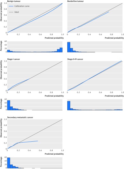
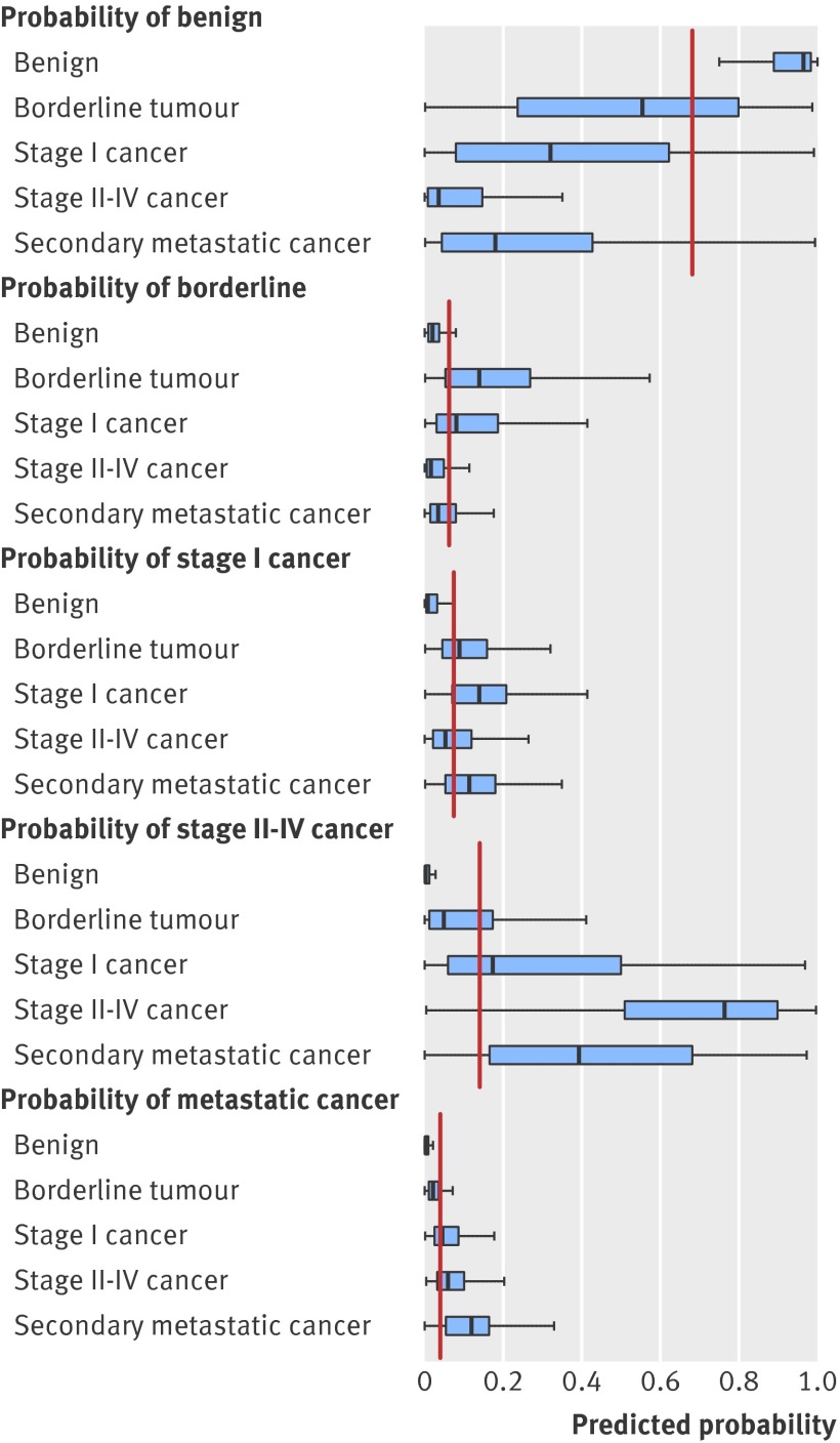
OBJECTIVES: To develop a risk prediction model to preoperatively discriminate between benign, borderline, stage I invasive, stage II-IV invasive, and secondary metastatic ovarian tumours. DESIGN: Observational diagnostic study using prospectively collected clinical and ultrasound data. SETTING: 24 ultrasound centres in 10 countries. PARTICIPANTS: Women with an ovarian (including para-ovarian and tubal) mass and who underwent a standardised ultrasound examination before surgery. The model was developed on 3506 patients recruited between 1999 and 2007, temporally validated on 2403 patients recruited between 2009 and 2012, and then updated on all 5909 patients. MAIN OUTCOME MEASURES: Histological classification and surgical staging of the mass. RESULTS: The Assessment of Different NEoplasias in the adneXa (ADNEX) model contains three clinical and six ultrasound predictors: age, serum CA-125 level, type of centre (oncology centres v other hospitals), maximum diameter of lesion, proportion of solid tissue, more than 10 cyst locules, number of papillary projections, acoustic shadows, and ascites. The area under the receiver operating characteristic curve (AUC) for the classic discrimination between benign and malignant tumours was 0.94 (0.93 to 0.95) on temporal validation. The AUC was 0.85 for benign versus borderline, 0.92 for benign versus stage I cancer, 0.99 for benign versus stage II-IV cancer, and 0.95 for benign versus secondary metastatic. AUCs between malignant subtypes varied between 0.71 and 0.95, with an AUC of 0.75 for borderline versus stage I cancer and 0.82 for stage II-IV versus secondary metastatic. Calibration curves showed that the estimated risks were accurate. CONCLUSIONS: The ADNEX model discriminates well between benign and malignant tumours and offers fair to excellent discrimination between four types of ovarian malignancy. The use of ADNEX has the potential to improve triage and management decisions and so reduce morbidity and mortality associated with adnexal pathology.
Overview
The claims and statistics of this paper haven’t been checked yet.
Run the check — free, no account needed. It reads the open-access text and checks the sources it cites.
Integrity signals
Registry
Cites
Statistics
Figures
Verification
Claims the paper makes
The claims and statistics of this paper haven’t been checked yet.
Run the check — free, no account needed. It reads the open-access text and checks the sources it cites.
Claims other papers make about this one
Each is checked against this paper’s own text: does it say what the citing paper says it does?
In the citing paper
“The ADNEX model, developed by the International Ovarian Tumor Analysis (IOTA) consortium (authors BVC and DT are part of this consortium), preoperatively estimates the probability of malignancy in women with an ovarian tumour who are scheduled for surgery. 31”
From Evaluation of performance measures in predictive artificial intelligence models to support medical decisions: overview and guidance (2025) · cites this paper as [31]
In the citing paper
“ADNEX was developed on the data from 5909 individuals recruited between 1999 and 2012 in 24 secondary and tertiary care centres from 10 countries (Italy, Belgium, Sweden, Czech Republic, Poland, France, United Kingdom, China, Spain, and Canada).”
From Evaluation of performance measures in predictive artificial intelligence models to support medical decisions: overview and guidance (2025) · cites this paper as [31]
Self-check
Percentages, sums and reported differences re-computed from the paper’s own numbers.
The self-check needs the paper’s full text.
Citations
Cites
(1990) · Choice Reviews Online · doi:10.5860/choice.27-3338 · Cited by 35,305
Hosmer, D. W., & Lemeshow, S. (2000) · doi:10.1002/0471722146 · Cited by 20,302
Siegel, R., Ma, J., Zou, Z., & Jemal, A. (2014) · CA: A Cancer Journal for Clinicians · doi:10.3322/caac.21208 · Cited by 11,499
SEER Cancer Statistics Review, 1975-2003
Ries, L. A. G., Harkins, D., Krapcho, M. F., Mariotto, A. B., Miller, B., Feuer, E. J., Clegg, L. X., Eisner, M. P., Horner, M., Howlader, N., et al. (2006) · Digital Archive @ GSU · Cited by 11,404
Regression Modeling Strategies
Harrell, F. E. (2001) · Springer series in statistics · doi:10.1007/978-1-4757-3462-1 · Cited by 9,270
Sterne, J. A. C., White, I. R., Carlin, J. B., Spratt, M., Royston, P., Kenward, M. G., Wood, A. M., & Carpenter, J. R. (2009) · BMJ · doi:10.1136/bmj.b2393 · Cited by 7,299
Signet-Ring Cell Carcinoma of the Colon: A Case Report and Review of the Literature
Park, P. Y., Goldin, T. A., Chang, J. C. S., Markman, M. M., & Kundranda, M. N. (2015) · Case Reports in Oncology · doi:10.1159/000441772 · Cited by 5,273
Clinical Prediction Models: A Practical Approach to Development, Validation, and Updating
Steyerberg, E. W. (2019) · Statistics for Biology and Health · doi:10.1007/978-3-030-16399-0 · Cited by 3,281
Rubin, M., & Stephen, C. D. (1993) · Cited by 3,149
Rao, S. J. (2003) · Journal of the American Statistical Association · doi:10.1198/jasa.2003.s263 · Cited by 1,967
Cited by
Sorted by citations
Items 401–500 of 510
Zeng, J., Cao, W., & Wang, L. (2021) · Journal of Shanghai Jiaotong University (Science) · doi:10.1007/s12204-021-2255-y
Multyparametric MRI in differential diagnosis of epithelial ovarian tumors
Nosova, Y. V., Solopova, A. E., & Khabas, G. N. (2020) · GYNECOLOGY · doi:10.26442/20795696.2020.6.200519
“новообразований придатков (ADNEX), проведенной группой Международного анализа опухолей яичников (IOTA): математическая модель, которая рассчитывает вероятность злокачественного потенциала новообразования с высокой положительной прогностической ценностью [13, 14].”
Çeliksoy, H. Y., Sözen, H., Baktıroğlu, M., Topuz, S., & Salihoğlu, M. Y. (2021) · Journal of Istanbul Faculty of Medicine / İstanbul Tıp Fakültesi Dergisi · doi:10.26650/iuitfd.2020.0038
“194 Clinicopathologic features of probably malignant adnexal masses İstanbul Tıp Fakültesi Dergisi • J Ist Faculty Med 2021;84(2):192-6”
BackgroundInfluentialCurva de aprendizaje para el diagnóstico ecográfico específico de masas anexiales
Guillermo Avilés, L., Cerva, H. V., Linder Díaz, C., Fernando Amor, L., Alberto Iturra, A., Mauricio León, A., Víctor Toledo, S., & Raimundo Avilés, D. (2021) · Revista chilena de obstetricia y ginecología · doi:10.4067/s0717-75262021000200137 · Cited by 1
Operatives Management von Adnextumoren
Schüler‐Toprak, S., & Ortmann, O. (2021) · Frauenheilkunde up2date · doi:10.1055/a-1372-7828
“Das ADNEX-Modell berücksichtigt zusätzlich zu sonografischen Kriterien auch klinische Parameter [19].”
BackgroundImaging in Gynaecological Practice
Amso, N. N. (2021) · Cambridge University Press eBooks · doi:10.1017/9781108582322.004
Riley, R. D., Tierney, J. F., & Stewart, L. A. (2021) · Wiley eBooks · doi:10.1002/9781119333784.refs5
Wang, J., He, P., Gao, L., Tian, Y., & Wu, Q. (2022) · Clinical and Experimental Obstetrics & Gynecology · doi:10.31083/j.ceog4904096
“The IOTA group has developed a number of diagnostic strategies, such as Easy Descriptors, Logistic Regression Models 1 and 2 (LR1 and LR2) [14], Simple Rules [15,16], Simple Rules risk assessment [17], and Assessment of Different Neoplasias in the adneXa (ADNEX) model [18].”
MethodSonography of adnexal lesion, “ADNEX”, and ovarian cancer: clinical utility of a predictive model
Jan, Ž., & Mörtl, M. (2022) · Geburtshilfe und Frauenheilkunde · doi:10.1055/s-0042-1746159
Sonographie in der Gynäkologie - Teil 2: suspekte und pathologische Befunde
Voigt, F., Faschingbauer, F., Dammer, U., Vasku, M., Maass, N., & Goecke, T. W. (2016) · Springer eBooks · doi:10.1007/978-3-662-49480-6_8
Kantelhardt, E. J., & Papendick, N. (2020) · Springer eBooks · doi:10.1007/978-3-662-59809-2_21
Horała, A., Plewa, S., Dereziński, P., Klupczyńska, A., Matysiak, J., Nowak‐Markwitz, E., & Kokot, Z. J. (2020) · Research Square · doi:10.21203/rs.3.rs-109758/v1
“Wiley-Blackwell; 2010 Oct;161(4):755–67.”
Germer, U. (2017) · Springer eBooks · doi:10.1007/978-3-662-53662-9_33
Borderline ovarian tumors: modern diagnostic criteria at the preoperative stage
Tsypurdeyeva, A. A., Protasova, A. E., Александровна, С. И., & Mikhelashvili, L. I. (2022) · Journal of obstetrics and women s diseases · doi:10.17816/jowd81798
Extramedullary haematopoiesis presenting as an adnexal mass in a patient with β-thalassaemia
Filippi, V., Reina, H., Monod, C., & Manegold‐Brauer, G. (2022) · BMJ Case Reports · doi:10.1136/bcr-2022-249422
Radiology Investigations and Interventions in Gynaeoncology
Ambadipudi, L. (2022) · Springer eBooks · doi:10.1007/978-3-030-94110-9_18
Gong, L., Dong, S., Li, X.-Y., Wu, Y., Zhang, S., Feng, Y.-N., Du, X.-S., Tian, J., & Sun, L.-T. (2022) · Research Square · doi:10.21203/rs.3.rs-2062886/v1
Aktuelle Aspekte zur künstlichen Intelligenz in der gynäkologischen Diagnostik
Scharf, J. L., Dracopoulos, C., Gembicki, M., Welp, A., & Weichert, J. (2022) · Die Gynäkologie · doi:10.1007/s00129-022-04993-8
Timmerman, D. (2015) · PubMed Central
The Female Urogenital System in Geriatric Patients
Cova, M. A., Bottaro, L., Marrocchio, C., & Bozzato, A. M. (2023) · Practical issues in geriatrics · doi:10.1007/978-3-031-14877-4_11
Mohamed, S. S. M., Elbehidy, T. A. E., Ahmed, R. A. E., & Ali, M. R. (2023) · The Egyptian Journal of Hospital Medicine · doi:10.21608/ejhm.2023.305559
“3% for a cut-off of 10% to predict malignancy ((28)).”
BackgroundPate, A., Riley, R. D., Sperrin, M., CALSTER, B. V., Sergeant, J. C., Peek, N. B., van Staa, T. P., Mamas, M. A., Lip, G. Y. H., O’Flaherty, M., et al. (2023) · Research Square · doi:10.21203/rs.3.rs-3121017/v1
Biziaev, T., Aktary, M. L., Wang, Q., Chekouo, T., Bhatti, P., Shack, L., Robson, P. J., & Kopciuk, K. A. (2023) · Cancers · doi:10.3390/cancers15143545 · Cited by 1
Ledger, A., Ceusters, J., Lil, V., Antonia, T., Holsbeke, C. V., Dorella, F., Tom, B., Froyman, W., Dirk, T., & CALSTER, B. V. (2023) · medRxiv · doi:10.1101/2023.07.25.23293141
Hernández, N. E., Escudero, J. F. D. L., Méndez, L. I. P., Quintana, D. R. B., Santana, E. B., Álvarez, J. A. P., Medina, N. R. S., Pérez, C. C., & Lopez-Fe, A. Q. (2023) · Clínica e Investigación en Ginecología y Obstetricia · doi:10.1016/j.gine.2023.100910
Sarmiento-Babiera, M. J. F., & Llamas-Clark, E. F. (2022) · Philippine Journal of Obstetrics and Gynecology · doi:10.4103/pjog.pjog_37_22
Beyond the AJR: Augmenting Adnexal Mass Evaluation Through Standardized Risk Models
Virarkar, M., & Bhosale, P. (2023) · American Journal of Roentgenology · doi:10.2214/ajr.23.30052
Singh, K., & Gupta, B. (2023) · Springer eBooks · doi:10.1007/978-3-031-36179-1_10
Li, Y., Shao, G., Wu, M., Zhang, F., Zhang, Y., & Shao, C. (2023) · Journal of obstetrics and gynaecology research · doi:10.1111/jog.15831
Applicabilité de l’Adnex score dans la prédiction de la malignité des kystes ovariens
Mekni, K., Baba, M., Haddad, I., Aaraar, M., Mejri, O., & ElFekih, C. (2023) · Gynécologie Obstétrique Fertilité & Sénologie · doi:10.1016/j.gofs.2023.12.001
Horta, M., & Cunha, T. M. (2023) · Springer eBooks · doi:10.1007/978-3-031-28524-0_17
Чекалова, М. А., Кедрова, А. Г., Cherkasov, E. Y., Kryazheva, V. S., & Greyan, T. A. (2023) · Tumors of female reproductive system · doi:10.17650/1994-4098-2023-19-4-114-122
El‐Tawab, S. S., Hamed, A. S., Tawfik, T. A. S., & Youssef, R. E. N. E. (2024) · Italian Journal of Gynaecology and Obstetrics · doi:10.36129/jog.2024.158
Differential Diagnosis and Detection of Malignancy of Ovarian Tumors
Kurjak, A., & Petanovski, Z. (2024) · Donald School Journal of Ultrasound in Obstetrics & Gynecology · doi:10.5005/jp-journals-10009-2010
Automatic segmentation of malignant and benign adnexal lesions on ultrasound scans
Whitney, H. M., Yoeli‐Bik, R., Abramowicz, J. S., Li, L., Li, H., Longman, R. E., Lengyel, E. R., & Giger, M. L. (2024) · SPIE Medical Imaging: Computer-Aided Diagnosis · doi:10.1117/12.3008273
Lourenço, M., Arrufat, T., Satorres-Pérez, E., Maderuelo, S., Álamo, B. N.-D., Guerriero, S., Orozco, R., & Alcázar, J. L. (2024) · Applied Sciences · doi:10.3390/app14072998
Nguyen, H. B., Tran, D. T., & Nguyễn, V. Q. H. (2024) · Tạp chí Y Dược Huế · doi:10.34071/jmp.2024.2.5
Kurniadi, A., Permadi, W., Kusuma, A. Y., Kireina, J., Andarini, M. Y., Winarno, G. N. A., & Harsono, A. B. (2024) · Indonesian Journal of Cancer · doi:10.33371/ijoc.v18i1.1044
The role of prediction models in the classification of adnexal mass
Vilendečić, Z., & Stefanović, A. (2024) · Medicinski podmladak · doi:10.5937/mp75-43210
Características ultrasonográficas del disgerminoma, a propósito de 3 casos
Paredes, R. O. L. (2024) · Revista Peruana de Ginecología y Obstetricia · doi:10.31403/rpgo.v70i2634
Bhaskar, S., Pasoria, S., & Manmohan, M. (2024) · International Journal of Science and Research (IJSR) · doi:10.21275/sr24613151035
Taşkıran, Ç., & Cantürk, M. M. (2024) · The Lancet Oncology · doi:10.1016/s1470-2045(24)00450-9
Pérez, A. R., Caruso, A., Garrido, M. P., Jiménez, I. R., Velasco, A. P., & Alba, J. J. F. (2024) · Clínica e Investigación en Ginecología y Obstetricia · doi:10.1016/j.gine.2024.101000
Barreñada, L., Ledger, A., Kotlarz, A., Dhiman, P., Collins, P. G. S., Wynants, L., Verbakel, J. Y. J., Valentin, L., Timmerman, D., & CALSTER, B. V. (2024) · medRxiv · doi:10.1101/2024.11.29.24318146
Sonografische Differenzialdiagnostik von Adnexbefunden
Hoopmann, M. (2024) · gynäkologie + geburtshilfe · doi:10.1007/s15013-024-5955-1
“Sowohl die IOTA-Ausgangsarbeit wie auch zahlreiche Bestätigungsstudien do-kumentieren eine gut funktionierende Unterscheidung zwischen benignen und bösartigen Befunden [19, 20, 21].”
Moro, F., Momi, M., Bertoldo, V., Ledger, A., Barreñada, L., Ceusters, J., Sturla, D., Ghezzi, F., Mor, E., Fornari, L., et al. (2024) · medRxiv · doi:10.1101/2024.12.23.24319517 · Cited by 1
Imaging Considerations for Adnexal Masses
Chawla, T. P. (2024) · Canadian Women s Health Today · doi:10.58931/cwht.2024.1315
Mixed germ cell tumor presenting with mixed sonographic appearance and unique clinical presentation
Dumont, S., Amant, F. C., Froyman, W., Timmerman, D., Rompuy, A. V., & den Bosch, T. V. (2025) · Ultrasound in Obstetrics and Gynecology · doi:10.1002/uog.29179
Dutta, L., Hoque, N. J., Chowdhury, R. A., & Nahar, N. (2025) · International Journal of Reproduction Contraception Obstetrics and Gynecology · doi:10.18203/2320-1770.ijrcog20250163
Germer, U., & Hoopmann, M. (2025) · Springer eBooks · doi:10.1007/978-3-662-67373-7_35
Geomini, P. M. A. J., Froyman, W., & Dreyer, K. (2024) · doi:10.1007/978-90-368-3035-5_5
Sonografische Differenzialdiagnostik von Adnexbefunden
Hoopmann, M. (2025) · Im Focus Onkologie · doi:10.1007/s15015-025-4190-5
Ovarian Malignance Risk Prediction from Medical Notes: a case study in Vietnam
Bui, H.-T., Nguyen, H.-L., Pham, C.-M., Vu, H., & Tran, T.-H. (2025) · International Conference on Multimedia Analysis and Pattern Recognition (MAPR) · doi:10.1109/mapr67746.2025.11133955
Badran, P. M., Stecca, C. M., de Castro, J. T. D. S., Ferreira, H. D. J., & Jales, R. M. (2025) · Clinical Radiology · doi:10.1016/j.crad.2025.107088
Lu, J., Chen, C., Cheng, M., Gao, Y., Xu, C., Ji, X., Fan, J., Huang, W., Sun, L., & Zhao, S. (2025) · Journal of Clinical Ultrasound · doi:10.1002/jcu.70092
Alcázar, J. L., Morales, C., Venturo, C., de la Maza, F., Lucio, L., Lozano, M., Jiménez, J. C. V., Orozco, R., & Ludovisi, M. (2025) · Onco · doi:10.3390/onco5040046
Sundar, S. S., Agarwal, R., Scandrett, K., Davenport, C., CALSTER, B. V., Johnson, S., Sengupta, P., Selvi-Vikram, R., Kwong, F. L., Mallett, S., et al. (2025) · medRxiv · doi:10.1101/2025.10.17.25338220
Bharwani, N., Mira, A. R., & Jeyarajah, A. (2025) · Elsevier eBooks · doi:10.1016/b978-0-443-27354-4.00007-7
Zhu, W., Xu, Y., & Yao, Q. (2025) · Frontiers in Medicine · doi:10.3389/fmed.2025.1685641
Chio, C.-F., Sin, L.-F., Loi, H.-S., Cheang, H.-K., Chan, I., Hong, S., Fong, W. I., Chan, K. A., & Wong, S. (2025) · Clinical Medicine Insights Oncology · doi:10.1177/11795549251388312
Giant ovarian cyst mimicking no-weight loss after sleeve gastrectomy
D’Amato, G., Musella, M., Bartolini, C., Borrelli, L., D’Ambrosio, A., Franzese, A., Schiavone, V., Bellantone, C., Caricato, C., & Ingenito, M. (2025) · Journal of Surgical Case Reports · doi:10.1093/jscr/rjaf872
“The application of the IOTA criteria (Simple Rules, ADNEX model) achiev es an accuracy > 90% in the benign/malignant distinction [11].”
Do, V., Crisp, H., Cummins, C., Kannangara, S., Smotra, G., Tarbuck, B., Duke, O. F., Salar, A., Jhita, N., Sai, V., et al. (2026) · BMJ Open Quality · doi:10.1136/bmjoq-2025-003909
Rethinking Adnexal Mass Diagnosis with Dynamic Contrast-enhanced US and Deep Learning
Huber, T. S., & Adams, L. C. (2025) · Radiology Artificial Intelligence · doi:10.1148/ryai.250977
Stecca, C. M., Badran, P. M., Mazzer, M. F. G., & Jales, R. M. (2025) · Sonography · doi:10.1002/sono.70029
Du, Y., Xiao, Y., Guo, W., Yao, J., Lan, T., Li, S., Wen, H., Zhu, W., He, G., Zheng, H., et al. (2024) · Figshare · doi:10.6084/m9.figshare.26704227
Du, Y., Xiao, Y., Guo, W., Yao, J., Lan, T., Li, S., Wen, H., Zhu, W., He, G., Zheng, H., et al. (2024) · Figshare · doi:10.6084/m9.figshare.25572533
Du, Y., Xiao, Y., Guo, W., Yao, J., Lan, T., Li, S., Wen, H., Zhu, W., He, G., Zheng, H., et al. (2024) · Figshare · doi:10.6084/m9.figshare.25572654
Ledger, A., Ceusters, J., Valentin, L., Antonia, T., Van Holsbeke, C., Franchi, D., Bourne, T., Froyman, W., Timmerman, D., & CALSTER, B. V. (2023) · Figshare · doi:10.6084/m9.figshare.24633313
Du, Y., Xiao, Y., Guo, W., Yao, J., Lan, T., Li, S., Wen, H., Zhu, W., He, G., Zheng, H., et al. (2024) · Figshare · doi:10.6084/m9.figshare.26704233
Du, Y., Xiao, Y., Guo, W., Yao, J., Lan, T., Li, S., Wen, H., Zhu, W., He, G., Zheng, H., et al. (2024) · Figshare · doi:10.6084/m9.figshare.26704224
Christodoulou, E., van Smeden, M., Edlinger, M., Timmerman, D., Wanitschek, M. M., Steyerberg, E. W., & CALSTER, B. V. (2021) · Figshare · doi:10.6084/m9.figshare.14256821
Du, Y., Xiao, Y., Guo, W., Yao, J., Lan, T., Li, S., Wen, H., Zhu, W., He, G., Zheng, H., et al. (2024) · Figshare · doi:10.6084/m9.figshare.26704230
Taliento, C., Nieuwenhuysen, E. V., Labie, P., Rompuy, A. V., Timmerman, D., & Froyman, W. (2026) · International Journal of Gynecological Cancer · doi:10.1016/j.ijgc.2026.104618
The diagnostic puzzle: Meigs' syndrome, pseudo‐Meigs' syndrome and ovarian cancer
Moro, F., Pellecchia, G., Perrone, F., D’Alessandris, N., Fagotti, A., & Testa, A. C. (2026) · Ultrasound in Obstetrics and Gynecology · doi:10.1002/uog.70195
Jeong, S., Cho, H., Kim, J. S., Lee, S., Song, J. Y., Hong, J. H., Lee, J. K., Kim, S. K., & Cho, H.-W. (2026) · Journal of Gynecologic Oncology · doi:10.3802/jgo.2026.37.e84
“Presence of solid component is one of the well-known important predictors of a malignant ovarian mass [9,25,30].”
Diagnostischer Einsatz bei Adnextumoren
Lengyel, D. (2026) · Springer eBooks · doi:10.1007/978-3-662-71396-9_27
Karlsen, N. M. S., Sakse, A., Høgdall, C. K., Høgdall, E. V. S., Karlsen, M. A., Gerds, T. A., Andrésdóttir, G., Thuesen, L. L., Forstholm, M. M., & Dreisler, E. (2026) · Journal of Gynecology Obstetrics and Human Reproduction · doi:10.1016/j.jogoh.2026.103193
Karlsen, N. M. S., Dreisler, E., Høgdall, C. K., Høgdall, E. V. S., Karlsen, M. A., Gerds, T. A., Andrésdóttir, G., Thuesen, L. L., Forstholm, M. M., & Sakse, A. (2026) · BJOG An International Journal of Obstetrics & Gynaecology · doi:10.1111/1471-0528.70253
Chiu, P.-C., Lee, C., Hsu, H., Tai, Y., Chiang, Y., & Lu, T. (2026) · Briefings in Bioinformatics · doi:10.1093/bib/bbag224
“…five benign and five malignant image features [11], and the Assessment of Different Neo-plasia in the AdneXa (ADNEX) model (https://www.evidencio.com/ models/show/946) integrates ultrasound features with clinical factors such as age [12] and CA-125 levels for risk c alculation of malignancy [13].”
Zhang, H.-P., Zhu, Q., Wu, J.-J., Yang, A.-Q., Chen, T.-T., Zhang, J., & Zhou, Y. (2026) · World Journal of Clinical Oncology · doi:10.5306/wjco.v17.i5.119512
Vargas, E., Torres, W., & Riaño-Moreno, J. C. (2026) · Frontiers in Oncology · doi:10.3389/fonc.2026.1839127
Sultana, N., Sultana, J., Nasrin, S., Parvin, S., & Akter, M. J. (2025) · Islami Bank Medical College Journal · doi:10.70945/ibmcj.v020i01.056
Zhang, L., Xiao, L., Yang, H., & Wang, H. (2026) · Frontiers in Medicine · doi:10.3389/fmed.2026.1779209
“However, apart from the ADNEX model, which incorporates three clinical indicators—patient age, serum CA125 level, and type of medical center—most models still rely solely on ultrasound imaging features (23–25), resulting in certain limitations for their broad clinical application (26).”
Marchetti, M., Spagnol, G., Panizzolo, E., Carollo, M., Vezzaro, T., Ferrari, J., Vio, C., Munerol, C., Tozzi, R., & Noventa, M. (2026) · International Journal of Gynecological Cancer · doi:10.1016/j.ijgc.2026.104824 · Cited by 1
O-RADS for assessment of adnexal lesions: current status, challenges and future directions
Guo, Y., Strachowski, L. M., Shinagare, A. B., Maturen, K. E., Patel, M. D., Sadowski, E. A., & Andreotti, R. F. (2026) · Cancer Imaging · doi:10.1186/s40644-026-01061-5
Djokovic, D., de Mattos, L. A., Keckstein, J., Kotlarz, A., Pinto, P., Reis, I. S., Linnamägi, A., Su, N., Gong, X., Laganà, A. S., et al. (2026) · European Journal of Obstetrics & Gynecology and Reproductive Biology · doi:10.1016/j.ejogrb.2026.115267
Wang, Y., Hu, L., Xue, Z., Li, Q., Li, J., & Kong, X. (2026) · Frontiers in Medicine · doi:10.3389/fmed.2026.1878351
Gong, J., Li, K., Liu, Y., & Ren, M. (2026) · BMC Medical Imaging · doi:10.1186/s12880-026-02560-4
Liu, M., Kong, Q., Su, E., Zhang, Q., Zhang, J.-Y., & Chen, P. (2026) · Journal of Ultrasound in Medicine · doi:10.1002/jum.70369
Wei, X., Yang, W., Li, F., Chang, L., Wang, L., Shi, C., Zhang, T., Zhu, J., & Wei, X. (2026) · European Journal of Radiology · doi:10.1016/j.ejrad.2026.113102
Wu, M., Chen, Y., Wu, S. S., Wang, R. J., Meng, X., Wen, H., Zhang, R., Mu, L., Xiao, L., Liu, T., et al. (2026) · BMC Women s Health · doi:10.1186/s12905-026-04700-y
Fertility-sparing surgery for ovarian clear cell carcinoma
Heredia, F. M., Pamela Villalobos, H., Galindo, P., Paredes, C., & Escalona, J. R. (2026) · International Journal of Gynecological Cancer · doi:10.1016/j.ijgc.2026.104895
Porcayo, M. A. T. (2026) · Springer eBooks · doi:10.1007/978-3-032-13454-7_34
Exosome-Associated Proteins as Mediators and Biomarkers of Ovarian Cancer Dissemination
Shefer, A., Dzhugashvili, E., Чернышова, А. Л., & TAMKOVICH, S. N. (2026) · Biomolecules · doi:10.3390/biom16081150
“In addition, the IOTA Assessment of Different NEoplasias in the adneXa (ADNEX) model combines clinical variables with ultrasound features to estimate the probabilities of a benign tumor, a borderline tumor, stage I invasive cancer, stage II–IV invasive cancer, or a secondary metastatic tumor [11].”
Efficient Variance Estimation for the Polytomous Discrimination Index
Feng, Q., Jia, T., Liu, P., CALSTER, B. V., & Li, J. (2026) · Statistics in Medicine · doi:10.1002/sim.70690
Almomani, S. Z., Almasaleha, A., Alfshikat, M., AL-Smadi, A. A., Alrawashdeh, M., Tarawneh, B. A., Sabayleh, M. A., & Al-Sharaia, I. (2026) · Cureus · doi:10.7759/cureus.114954
“These findings address key limitations of standalone imaging and biomarker testing, particularly the variable performance across different histologic subtypes and the persistent challenge of false positives [6,8].”
Erdodi, B., Szöllösi, G. J., Torok, F., Váradi, L., Krasznai, Z. T., & Jakab, A. (2026) · Diagnostics · doi:10.3390/diagnostics16172719
“…three clinical variables (patient age, oncology center status, and CA125 value) together with six ultrasound variables (maximum lesion diameter, maximum diameter of the solid component, presence of more than 10 locules, acoustic shadows, number of papillary projections, and ascites) [10].”
InfluentialRisk-Adapted Surveillance in Borderline Ovarian Tumours (BOTs): The “Barts” Evidence-Based Framework
Lekka, S., Haran, S. S., Seksaf, N. A., Arora, I., Jeyarajah, A., Phadnis, S., Lawrence, A. C., Brockbank, E. C., Manchanda, R., & Sideris, M. C. (2026) · Cancers · doi:10.3390/cancers18172764
“Standardised ultrasound reporting [34–36] is the principal prerequisite for delivering this safely outside a tertiary centre.”
Alvarez, M., Herrera, S., Cruz, D., & Castro, A. (2026) · Interdisciplinary Research in Medical Sciences Specialty · doi:10.51847/csbeqzi731
Metadata
- Authors
- Van Calster, B., Van Hoorde, K., Valentin, L., Testa, A. C., Fischerova, D., Van Holsbeke, C., Savelli, L., Franchi, D., Epstein, E., Kaijser, J., Van Belle, V., Czekierdowski, A., Guerriero, S., Fruscio, R., Lanzani, C., Scala, F., Bourne, T., Timmerman, D., & International Ovarian Tumour Analysis (IOTA) group
- Affiliations
- Lund University (SE)
- Università Cattolica del Sacro Cuore (IT)
- IRCCS Azienda Ospedliero-Universitaria di Bologna Policlinico di Sant'Orsola (IT)
- European Institute of Oncology (IT)
- Karolinska University Hospital (SE)
- Universitair Ziekenhuis Leuven (BE)
- Medical University of Lublin (PL)
- Azienda Ospedaliero-Universitaria Cagliari (IT)
- University of Milan (IT)
- Istituto Nazionale Tumori IRCCS "Fondazione G. Pascale" (IT)
- ORCID
- Venue
- BMJ
- Publisher
- BMJ
- Published
- 2014-10-15
- Volume
- 349
- Issue
- oct07 3
- Pages
- g5920-g5920
- ISSN
- 1756-1833 (ISSN-L)
- PMC
- PMC4198550
- OpenAlex
- W2099878111
- Cited by
- 510
- References
- 57
- Citations by year
- 2026: 52 · 2025: 60 · 2024: 70 · 2023: 54 · 2022: 55 · 2021: 59 · 2020: 36 · 2019: 24 · 2018: 30 · 2017: 19 · 2016: 27 · 2015: 19 · 2014: 4
- FWCI
- 27.87
- Type
- journal article
- Language
- en
- Open access
- hybrid
- doi.org
- Licence
- cc-by-nc
- Locations
- BMJ (publishedVersion, cc-by-nc)
- PubMed Central (publishedVersion, cc-by-nc)
- PubMed (publishedVersion)
- BOA (University of Milano-Bicocca) (submittedVersion, cc-by-nc)
- Lirias (KU Leuven) (submittedVersion, cc-by-nc)
- UNICA IRIS Institutional Research Information System (University of Cagliari) (submittedVersion, other-oa)
- Lund University Publications (Lund University) (submittedVersion, other-oa)
- HighWire Press Open Archive (submittedVersion)
- PubliCatt (Università Cattolica del Sacro Cuore) (submittedVersion, cc-by-nc)
- Spiral (Imperial College London) (submittedVersion, other-oa)
- Funders
- National Institute for Health and Care Research
- Imperial College London
- Imperial College Healthcare NHS Trust
- Fonds Wetenschappelijk Onderzoek (G049312N)
- iMinds
- Malmö Högskola
- Skånes universitetssjukhus
- Vlaamse regering (G049312N)
- Medical Research Council
- Topics
- Health Sciences › Medicine › Oncology › Ovarian cancer diagnosis and treatment
- Life Sciences › Biochemistry, Genetics and Molecular Biology › Genetics › BRCA gene mutations in cancer
- Health Sciences › Medicine › Pathology and Forensic Medicine › Breast Lesions and Carcinomas
- Keywords
- ovarian cancer, ADNEX model, preoperative risk prediction, ovarian ultrasonography, borderline ovarian tumors, CA125, surgical staging, adnexal mass, triage
- MeSH terms
- Adnexal Diseases, Adult, Female, Humans, Neoplasm Staging, Ovarian Neoplasms, Predictive Value of Tests, Prospective Studies, Ultrasonography, Models, Statistical, Risk Assessment
About these checks
Registry, retraction, Cites and statistics results are computed, not judged, with one exception: a printed reference that no index lists is read once by a model before it counts as a citation error. Claims are extracted by a model and kept only when their quote appears verbatim in the text; each cited source is then checked, with a model’s help, for whether it says what the claim relies on. Those verdicts can be wrong — read the claim’s quote and the cited source before relying on one.
Statistics and arithmetic are recomputed, not judged.
Confidence bands are shown only for fields calibrated on our annotated test set. None is calibrated yet, so no band is shown.
What counts as a claim ›How we measure confidence ›How we check this ›
Are you an author, or do you see a problem with this page? Request a review
