

The DOI matches its Crossref record; 57 of the 57 references it cites were found in a registry or an index.
Evaluating the risk of ovarian cancer before surgery using the ADNEX model to differentiate between benign, borderline, early and advanced stage invasive, and secondary metastatic tumours: prospective multicentre diagnostic study
Open accessAbstract
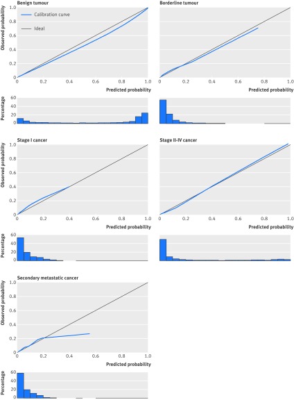
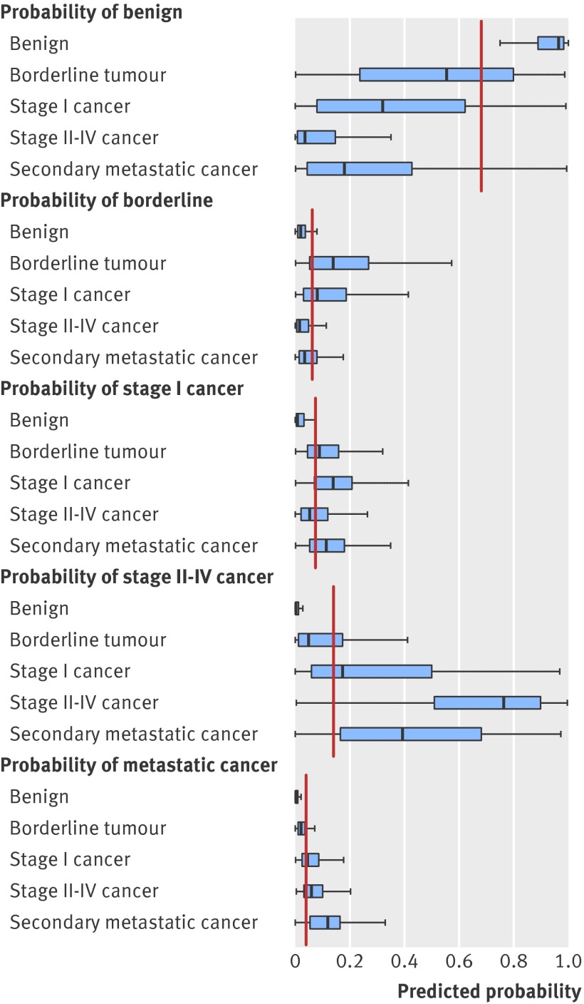
OBJECTIVES: To develop a risk prediction model to preoperatively discriminate between benign, borderline, stage I invasive, stage II-IV invasive, and secondary metastatic ovarian tumours. DESIGN: Observational diagnostic study using prospectively collected clinical and ultrasound data. SETTING: 24 ultrasound centres in 10 countries. PARTICIPANTS: Women with an ovarian (including para-ovarian and tubal) mass and who underwent a standardised ultrasound examination before surgery. The model was developed on 3506 patients recruited between 1999 and 2007, temporally validated on 2403 patients recruited between 2009 and 2012, and then updated on all 5909 patients. MAIN OUTCOME MEASURES: Histological classification and surgical staging of the mass. RESULTS: The Assessment of Different NEoplasias in the adneXa (ADNEX) model contains three clinical and six ultrasound predictors: age, serum CA-125 level, type of centre (oncology centres v other hospitals), maximum diameter of lesion, proportion of solid tissue, more than 10 cyst locules, number of papillary projections, acoustic shadows, and ascites. The area under the receiver operating characteristic curve (AUC) for the classic discrimination between benign and malignant tumours was 0.94 (0.93 to 0.95) on temporal validation. The AUC was 0.85 for benign versus borderline, 0.92 for benign versus stage I cancer, 0.99 for benign versus stage II-IV cancer, and 0.95 for benign versus secondary metastatic. AUCs between malignant subtypes varied between 0.71 and 0.95, with an AUC of 0.75 for borderline versus stage I cancer and 0.82 for stage II-IV versus secondary metastatic. Calibration curves showed that the estimated risks were accurate. CONCLUSIONS: The ADNEX model discriminates well between benign and malignant tumours and offers fair to excellent discrimination between four types of ovarian malignancy. The use of ADNEX has the potential to improve triage and management decisions and so reduce morbidity and mortality associated with adnexal pathology.
Overview
The claims and statistics of this paper haven’t been checked yet.
Run the check — free, no account needed. It reads the open-access text and checks the sources it cites.
Integrity signals
Registry
Cites
Statistics
Figures
Verification
Claims the paper makes
The claims and statistics of this paper haven’t been checked yet.
Run the check — free, no account needed. It reads the open-access text and checks the sources it cites.
Claims other papers make about this one
Each is checked against this paper’s own text: does it say what the citing paper says it does?
In the citing paper
“The ADNEX model, developed by the International Ovarian Tumor Analysis (IOTA) consortium (authors BVC and DT are part of this consortium), preoperatively estimates the probability of malignancy in women with an ovarian tumour who are scheduled for surgery. 31”
From Evaluation of performance measures in predictive artificial intelligence models to support medical decisions: overview and guidance (2025) · cites this paper as [31]
In the citing paper
“ADNEX was developed on the data from 5909 individuals recruited between 1999 and 2012 in 24 secondary and tertiary care centres from 10 countries (Italy, Belgium, Sweden, Czech Republic, Poland, France, United Kingdom, China, Spain, and Canada).”
From Evaluation of performance measures in predictive artificial intelligence models to support medical decisions: overview and guidance (2025) · cites this paper as [31]
Self-check
Percentages, sums and reported differences re-computed from the paper’s own numbers.
The self-check needs the paper’s full text.
Citations
Cites
(1990) · Choice Reviews Online · doi:10.5860/choice.27-3338 · Cited by 35,305
Hosmer, D. W., & Lemeshow, S. (2000) · doi:10.1002/0471722146 · Cited by 20,302
Siegel, R., Ma, J., Zou, Z., & Jemal, A. (2014) · CA: A Cancer Journal for Clinicians · doi:10.3322/caac.21208 · Cited by 11,499
SEER Cancer Statistics Review, 1975-2003
Ries, L. A. G., Harkins, D., Krapcho, M. F., Mariotto, A. B., Miller, B., Feuer, E. J., Clegg, L. X., Eisner, M. P., Horner, M., Howlader, N., et al. (2006) · Digital Archive @ GSU · Cited by 11,404
Regression Modeling Strategies
Harrell, F. E. (2001) · Springer series in statistics · doi:10.1007/978-1-4757-3462-1 · Cited by 9,270
Sterne, J. A. C., White, I. R., Carlin, J. B., Spratt, M., Royston, P., Kenward, M. G., Wood, A. M., & Carpenter, J. R. (2009) · BMJ · doi:10.1136/bmj.b2393 · Cited by 7,299
Signet-Ring Cell Carcinoma of the Colon: A Case Report and Review of the Literature
Park, P. Y., Goldin, T. A., Chang, J. C. S., Markman, M. M., & Kundranda, M. N. (2015) · Case Reports in Oncology · doi:10.1159/000441772 · Cited by 5,273
Clinical Prediction Models: A Practical Approach to Development, Validation, and Updating
Steyerberg, E. W. (2019) · Statistics for Biology and Health · doi:10.1007/978-3-030-16399-0 · Cited by 3,281
Rubin, M., & Stephen, C. D. (1993) · Cited by 3,149
Rao, S. J. (2003) · Journal of the American Statistical Association · doi:10.1198/jasa.2003.s263 · Cited by 1,967
Cited by
Sorted by citations
Items 201–300 of 510
Stukan, M., Alcázar, J. L., Gębicki, J., Epstein, E., Liro, M., Sufliarska, A., Szubert, S., Guerriero, S., Braicu, E. I., Szajewski, M., et al. (2019) · Diagnostics · doi:10.3390/diagnostics9040210 · Cited by 9
“In the largest published cohort of patients with ovarian metastatic cancers who underwent comprehensive ultrasound examination, the median tumor diameter was 86 mm; the majority (95%) had a solid component, with a median proportion of solid tissue of 100% (range 64–100) [14].”
BackgroundComparison of Ultrasound and Tumor Marker CA125 in Diagnosis of Adnexal Mass Malignancies
Behnamfar, F., Esmaeilian, F., Adibi, A., & Rouholamin, S. (2022) · Advanced Biomedical Research · doi:10.4103/abr.abr_164_20 · Cited by 9
“All of the women in this set were initially evaluated for an adnexal mass and CA‐125 results would be expected to receive priority consideration in patients in this age group, for which malignancy is more uncommon.[12] In order to investigate the predictive role of ultrasound in malignancy of ovarian tumors, this stud…”
BackgroundOvarian Masses: The Value of Acoustic Shadowing on Ultrasound Examination
Yoeli‐Bik, R., Lengyel, E. R., Mills, K. A., & Abramowicz, J. S. (2022) · Journal of Ultrasound in Medicine · doi:10.1002/jum.16100 · Cited by 9
“Modified from Van Calster et al.50 *The ADNEX model is the first risk model to differentiate between benign and malignant tumors while offering sub-classification of malignancies into borderline tumors, Stage I and Stage II–IV primary cancers, and secondary metastatic tumors.”
MethodFlawed external validation study of the ADNEX model to diagnose ovarian cancer
CALSTER, B. V., Steyerberg, E. W., Bourne, T., Timmerman, D., & Collins, P. G. S. (2016) · Gynecologic Oncology Reports · doi:10.1016/j.gore.2016.09.003 · Cited by 9
“It is useful to observe that the AUCs were 0.955 and 0.907, since this is in line with the center-specific values reported in the original publication describing the ADNEX model (Van Calster et al., 2014).”
BackgroundMethodResultInfluentialYang, Y., Wang, H., Liu, Z., Su, N., Gao, L., Tao, X., Zhang, R., Gu, Y., Ma, L., Wang, R., et al. (2023) · Journal of Cancer Research and Clinical Oncology · doi:10.1007/s00432-023-05108-z · Cited by 8
Moro, F., Ciancia, M., Maria, S., Baldassari, G., Tran, H. E., Carcagnì, A., Fagotti, A., & Testa, A. C. (2025) · Acta Obstetricia Et Gynecologica Scandinavica · doi:10.1111/aogs.15146 · Cited by 9
“In particular, the IOTA-Assessment of Different Neoplasia in the adneXa (ADNEX) model, which combines clinical and simple ultrasound variables, 4 has been proposed in international guidelines for the diagnosis and management of patients with adnexal masses.”
A multimodal uncertainty-aware AI system optimizes ovarian cancer risk assessment workflow
Wang, X., Lv, X., Wang, J., Zou, L., Chen, Z., Zhao, R., Zhao, L., Zhao, M., Zhang, X., Zhang, B., et al. (2025) · npj Digital Medicine · doi:10.1038/s41746-025-01986-4 · Cited by 8
Malignant epithelioid neoplasm of the ileum with ACTB-GLI1 fusion mimicking an adnexal mass
Ambrosio, M., Virgilio, A., Raffone, A., Arena, A., Raimondo, D., Alletto, A., Seracchioli, R., & Casadio, P. (2022) · BMC Women s Health · doi:10.1186/s12905-022-01679-0 · Cited by 8
Evaluation of Adnexal Masses: The IOTA Concept
Manegold‐Brauer, G., Timmerman, D., & Hoopmann, M. (2022) · Ultraschall in der Medizin - European Journal of Ultrasound · doi:10.1055/a-1912-5361 · Cited by 8
Thakur, M., & Timmerman, D. (2016) · Clinical Obstetrics & Gynecology · doi:10.1097/grf.0000000000000261 · Cited by 8
“In a multicenter prospective cohort study, the ADNEX model discriminated well between benign and malignant tumors and offered a fair to excellent discrimination between the 4 types of malignancy.(23) Clinicians who do not perform diagnostic gynecological ultrasound scans themselves, should establish referral services…”
MethodImmunological parameters as a new lead in the diagnosis of ovarian cancer.
Baert, T., Timmerman, D., Vergote, I. B., & Coosemans, A. (2015) · PubMed Central · Cited by 8
Wu, M., Wang, Q., Zhang, M., Cao, J., Chen, Y., Zheng, J., Luo, L., Su, M., Lin, X., Kuang, X., et al. (2022) · Journal of Ultrasound in Medicine · doi:10.1002/jum.16065 · Cited by 8
“88.(13,24,25) Nevertheless, the preoperative diagnosis of these three tumor subtypes still remains a challenge in current clinical practice.”
BackgroundPsychological impact of referral to an oncology hospital on patients with an ovarian mass
Lof, P., Engelhardt, E., van Gent, M., Mom, C., Dunné, F. R. R.-V., van Baal, W. V., Verhoeve, H., Hermsen, B., Verbruggen, M., Hemelaar, M., et al. (2022) · International Journal of Gynecological Cancer · doi:10.1136/ijgc-2022-003753 · Cited by 6
Velayo, C. L., Reforma, K. N., Sicam, R. V. G., Diwa, M. H., Sy, A. D. R., & Tantengco, O. A. G. (2022) · Healthcare · doi:10.3390/healthcare11010008 · Cited by 8
“According to the results of the IOTA group’s recent studies, a cutoff risk set at 20% was related to the best balance between the ADNEX model’s sensitivity and specificity [14], while the cutoff for LR1 and LR2 was set at 0.”
ResultWang, R., & Yang, Z. (2023) · Ginekologia Polska · doi:10.5603/gp.a2023.0019 · Cited by 8
“The ADNEX model [10] includes nine variables: age (years), serum carbohydrate antigen 125 (CA125) level (U/mL), type of center (oncology center/other hospital), maximum diameter of the lesion (mm), maximal diameter of the largest solid part (mm), more than 10 locules (yes/no), number of papillary projections (0/1/2/3/…”
BackgroundMethodNam, G. E., Lee, S. R., Jeong, K., Kim, S. H., Moon, H. S., & Chae, H. D. (2021) · Obstetrics & Gynecology Science · doi:10.5468/ogs.21012 · Cited by 8
“The general malignancy rate in a previous report was 27% [2].”
BackgroundMethodInfluentialDiagnostic imaging in gynecology
Łuczyńska, E., & Zbigniew, K. (2022) · Ginekologia Polska · doi:10.5603/gp.a2021.0209 · Cited by 8
Gong, L., Li, X.-Y., Wu, Y., Dong, S., Zhang, S., Feng, Y.-N., Lv, Y.-E., Guo, X.-J., Peng, Y., Du, X.-S., et al. (2023) · Journal of Ovarian Research · doi:10.1186/s13048-023-01133-1 · Cited by 8
“Acoustic shadowing was included in IOTA SR and ADNEX model as one of the key benign features [8, 10], but it has not been included in the O-RADS classification system.”
BackgroundMethodPatel-Lippmann, K. K., Gupta, A., Martin, M. F., Phillips, C. H., Maturen, K. E., Jha, P., Sadowski, E. A., & Stein, E. B. (2024) · Radiology · doi:10.1148/radiol.233332 · Cited by 7
Ronsini, C., Restaino, S., Vizzielli, G., Donna, M. C. D., Cucinella, G., Solazzo, M. C., Scaffa, C., FRANCISCIS, P. D., & Chiantera, V. (2025) · Medicina · doi:10.3390/medicina61050777 · Cited by 7
“Future prospective studies involving patients without suspected disease will be needed to improve the usability of these results.”
Ronsini, C., Restaino, S., Ludovisi, M., Vizzielli, G., Donna, M. C. D., Cucinella, G., Solazzo, M. C., Scaffa, C., FRANCISCIS, P. D., Fordellone, M., et al. (2025) · Journal of Personalized Medicine · doi:10.3390/jpm15090426 · Cited by 7
Guo, X., Liu, Z., Li, X., Zhou, B., & Chen, J. (2026) · Journal of Advanced Research · doi:10.1016/j.jare.2026.01.047 · Cited by 7
Pelayo, M., Saúco, J. S., Sánchez-Zurdo, J., Perez‐Mies, B., Abarca-Martínez, L., Hidalgo, M. J. C., Sainz‐Bueno, J. A., Alcázar, J. L., & Pelayo‐Delgado, I. (2023) · Diagnostics · doi:10.3390/diagnostics13172785 · Cited by 7
Ovarian cancer risk among older patients with stable adnexal masses
Suh‐Burgmann, E., Hung, Y., & Schmittdiel, J. A. (2024) · American Journal of Obstetrics and Gynecology · doi:10.1016/j.ajog.2024.04.019 · Cited by 7
ACR Appropriateness Criteria® Clinically Suspected Adnexal Mass, No Acute Symptoms: 2023 Update
Patel-Lippmann, K. K., Wasnik, A. P., Akin, E. A., Andreotti, R. F., Ascher, S. M., Brook, O. R., Eskander, R. N., Feldman, M., Jones, L. P., Martino, M. A., et al. (2024) · Journal of the American College of Radiology · doi:10.1016/j.jacr.2024.02.017 · Cited by 8
ACR Appropriateness Criteria® Postmenopausal Acute Pelvic Pain
Henrichsen, T. L., Maturen, K. E., Robbins, J. B., Akin, E. A., Ascher, S. M., Brook, O. R., Dassel, M., Friedman, L., Learman, L. A., Patlas, M. N., et al. (2021) · Journal of the American College of Radiology · doi:10.1016/j.jacr.2021.02.003 · Cited by 7
Brito, M., Borges, A. L., Rodrigues, S., Ambrósio, P., Condeço, R., Lacerda, A., Bernardo, M. J., Pinto, P., & Djokovic, D. (2021) · Diagnostics · doi:10.3390/diagnostics11111992 · Cited by 8
“Furthermore, the IOTA ADNEX model is the first prediction model that also provides the risk distribution between four malignancy categories (borderline tumors, stage I, stage II-IV primary cancers and secondary metastatic tumors in adnexal lesions) [16].”
MethodZhou, S., Guo, Y., Wen, L., Zhao, B., & Liu, M. (2021) · ULTRASONOGRAPHY · doi:10.14366/usg.21158 · Cited by 8
“org improve the malignancy risk estimate and the management of AMs, many guidelines and structured reporting systems have been established, using subjective assessments, simple scoring, or statistically derived scoring [2-10].”
BackgroundDeng, Y.-Y., Jiang, D., Zhu, P.-F., Lu, H., Liu, Q., Zhang, X., Pan, S. L., Chen, Z.-L., & Yang, L. (2023) · World Journal of Surgical Oncology · doi:10.1186/s12957-023-02973-3 · Cited by 7
“integrated clinical predictors (age, serum CA-125 level, type of center) and ultrasound predictors to distinguish primary and metastatic ovarian tumors [27].”
BackgroundBARD1 Autoantibody Blood Test for Early Detection of Ovarian Cancer
Pilyugin, M., Ratajska, M., Stukan, M., Concin, N., Zeillinger, R., & Irminger‐Finger, I. (2021) · Genes · doi:10.3390/genes12070969 · Cited by 7
“There have also been attempts to simplify the IOTA algorithm [13,14].”
BackgroundLiu, C., Zhu, Y., Dai, K.-J., Tan, B., Dong, H., Lin, J., He, R. L., Lu, M., & Li, Y. (2025) · Journal of Ovarian Research · doi:10.1186/s13048-025-01707-1 · Cited by 6
Taliento, C., Bruyn, C. D., Ceusters, J., Timmerman, D., den Bosch, T. V., & Coosemans, A. (2025) · Maturitas · doi:10.1016/j.maturitas.2025.108634 · Cited by 6
Serous Papillary Adenofibroma Cyst of the Ovary in a Young Woman: Case Report and Literature Review
Barbu, L. A., Cercelaru, L., Şurlin, V., Mogoantă, S. Ş., Cojan, T. Ș. Ț., Mărgăritescu, N.-D., Cojan, A.-M. Ț., Popescu, M., Căluianu, V., Mogoș, G. F. R., et al. (2025) · Life · doi:10.3390/life15101601 · Cited by 6
Wang, Y., Zhang, J., He, Y., Wang, X., Wu, X., Zhang, W., Gong, M., Gao, D., Liu, S., Liu, P., et al. (2025) · Insights into Imaging · doi:10.1186/s13244-025-02112-4 · Cited by 7
Buranaworathitikul, P., Wisanumahimachai, V., Phoblap, N., Porngasemsart, Y., Rugfoong, W., Yotchana, N., Uthaichalanont, P., Jiampochaman, T., Kunanukulwatana, C., Thiamkaew, A., et al. (2024) · Cancers · doi:10.3390/cancers16223820 · Cited by 6
Khastgir, G., Siwatch, S., Singh, T., Jain, V., Sikka, P., & Srinivasan, R. (2025) · European Journal of Obstetrics & Gynecology and Reproductive Biology · doi:10.1016/j.ejogrb.2025.114585 · Cited by 6
Adnexal tumors in menopausal women: surgery or surveillance?
Burgess, B. T., & Ueland, F. R. (2019) · Menopause The Journal of The North American Menopause Society · doi:10.1097/gme.0000000000001330 · Cited by 6
Rashmi, N., Singh, S., Begüm, J., & Sable, M. (2023) · Women s Health Reports · doi:10.1089/whr.2022.0072 · Cited by 6
Rai, N., Champaneria, R., Snell, K. I. E., Mallett, S., Bayliss, S. E., Neal, R. D., Balogun, M., Kehoe, S. T., Deeks, J. J., Sundar, S. S., et al. (2015) · Cochrane Database of Systematic Reviews · doi:10.1002/14651858.cd011964 · Cited by 6
Abramowicz, J. S., Condous, G. S., & Timmerman, D. (2018) · Australasian Journal of Ultrasound in Medicine · doi:10.1002/ajum.12108 · Cited by 6
Characteristics and prognosis of borderline ovarian tumors in pre and postmenopausal patients
Tal, O., Herman, H. G., Gluck, O., Levy, T., Kerner, R., Bar, J., & Sagiv, R. (2020) · Archives of Gynecology and Obstetrics · doi:10.1007/s00404-020-05652-w · Cited by 6
“In 2014 Van Calster and colleagues [26] demonstrated that the combination of the following parameters can be used for successful preoperative discrimination between benign and malignant ovarian masses: age, serum CA-125 levels and different sonographic parameters (lesion diameter, solid tissue, cyst locules, papillary…”
BackgroundBatra, N., & Gupta, B. (2023) · Springer eBooks · doi:10.1007/978-3-031-36179-1_1 · Cited by 6
Zhong, D., Gao, X.-Q., Li, H., Wang, H., & Liu, Y. (2024) · Journal of Imaging Informatics in Medicine · doi:10.1007/s10278-024-01170-2 · Cited by 5
“The use of a single, fixed cutoff value should be avoided according to the ADNEX model application guidance [6], because it may lead to a non-patient-centered diagnostic decision.”
InfluentialClinical value of ACR O-RADS combined with CA125 in the risk stratification of adnexal masses
Pan, R., Zhang, S., Zhang, X., Xu, T., Cui, X., Li, R., Yu, M., & Zhang, B. (2024) · Frontiers in Oncology · doi:10.3389/fonc.2024.1369900 · Cited by 5
Wu, C.-I., Wu, L.-H., Shang, J., Xie, H., & Peng, R. (2025) · Journal of Ultrasound in Medicine · doi:10.1002/jum.16644 · Cited by 5
Liu, L., Cai, W., Zheng, F., Tian, H., Li, Y., Wang, T., Chen, X., & Zhu, W. (2025) · Insights into Imaging · doi:10.1186/s13244-024-01874-7 · Cited by 7
Mascilini, F., Moro, F., Pasciuto, T., Sladkevicius, P., Froyman, W., Jokubkiene, L., Holsbeke, C. V., Franchi, D., Epstein, E., Guerriero, S., et al. (2025) · Ultrasound in Obstetrics and Gynecology · doi:10.1002/uog.29248 · Cited by 5
Adnexal Mass Imaging: Contemporary Guidelines for Clinical Practice
Roseland, M. E., Maturen, K. E., Shampain, K. L., Wasnik, A. P., & Stein, E. B. (2023) · Radiologic Clinics of North America · doi:10.1016/j.rcl.2023.02.002 · Cited by 5
Prediction of ovarian cancer prognosis using statistical radiomic features of ultrasound images
Zuo, R., Li, X., Hu, J., Wang, W., Lü, B., Zhang, H., Cheng, X., Lu, W., Qin, J., Liu, P., et al. (2024) · Physics in Medicine and Biology · doi:10.1088/1361-6560/ad4a02 · Cited by 5
“…for example [based on three factors namely age, Cancer Antigen 125 (CA125), and a combination of six ultrasound features including diameter of lesion, blood flow and proportion of solid tissue], provides excellent discrimination between benign and malignant ovarian tumors (Van Calster et al 2014).”
BackgroundMethodPetrocelli, R., Doshi, A. M., Slywotzky, C. M., Savino, M., Melamud, K., Tong, A. Y., & Hindman, N. M. (2024) · Journal of Computer Assisted Tomography · doi:10.1097/rct.0000000000001629 · Cited by 5
Su, C., Miao, K., Zhang, L., Yu, X., Guo, Z., Li, D., Xu, M., Zhang, Q., & Dong, X. (2025) · Journal of Imaging Informatics in Medicine · doi:10.1007/s10278-025-01566-8 · Cited by 7
Algorithmic Approach to Sonography of Adnexal Masses: An Evolving Paradigm
Bhalla, D., Manchanda, S., & Vyas, S. (2020) · Current Problems in Diagnostic Radiology · doi:10.1067/j.cpradiol.2020.08.008 · Cited by 5
Ovarian-Adnexal Reporting and Data Systems MR Imaging
Melamud, K., Hindman, N. M., & Sadowski, E. A. (2022) · Magnetic Resonance Imaging Clinics of North America · doi:10.1016/j.mric.2022.06.004 · Cited by 5
Strumal Carcinoid Tumor of the Ovary: Report of Rare Occurrence with Review of Literature
Shen, L., Yang, A.-Q., & Jin, L. (2022) · Diagnostics · doi:10.3390/diagnostics12112706 · Cited by 5
“The probabilities provided by the ADNEX model [25] were as follows: The incidence rate of benign tumors, malignant tumors, and borderline tumors was 64.”
MethodShang, J., Lei, T., Wu, L., Lin, M., & Xie, H. (2023) · Archives of Gynecology and Obstetrics · doi:10.1007/s00404-022-06903-8 · Cited by 4
Massobrio, R., Mariani, L. L., Conti, D., Grandis, T. D., Buonomo, F., Badellino, E., Novara, L., Bounous, V. E., Perotto, S., Mancarella, M., et al. (2023) · Archives of Gynecology and Obstetrics · doi:10.1007/s00404-023-07233-z · Cited by 5
Clinical practice guidelines for molecular tumor marker, 2nd edition review part 2
Kikuchi, Y., Shimada, H., Yamasaki, F., Yamashita, T., Araki, K., Horimoto, K., Yajima, S., Yashiro, M., Yokoi, K., Cho, H., et al. (2024) · International Journal of Clinical Oncology · doi:10.1007/s10147-024-02497-0 · Cited by 5
ReClassification of Patients with Ambiguous CA125 for Optimised Pre-Surgical Triage
Stephens, A. N., Hobbs, S. J., Kang, S.-W., Oehler, M. K., Jobling, T. W., & Allman, R. (2024) · Diagnostics · doi:10.3390/diagnostics14070671 · Cited by 5
Adenomatoid tumor of the fallopian tube: A case report and a review of the literature
Mitta, K., Michos, G., Athanasiadou, E. A., Papanikolaou, E. G. E., Tsakiridis, I. G., & Mamopoulos, A. M. (2024) · International Journal of Surgery Case Reports · doi:10.1016/j.ijscr.2024.110268 · Cited by 5
“The calculated risk for a malignant ovarian tumor was determined to be 9.1 % [13].”
BackgroundInfluentialTomasińska, A., Stukan, M., Badocha, M., & Myszewska, A. (2021) · Diagnostics · doi:10.3390/diagnostics11091600 · Cited by 5
“Prediction of malignancy was based on the examiner’s subjective assessment, or if ambiguous in appearance, multivariable predictive models described elsewhere were used [20,21].”
MethodBuţureanu, T., Socolov, D. G., Matasariu, D. R., Ursache, A., Apetrei, A.-M., Dumitraşcu, I., Vasilache, I.-A., Rudisteanu, D., Boiculese, V. L., & Lozneanu, L. (2021) · Applied Sciences · doi:10.3390/app112210789 · Cited by 5
“It includes three clinical criteria (age, CA 125 serum level, and the type of center: oncological/other types of hospitals) and six ultrasound features (the maximum tumoral diameter in mm, the maximum diameter of the solid component in mm; more than 10 loculus, number of papillary projections, presence/absence of acou…”
BackgroundReservations Regarding O-RADS Recommendations
Suh‐Burgmann, E., FLANAGAN, T. A., & Brasic, N. (2020) · Radiology · doi:10.1148/radiol.2020192559 · Cited by 5
Maternal adnexal masses in pregnancy
Djokovic, D., Pinto, P., & Reis, I. S. (2025) · Best Practice & Research Clinical Obstetrics & Gynaecology · doi:10.1016/j.bpobgyn.2025.102643 · Cited by 4
Clustered flexible calibration plots for binary outcomes using random effects modeling
Barreñada, L., Cock, B. D., Wynants, L., & CALSTER, B. V. (2025) · Research Synthesis Methods · doi:10.1017/rsm.2025.10046 · Cited by 5
Eriksson, J., Puglisi, D., Herbst, F., Dobilas, A., Shtepliuk, I., Joneborg, U., Falconer, H., Rådestad, A. F., & Borgfeldt, C. (2025) · EBioMedicine · doi:10.1016/j.ebiom.2025.106027 · Cited by 4
Geysels, A., Garofalo, G., Timmerman, S., Ceusters, J., Buonomo, F., Fischerová, D., Testa, A. C., Moro, F., Sladkevicius, P., Jokubkiene, L., et al. (2025) · npj Precision Oncology · doi:10.1038/s41698-025-01215-x · Cited by 4
“TheAssessmentofDifferentNEoplasias in the adneXa (ADNEX) model, developed by the International Ovarian TumourAnalysis(IOTA)group,isthebestperformingmodelandhasbeen externallyvalidatedinnumerousstudies 6 – 8 .”
Salomón, C., Razo-Azamar, M., Gentry‐Maharaj, A., Rice, G. E., & Menon, U. (2026) · Expert Review of Molecular Diagnostics · doi:10.1080/14737159.2025.2608820 · Cited by 4
Moro, F., Ciancia, M., Berardino, S. D., Baldassari, G., Mascilini, F., Ciccarone, F., Nero, C., Tran, H. E., Pasciuto, T., Boldrini, L., et al. (2026) · Ultrasound in Obstetrics and Gynecology · doi:10.1002/uog.70203 · Cited by 4
Timmerman, D., Valentin, L., Testa, A. C., Froyman, W., Landolfo, C., Kotlarz, A., Fischerová, D., CALSTER, B. V., Bourne, T., & Collaborators (2026) · Ultrasound in Obstetrics and Gynecology · doi:10.1002/uog.70191 · Cited by 4
Laparoscopic Surgery for Ovarian Neoplasms – What is Possible, What is Useful?
Droste, A., Anić, K., & Hasenburg, A. (2022) · Geburtshilfe und Frauenheilkunde · doi:10.1055/a-1787-9144 · Cited by 4
Tantipalakorn, C., Tinnangwattana, D., Lerthiranwong, T., Luewan, S., & Tongsong, T. (2023) · International Journal of Environmental Research and Public Health · doi:10.3390/ijerph20010888 · Cited by 6
Kotlarz, A., Froyman, W., Valentin, L., Testa, A. C., Hove, M. V., CALSTER, B. V., Bourne, T., & Timmerman, D. (2024) · Ultrasound in Obstetrics and Gynecology · doi:10.1002/uog.27675 · Cited by 4
AIUM Practice Parameter for the Performance of Ultrasound of the Female Pelvis, 2024 Revision
(2024) · Journal of Ultrasound in Medicine · doi:10.1002/jum.16556 · Cited by 4
Lems, E., Mongula, J. E., Muntinga, C. L. P., Leemans, J. C., Evert, J. S. H., Smit, R. A., van der Steeg, J. W., Pijlman, B. M., Coppus, S. F. P. J., Bongers, M. Y., et al. (2025) · International Journal of Gynecological Cancer · doi:10.1016/j.ijgc.2025.101917 · Cited by 4
Kashyap, P. (2021) · International Journal of Reproduction Contraception Obstetrics and Gynecology · doi:10.18203/2320-1770.ijrcog20213510 · Cited by 4
“The change in CA125 level overtime may be more helpful than a single value and therefore it is useful for follow up of patients of ovarian cancer after treatment.(23)”
BackgroundFiliz, A. A., Kahyaoğlu, S., & Atalay, C. (2023) · Obstetrics & Gynecology Science · doi:10.5468/ogs.23061 · Cited by 4
“After applying the ADNEX model to a patient with an adnexal mass, the lesion was classified into five categories: benign, borderline, stage I invasive, stage II-IV invasive, and secondary metastatic tumors [9,10].”
MethodCurrent and Emerging Strategies for Tubo-Ovarian Cancer Diagnostics
Brincat, M., Mira, A. R., & Lawrence, A. C. (2023) · Diagnostics · doi:10.3390/diagnostics13213331 · Cited by 4
Perez, M., Meseguer, A., Vara, J., Jiménez, J. C. V., Garcia, I. B., Lozano, M., Orozco, R., & Alcázar, J. L. (2024) · ULTRASONOGRAPHY · doi:10.14366/usg.24105 · Cited by 4
Erdodi, B., Szöllösi, G. J., Tóth, Z., Krasznai, Z. T., & Jakab, A. (2025) · Cancers · doi:10.3390/cancers17081370 · Cited by 4
“Nonetheless, CA-125 remains an integral component of advanced diagnostic models such as the ADNEX model, where it contributes to the differentiation of malignant subtypes [18].”
InfluentialSandal, K., Polat, M., Yassa, M., Günay, T., Erdem, G., & Güzin, K. (2018) · Zeynep Kamil Tıp Bülteni · doi:10.16948/zktipb.445755 · Cited by 4
Bullock, R. G., Smith, A., Munroe, D. G., Ueland, F. R., Goodrich, S. T., Pappas, T. C., Fredericks, T. I., & Bonato, V. (2019) · Journal of Surgical Oncology · doi:10.31487/j.jso.2019.03.04 · Cited by 4
Ultrasound differentiation between benign versus malignant adnexal masses in pregnant patients
Rabiej-Wronska, E., Wiechec, M., Pitynski, K., Wiercinska, E., & Kotlarz, A. (2021) · Ginekologia Polska · doi:10.5603/gp.a2021.0176 · Cited by 4
Quaranta, M., Nath, R., Mehra, G., Diab, Y. M. S. K., & Sayasneh, A. (2020) · Cureus · doi:10.7759/cureus.9201 · Cited by 4
“The IOTA ADNEX model was calculated when the required parameters were retrievable retrospectively [2].”
BackgroundMethodBenacerraf, B. R. (2016) · American Journal of Obstetrics and Gynecology · doi:10.1016/j.ajog.2015.12.045 · Cited by 4
Conservative management of adnexal tumors: how to tell good from bad
Jurkovic, D. (2023) · Ultrasound in Obstetrics and Gynecology · doi:10.1002/uog.26158 · Cited by 4
Barcroft, J., Linton‐Reid, K., Landolfo, C., Al‐Memar, M., Parker, N., Kyriacou, C., Munaretto, M., Fantauzzi, M., Cooper, N., Yazbek, J., et al. (2023) · medRxiv · doi:10.1101/2023.04.26.23289150 · Cited by 4
“Various ultrasound-based diagnostic models, using a combination of ultrasound features with or without serological markers (CA125) exist to support the classification of adnexal masses, including the Risk of malignancy index (RMI)(6), International Ovarian Tumour Analysis (IOTA) Simple Rules (SR) (7), the IOTA Assessm…”
MethodAdnexal Lesion Discrimination Using Deep Learning Analysis of Dynamic Contrast-enhanced US Images
Wu, M., Yang, H., Chen, Y., Wu, S., Liang, T., Zhang, M., Qu, E., Sun, X., Zhang, R., Mu, L., et al. (2025) · Radiology Artificial Intelligence · doi:10.1148/ryai.240786 · Cited by 3
Lems, E., Koch, A. H., Delvaux, E. J. L. G., Leemans, J. C., Bongers, M. Y., Lok, C. A. R., Ramaekers, B. L. T., & Geomini, P. M. A. J. (2025) · Ultrasound in Obstetrics and Gynecology · doi:10.1002/uog.70135 · Cited by 3
“…Ovarian Tumor Analysis (IOTA) group have developed several ultrasound models, including the Logistic Regression model 2 (LR2) 12 , Simple ultrasound-based Rules (SR) 13 and Assessment of Different NEoplasias in the adneXa (ADNEX) model 14 , all of which outperform the RMI in study settings 9,15 .”
Ryu, L., Han, K., Jung, I., Park, Y. W., Ahn, S. S., & Hwang, D. (2026) · Korean Journal of Radiology · doi:10.3348/kjr.2025.1447 · Cited by 3
“Several studies have employed the PDI to evaluate model performance in predicting ovarian tumor subclasses [29-31].”
A new risk-scoring system to predict Xp11.2 translocation renal cell carcinoma in adults
Shi, Q., Liu, N., Zhu, Y., Qu, F., Xu, L., Li, X., Zhang, G., Guo, H., Li, D., & Gan, W. (2021) · Journal of International Medical Research · doi:10.1177/0300060521997661 · Cited by 3
“3% specificity.(21) Scoring systems for identification of endometrial cancer or atypical hyperplasia can likewise provide a high diagnostic value.”
BackgroundLuo, H., Lin, Z., Wu, L., Wang, Y., Ning, H., Feng, Y., Cheng, Y., Wen, X., & Liu, X. (2022) · BioMed Research International · doi:10.1155/2022/7187334 · Cited by 4
“…preoperative diagnostic accuracy of ovarian tumors, many ultrasound-based integrated scoring systems have been proposed and used, such as Risk of Malignancy Index, gynecologic imaging-RADS, International Ovarian Tumor Analysis logistic regression models, simpli fi ed rules, and ADNEX mode [6 – 9].”
Qi, L., Li, X., Yang, Y. S., Zhao, M., Lin, A., & Ma, L. (2024) · Clinical Radiology · doi:10.1016/j.crad.2024.02.012 · Cited by 3
Takkar, N., Arora, P., Goel, B., Kaur, R., Kaur, N., & Aggarwal, P. (2025) · International Journal of Reproduction Contraception Obstetrics and Gynecology · doi:10.18203/2320-1770.ijrcog20250197 · Cited by 3
Yu, J., Hu, P., Dai, R., Liu, X., Zhang, S., Xu, Y., Gao, Y., Lü, B., Wang, W., Niu, Y., et al. (2026) · npj Digital Medicine · doi:10.1038/s41746-026-02875-0 · Cited by 3
“27,47 .”
Tangjanyatham, P., & Chaowawanit, W. (2025) · International Journal of Gynecological Cancer · doi:10.1016/j.ijgc.2025.101827 · Cited by 3
Barreñada, L., Ledger, A., Kotlarz, A., Dhiman, P., Collins, P. G. S., Wynants, L., Verbakel, J. Y. J., Valentin, L., Timmerman, D., & CALSTER, B. V. (2025) · BMJ Open · doi:10.1136/bmjopen-2025-104141 · Cited by 3
Case Report: Borderline tumor and primary peritoneal carcinoma - a rare synchronism
Rei, M., Raposo, S., Figueiredo, P., Sousa, R. G., & Sá, L. (2019) · F1000Research · doi:10.12688/f1000research.20420.1 · Cited by 3
Ultrasound features of polypoid endometriosis: a case report and a short review of Literature
Mancarella, M., Sgrò, L. G., Fuso, L., Campisi, P., Biglia, N., & Mariani, L. L. (2021) · Journal of Endometriosis and Pelvic Pain Disorders · doi:10.1177/22840265211053093 · Cited by 3
Pereira, P. N., Derchain, S., Yoshida, A., Barros, R. H. D. O., Jales, R. M., & Sarian, L. O. (2023) · PLoS ONE · doi:10.1371/journal.pone.0283212 · Cited by 3
“The ultrasound (US) and cancer antigen 125 (CA125)-based malignancy prediction model of the International Ovarian Tumor Analysis (IOTA) group called ADNEX has been designed to categorize adnexal masses as benign, borderline, malignant (initial, advanced) or metastatic [7].”
BackgroundPozzati, F., Sassu, C. M., Marini, G., Mascilini, F., Biscione, A., Giannarelli, D., Garganese, G., Fragomeni, S. M., Scambia, G., Testa, A. C., et al. (2023) · Ultrasound in Obstetrics and Gynecology · doi:10.1002/uog.26253 · Cited by 3
Metadata
- Authors
- Van Calster, B., Van Hoorde, K., Valentin, L., Testa, A. C., Fischerova, D., Van Holsbeke, C., Savelli, L., Franchi, D., Epstein, E., Kaijser, J., Van Belle, V., Czekierdowski, A., Guerriero, S., Fruscio, R., Lanzani, C., Scala, F., Bourne, T., Timmerman, D., & International Ovarian Tumour Analysis (IOTA) group
- Affiliations
- Lund University (SE)
- Università Cattolica del Sacro Cuore (IT)
- IRCCS Azienda Ospedliero-Universitaria di Bologna Policlinico di Sant'Orsola (IT)
- European Institute of Oncology (IT)
- Karolinska University Hospital (SE)
- Universitair Ziekenhuis Leuven (BE)
- Medical University of Lublin (PL)
- Azienda Ospedaliero-Universitaria Cagliari (IT)
- University of Milan (IT)
- Istituto Nazionale Tumori IRCCS "Fondazione G. Pascale" (IT)
- ORCID
- Venue
- BMJ
- Publisher
- BMJ
- Published
- 2014-10-15
- Volume
- 349
- Issue
- oct07 3
- Pages
- g5920-g5920
- ISSN
- 1756-1833 (ISSN-L)
- PMC
- PMC4198550
- OpenAlex
- W2099878111
- Cited by
- 510
- References
- 57
- Citations by year
- 2026: 52 · 2025: 60 · 2024: 70 · 2023: 54 · 2022: 55 · 2021: 59 · 2020: 36 · 2019: 24 · 2018: 30 · 2017: 19 · 2016: 27 · 2015: 19 · 2014: 4
- FWCI
- 27.87
- Type
- journal article
- Language
- en
- Open access
- hybrid
- doi.org
- Licence
- cc-by-nc
- Locations
- BMJ (publishedVersion, cc-by-nc)
- PubMed Central (publishedVersion, cc-by-nc)
- PubMed (publishedVersion)
- BOA (University of Milano-Bicocca) (submittedVersion, cc-by-nc)
- Lirias (KU Leuven) (submittedVersion, cc-by-nc)
- UNICA IRIS Institutional Research Information System (University of Cagliari) (submittedVersion, other-oa)
- Lund University Publications (Lund University) (submittedVersion, other-oa)
- HighWire Press Open Archive (submittedVersion)
- PubliCatt (Università Cattolica del Sacro Cuore) (submittedVersion, cc-by-nc)
- Spiral (Imperial College London) (submittedVersion, other-oa)
- Funders
- National Institute for Health and Care Research
- Imperial College London
- Imperial College Healthcare NHS Trust
- Fonds Wetenschappelijk Onderzoek (G049312N)
- iMinds
- Malmö Högskola
- Skånes universitetssjukhus
- Vlaamse regering (G049312N)
- Medical Research Council
- Topics
- Health Sciences › Medicine › Oncology › Ovarian cancer diagnosis and treatment
- Life Sciences › Biochemistry, Genetics and Molecular Biology › Genetics › BRCA gene mutations in cancer
- Health Sciences › Medicine › Pathology and Forensic Medicine › Breast Lesions and Carcinomas
- Keywords
- ovarian cancer, ADNEX model, preoperative risk prediction, ovarian ultrasonography, borderline ovarian tumors, CA125, surgical staging, adnexal mass, triage
- MeSH terms
- Adnexal Diseases, Adult, Female, Humans, Neoplasm Staging, Ovarian Neoplasms, Predictive Value of Tests, Prospective Studies, Ultrasonography, Models, Statistical, Risk Assessment
About these checks
Registry, retraction, Cites and statistics results are computed, not judged, with one exception: a printed reference that no index lists is read once by a model before it counts as a citation error. Claims are extracted by a model and kept only when their quote appears verbatim in the text; each cited source is then checked, with a model’s help, for whether it says what the claim relies on. Those verdicts can be wrong — read the claim’s quote and the cited source before relying on one.
Statistics and arithmetic are recomputed, not judged.
Confidence bands are shown only for fields calibrated on our annotated test set. None is calibrated yet, so no band is shown.
What counts as a claim ›How we measure confidence ›How we check this ›
Are you an author, or do you see a problem with this page? Request a review
