

The DOI matches its Crossref record; 32 of the 32 references it cites were found in a registry or an index.
Extensive Proliferation of a Subset of Differentiated, yet Plastic, Medial Vascular Smooth Muscle Cells Contributes to Neointimal Formation in Mouse Injury and Atherosclerosis Models
Open accessAbstract
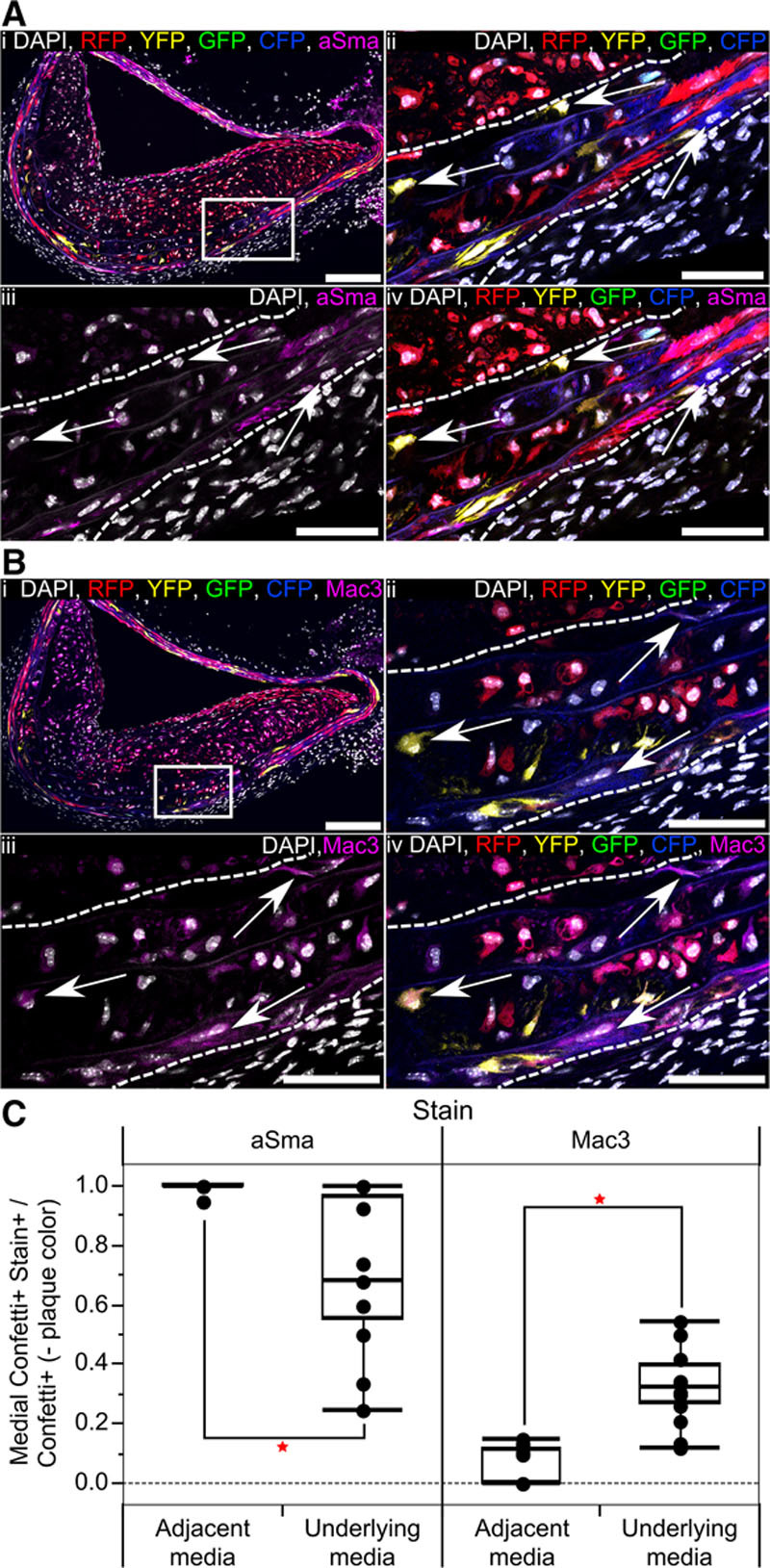
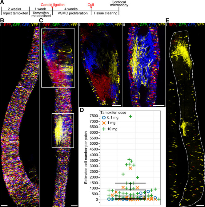
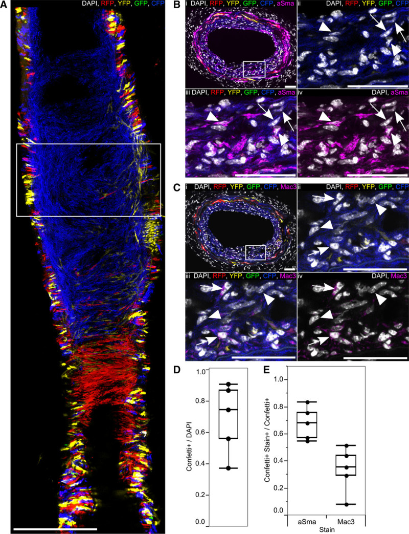
Rationale: Vascular smooth muscle cell (VSMC) accumulation is a hallmark of atherosclerosis and vascular injury. However, fundamental aspects of proliferation and the phenotypic changes within individual VSMCs, which underlie vascular disease, remain unresolved. In particular, it is not known whether all VSMCs proliferate and display plasticity or whether individual cells can switch to multiple phenotypes. Objective: To assess whether proliferation and plasticity in disease is a general characteristic of VSMCs or a feature of a subset of cells. Methods and Results: Using multicolor lineage labeling, we demonstrate that VSMCs in injury-induced neointimal lesions and in atherosclerotic plaques are oligoclonal, derived from few expanding cells. Lineage tracing also revealed that the progeny of individual VSMCs contributes to both alpha smooth muscle actin (aSma)–positive fibrous cap and Mac3-expressing macrophage-like plaque core cells. Costaining for phenotypic markers further identified a double-positive aSma+ Mac3+ cell population, which is specific to VSMC-derived plaque cells. In contrast, VSMC-derived cells generating the neointima after vascular injury generally retained the expression of VSMC markers and the upregulation of Mac3 was less pronounced. Monochromatic regions in atherosclerotic plaques and injury-induced neointima did not contain VSMC-derived cells expressing a different fluorescent reporter protein, suggesting that proliferation-independent VSMC migration does not make a major contribution to VSMC accumulation in vascular disease. Conclusions: We demonstrate that extensive proliferation of a low proportion of highly plastic VSMCs results in the observed VSMC accumulation after injury and in atherosclerotic plaques. Therapeutic targeting of these hyperproliferating VSMCs might effectively reduce vascular disease without affecting vascular integrity.
Overview
The claims and statistics of this paper haven’t been checked yet.
Run the check — free, no account needed. It reads the open-access text and checks the sources it cites.
Integrity signals
Registry
Cites
Statistics
Figures
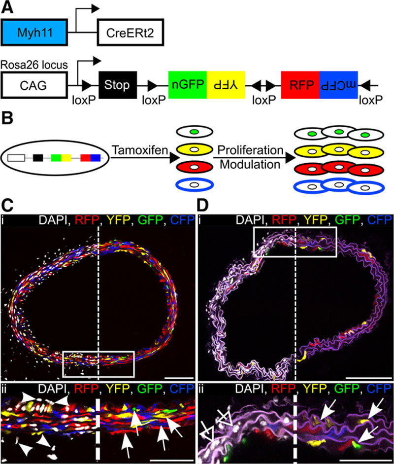
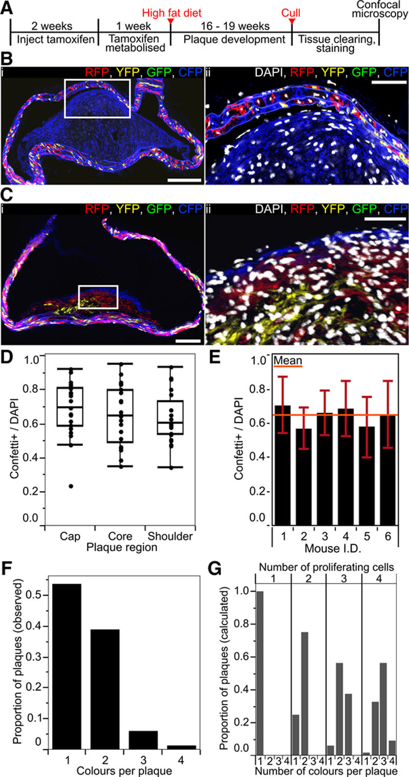
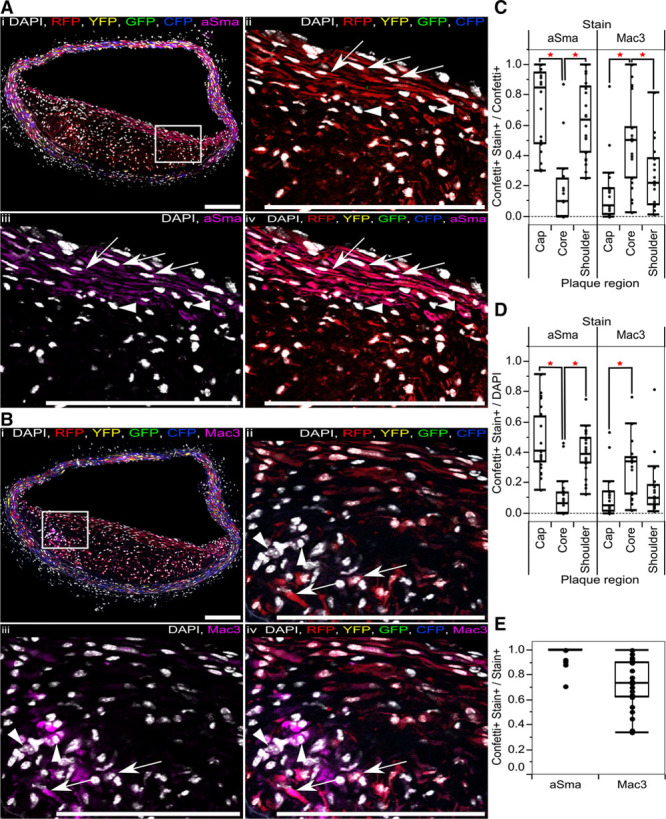
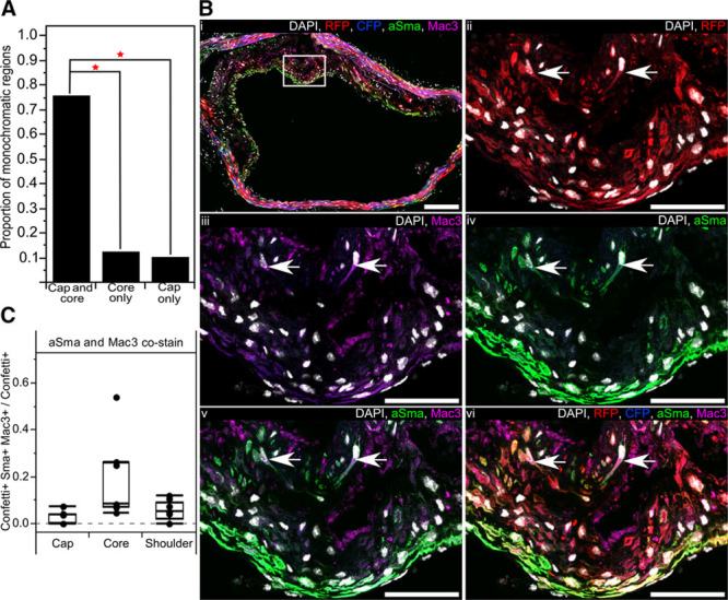
Verification
Claims the paper makes
The claims and statistics of this paper haven’t been checked yet.
Run the check — free, no account needed. It reads the open-access text and checks the sources it cites.
Claims other papers make about this one
Each is checked against this paper’s own text: does it say what the citing paper says it does?
In the citing paper
“Lineage tracing studies have unambiguously demonstrated that VSMCs contribute substantially to plaque formation in murine models of atherosclerosis, generating 30-70% of all plaque cells 11,18-20,22,23 .”
From Vascular smooth muscle cells in atherosclerosis (2019) · cites this paper as [20]
In the citing paper
“Within plaques a large proportion of reporter-expressing VSMC-derived cells do not have detectable levels of the contractile smooth muscle cell marker α SMA 11,20 .”
From Vascular smooth muscle cells in atherosclerosis (2019) · cites this paper as [20]
In the citing paper
“Instead, some plaque reporter-expressing cells were positive for Mac-3 20 , Lgals3 11 and CD68 17 - markers that have been previously used to study macrophages in atherosclerosis.”
From Vascular smooth muscle cells in atherosclerosis (2019) · cites this paper as [20]
Self-check
Percentages, sums and reported differences re-computed from the paper’s own numbers.
The self-check needs the paper’s full text.
Citations
Cites
Vascular smooth muscle cells in atherosclerosis
Bennett, M. R., Sinha, S., & Owens, G. K. (2016) · Circulation Research · doi:10.1161/circresaha.115.306361 · Cited by 2,179
Self-citationSnippert, H. J., van der Flier, L. G., Sato, T., van Es, J. H., van den Born, M., Kroon-Veenboer, C., Barker, N., Klein, A. M., van Rheenen, J., Simons, B. D., et al. (2010) · Cell · doi:10.1016/j.cell.2010.09.016 · Cited by 1,951
Self-citationClowes, A. W., Reidy, M. A., & Mm, C. (1983) · PubMed · Cited by 1,439
Shankman, L. S., Gomez, D., Cherepanova, O. A., Salmon, M., Alencar, G. F., Haskins, R. M., Swiatlowska, P., Newman, A. A. C., Greene, E. S., Straub, A. C., et al. (2015) · Nature Medicine · doi:10.1038/nm.3866 · Cited by 1,218
Piedrahita, J. A., Zhang, S. H., Hagaman, J. R., Oliver, P. M., & Maeda, N. (1992) · Proceedings of the National Academy of Sciences · doi:10.1073/pnas.89.10.4471 · Cited by 944
Regulation and characteristics of vascular smooth muscle cell phenotypic diversity
Rensen, S. S., Doevendans, P. A., & van Eys, G. J. J. M. (2007) · Netherlands Heart Journal · doi:10.1007/bf03085963 · Cited by 917
Evidence for a Monoclonal Origin of Human Atherosclerotic Plaques
Benditt, E. P., & Benditt, J. M. (1973) · Proceedings of the National Academy of Sciences · doi:10.1073/pnas.70.6.1753 · Cited by 913
Alexander, M. R., & Owens, G. K. (2011) · Annual Review of Physiology · doi:10.1146/annurev-physiol-012110-142315 · Cited by 796
Shankman, L. S., Gomez, D. A., Cherepanova, O. A., Salmon, M., Alencar, G. F., Haskins, R. M., Swiatlowska, P., Newman, A. A. C., Greene, E. S., Straub, A. C., et al. (2016) · Nature Medicine · doi:10.1038/nm0216-217a · Cited by 775
G12-G13–LARG–mediated signaling in vascular smooth muscle is required for salt-induced hypertension
Wirth, A., Benyó, Z., Lukasova, M., Leutgeb, B., Wettschureck, N., Gorbey, S., Örsy, P., Horváth, B., Maser‐Gluth, C., Greiner, E. F., et al. (2007) · Nature Medicine · doi:10.1038/nm1666 · Cited by 692
Research works printed but not linked: 1
- 10.1002/1098-2280 · DOI
Cited by
Sorted by citations
Items 301–400 of 440
Application of genetic cell-lineage tracing technology to study cardiovascular diseases
Sun, X., Lyu, L., Zhong, X., Ni, Z., & Xu, Q. (2021) · Journal of Molecular and Cellular Cardiology · doi:10.1016/j.yjmcc.2021.03.006 · Cited by 5
Quan, W., Huo, Y., Chen, Y., Yang, D., Xie, J., Shi, Z., Liao, D., & Tuo, Q. (2022) · Frontiers in Bioscience-Landmark · doi:10.31083/j.fbl2711299 · Cited by 5
Clonal Smooth Muscle Cell Expansion, Autophagy, and Vascular Integrity in Aortic Aneurysm Disease
Wang, Y., Nanda, V., & Leeper, N. J. (2019) · Arteriosclerosis Thrombosis and Vascular Biology · doi:10.1161/atvbaha.119.312739 · Cited by 5
Wihartady, H. (2009) · Research Repository (Delft University of Technology) · Cited by 5
Wang, Y., Moura, A. K., Zuo, R. M., Guan, Y., Lee, E. A., Hu, J. Z., Roudbari, K., Wang, Z., Wang, M., Li, P., et al. (2025) · American Journal of Physiology-Cell Physiology · doi:10.1152/ajpcell.00226.2025 · Cited by 5
Li, W., Liao, Y., Liu, Z., Niu, L., Huang, J., Jia, Y., Xu, R., Guan, S., Liang, Z., Li, Y., et al. (2025) · Nature Communications · doi:10.1038/s41467-025-63876-3 · Cited by 4
Natalí, L., de la Cruz-Thea, B., Godino, A., Conde, C. B., Peinado, V. I., & Musri, M. M. (2026) · Biomolecules · doi:10.3390/biom16010173 · Cited by 5
Mu, W.-J., Wei, S.-Q., Peng, W.-Q., & Guo, L. (2026) · Ageing Research Reviews · doi:10.1016/j.arr.2026.103141 · Cited by 4
Wang, Y., Yang, K., Jiang, G., Zheng, X., Cheng, F., & Li, W. (2025) · Atherosclerosis · doi:10.1016/j.atherosclerosis.2025.120496 · Cited by 4
Zhao, S., Deslarzes-Dubuis, C., Urfer, S., Lambelet, M., Déglise, S., & Allagnat, F. (2023) · Antioxidants · doi:10.3390/antiox12091731 · Cited by 4
Larsen, J. H., Hegelund, J. S., Pedersen, M. K., Andersson, C. M., Lindegaard, C. A., Hansen, D. R., Stubbe, J., Lindholt, J. S., Hansen, C. S., Grentzmann, A., et al. (2024) · Cardiovascular Research · doi:10.1093/cvr/cvae174 · Cited by 5
MicroRNAs in the progression of atherosclerosis: rise and fall of the atherosclerotic plaque
Rampin, A., Mutoli, M., Martelli, M., Settembrini, A. M., Martelli, F., Mitić, T., Sopić, M., Bruno, A., Spinetti, G., & CA, A. C. A. (2024) · European Atherosclerosis Journal · doi:10.56095/eaj.v3i2.63 · Cited by 5
The Dichotomy of Vascular Smooth Muscle Differentiation/De- Differentiation in Health and Disease
Luca, M. D., Hakimjavadi, R., Burtenshaw, D., Fitzpatrick, E., Mathiue, P., Lally, C., Redmond, E. M., & Cahill, P. A. (2018) · InTech eBooks · doi:10.5772/intechopen.76812 · Cited by 4
Rusanov, S. (2022) · Medical Research Archives · doi:10.18103/mra.v10i4.2760 · Cited by 4
Developmental heterogeneity of vascular cells: Insights into cellular plasticity in atherosclerosis?
Lin, A., Ramaswamy, Y., & Misra, A. (2023) · Seminars in Cell and Developmental Biology · doi:10.1016/j.semcdb.2023.06.002 · Cited by 4
Shao, Y., Luo, J., Liu, Y., Ran, H.-Y., Shi, H., Zhang, C., & Wu, Q. (2021) · International Journal of General Medicine · doi:10.2147/ijgm.s335082 · Cited by 4
CD98 promotes vascular smooth muscle cell accumulation in atherosclerosis to confer plaque stability
Zernecke, A. (2016) · Atherosclerosis · doi:10.1016/j.atherosclerosis.2016.11.028 · Cited by 4
Vascular Progenitors and Smooth Muscle Cells Chicken and Egg?
Zhang, L., & Xu, Q. (2017) · Circulation Research · doi:10.1161/circresaha.116.310341 · Cited by 4
Cheng, P. P., Wirka, R. C., Kim, J. B., Nguyen, T., Kundu, R. K., Zhao, Q., Pedroza, A. J., Nagao, M., Iyer, D., Fischbein, M. P., et al. (2020) · bioRxiv (Cold Spring Harbor Laboratory) · doi:10.1101/2020.09.15.299131 · Cited by 4
Somatic mutations reveal clonal cell populations in atherosclerotic plaquesPreprint
Steffensen, L. B., Kavan, S., Jensen, P., Møller‐Larsen, S., Larsen, M. L., Dembić, M., Andersen, L., Lindholt, J. S., Houlind, K. C., Thomassen, M., et al. (2022) · medRxiv · doi:10.1101/2022.05.16.22275001 · Cited by 4
“Cre”ating New Tools for Smooth Muscle Analysis
O'Brien, B., Martin, K. A., & Offermanns, S. (2023) · Arteriosclerosis Thrombosis and Vascular Biology · doi:10.1161/atvbaha.122.318855 · Cited by 4
Steegen, L., & Grootaert, M. O. J. (2026) · Cells · doi:10.3390/cells15020114 · Cited by 4
Park, J. K., Jung, W. B., & Yoon, J.-H. (2021) · Vascular Health and Risk Management · doi:10.2147/vhrm.s287194 · Cited by 3
Dave, J. M., Saito, J., Mottola, G., & Greif, D. M. (2022) · Elsevier eBooks · doi:10.1016/b978-0-12-822546-2.00015-0 · Cited by 3
Pløen, G. G., Sørensen, C. B., & Bentzon, J. F. (2023) · Atherosclerosis · doi:10.1016/j.atherosclerosis.2023.117341 · Cited by 3
Bolanle, I. O., & Palmer, T. M. (2025) · International Journal of Molecular Sciences · doi:10.3390/ijms26073303 · Cited by 3
Huynh, D. T. N., & Heo, K. (2022) · CardioMetabolic Syndrome Journal · doi:10.51789/cmsj.2022.2.e16 · Cited by 3
Mathieu, P. S., Fitzpatrick, E., Luca, M. D., Cahill, P. A., & Lally, C. (2021) · Biochemistry and Biophysics Reports · doi:10.1016/j.bbrep.2021.101183 · Cited by 3
Branch Point Smooth Muscle Cells Highlighted by Novel Lineage Tracking Approach
Brewer, C. M., & Majesky, M. W. (2018) · Circulation Research · doi:10.1161/circresaha.117.312444 · Cited by 3
Rebello, K. R., LeMaire, S. A., & Shen, Y. H. (2021) · Arteriosclerosis Thrombosis and Vascular Biology · doi:10.1161/atvbaha.121.317021 · Cited by 3
The caloric restriction fallacy, rodent diet aids and the tumor suppression theory of agingPreprint
Wolf, A. M. (2021) · OSF Preprints (OSF Preprints) · doi:10.31219/osf.io/a2689 · Cited by 2
Jacob, A. G., Moutsopoulous, I., Petchey, A., Mohorianu, I. I., Sinha, S., & Smith, C. W. J. (2022) · bioRxiv (Cold Spring Harbor Laboratory) · doi:10.1101/2022.11.27.516868 · Cited by 3
Epsins oversee smooth muscle cell reprograming by influencing master regulators KLF4 and OCT4Preprint
Wang, B., Cui, K., Zhu, B., Dong, Y., Wang, D., Singh, B., Wu, H., Li, K. S., Eisa-Beygi, S., Sun, Y., et al. (2024) · bioRxiv (Cold Spring Harbor Laboratory) · doi:10.1101/2024.01.08.574714 · Cited by 3
Gunnersen, S., Albarrán-Juárez, J., Jensen, L. F., Alonso-Herranz, L., Shim, J. T., Sørensen, C. B., & Bentzon, J. F. (2025) · Atherosclerosis · doi:10.1016/j.atherosclerosis.2025.120573 · Cited by 2
Yao, Q., Liu, H., Wang, X., Zhang, Z., Xie, H., Xie, C., & Gao, H. (2026) · Biological Research · doi:10.1186/s40659-025-00665-8 · Cited by 5
The nonexcitable smooth muscle: remodeling the smooth muscle ion transport toolkit in disease
Johnson, M. T., & Trebak, M. (2026) · Physiological Reviews · doi:10.1152/physrev.00031.2025 · Cited by 3
Parker, S. J., Mao, C., Caudell, D. L., Seals, A. L., Wang, Y., Green, T. D., McClung, J. M., Maxwell, J. T., Roddey, J. T., Shakeriastani, K., et al. (2026) · European Heart Journal · doi:10.1093/eurheartj/ehag166 · Cited by 2
Lu, Y., Ni, H., Shen, J., Wu, S., Zhu, S., Liu, C., Su, S., Ma, H., Zhou, B., Xiang, M., et al. (2026) · Cell stem cell · doi:10.1016/j.stem.2026.04.023 · Cited by 2
Mahan, S., Liu, M., Baylis, R. A., & Gomez, D. A. (2019) · Journal of Visualized Experiments · doi:10.3791/59139 · Cited by 2
Xiong, T., Qian, A. S., & Trigatti, B. L. (2021) · Vessel Plus · doi:10.20517/2574-1209.2020.71 · Cited by 2
Haybar, H., Behzad, M. M., Shahrabi, S., Ansari, N., & Saki, N. (2019) · Laboratory Medicine · doi:10.1093/labmed/lmz049 · Cited by 2
Circulation Research “In This Issue” Anthology
Williams, R. R. E., & Editors, T. (2017) · Circulation Research · doi:10.1161/res.0000000000000152 · Cited by 2
Swiatlowska, P., Sit, B. H. M., Feng, Z., Marhuenda, E., Xanthis, I., Zingaro, S., Ward, M. J., Zhou, X., Xiao, Q., Shanahan, C. M., et al. (2020) · bioRxiv (Cold Spring Harbor Laboratory) · doi:10.1101/2020.12.27.424498 · Cited by 2
Extracellular vesicles stimulate smooth muscle cell migration by presenting collagen VIPreprint
Kapustin, A. N., Tsakali, S. S., Whitehead, M., Chennell, G., Wu, M., Molenaar, C., Kutikhin, A. G., Bogdanov, L. A., Sinitsky, M. Y., Rubina, K. A., et al. (2023) · eLife · doi:10.7554/elife.90375.1 · Cited by 2
Single-cell/nucleus transcriptomic and muscle pathologies
Chemello, F., Sales, G., & Cagnin, S. (2022) · Elsevier eBooks · doi:10.1016/b978-0-323-91810-7.00011-x · Cited by 1
Petrovič, D., Letonja, J., & Petrovič, D. (2024) · Biomedicines · doi:10.3390/biomedicines12051103 · Cited by 1
Mesenchymal stem cell therapy in atherosclerosis: A bibliometric and visual analysis
Cheng, X., Li, Y., Wang, H., Zhang, R., Fan, K., Qi, X., Zheng, G., & Dong, H.-L. (2024) · World Journal of Stem Cells · doi:10.4252/wjsc.v16.i12.1062 · Cited by 1
Dou, Y., Zhang, X., Guo, R., Huang, X., Song, Y., Liu, X., Shi, J., Li, F., Zhang, D., Kong, P., et al. (2025) · MedComm · doi:10.1002/mco2.70167 · Cited by 1
Mouse, Pig, and Human Atherosclerotic Lesions Have Common and Distinct Mesenchymal Cell Populations
Sharysh, D., Nogales, P., Morales‐Cano, D., Markov, A., Izquierdo-Serrano, R., Carramolino, L., Albarrán-Juárez, J., & Bentzon, J. F. (2026) · Arteriosclerosis Thrombosis and Vascular Biology · doi:10.1161/atvbaha.125.323640 · Cited by 1
Zhao, L., Lv, X., Chen, W., Li, X., Zhou, J., Ai, Q., & Tuo, Q. (2025) · Frontiers in Immunology · doi:10.3389/fimmu.2025.1616096 · Cited by 1
Dong, C., Finigan, A. J., Figg, N. L., Jenkins, B., Koulman, A., Chowdhury, S. R., Tricolici, A.-M., Lambert, J., Oc, S., Jørgensen, H. F., et al. (2025) · Nature Communications · doi:10.1038/s41467-025-65873-y · Cited by 1
Self-citationExploring immune-inflammatory cross-talk in atherosclerosis using AI-enhanced multi-omics approaches
Zhao, Z., Zhao, Z., Jia, H., Qin, Y., Xiahou, Z., & Li, W. (2026) · Clinical and Experimental Medicine · doi:10.1007/s10238-026-02062-1 · Cited by 1
Wang, J., Shi, X., Wang, D., Gao, J., Huang, K., Li, J., Dong, W., Li, Y., Guo, H., Wang, Y., et al. (2026) · Cellular and Molecular Life Sciences · doi:10.1007/s00018-026-06094-4 · Cited by 2
Zhao, X., Yu, C., Adamson, A. D., Zhao, A., Zhou, H., Sharma, P., & Wang, T. (2026) · Science Advances · doi:10.1126/sciadv.aeb1134 · Cited by 1
An, W., Reyes, C., Espinosa‐Díez, C., Patel, P., Guimaraes, D., Villareal, F., Jessup, M., Croix, C. S., Gomez, D. A., Rao, K., et al. (2026) · Redox Biology · doi:10.1016/j.redox.2026.104169 · Cited by 1
Smooth muscle contractile cytoskeleton in health and disease
Carrington, G., Zahedi, M., & Peckham, M. (2026) · Journal of Muscle Research and Cell Motility · doi:10.1007/s10974-026-09731-4 · Cited by 1
Basak, S. C., & Gomez, D. A. (2026) · Biomolecules · doi:10.3390/biom16060825 · Cited by 1
Merino, L. G., Revêchon, G., Subhash, S., Stefani, F., Whisenant, D., Skipitari, M., Giraud, Q., Muhl, L., Mocci, G., Björkegren, J., et al. (2026) · Genome Medicine · doi:10.1186/s13073-026-01719-6 · Cited by 1
Salem, A. R., Kumar, A., Doja, J., Wally, A., Ge, C., Robichaud, S., Slivano, O. J., Griffin, S. H., Marshall, B., Perry, E., et al. (2026) · Circulation · doi:10.1161/circulationaha.126.080105 · Cited by 3
Deaton, R. A., Raihan, T., Milosek, V. M., Allaham, F., Krinsky, A. L., Shankman, L. S., Como, A. M., Alencar, G. F., Salamon, A., Moradinasab, N., et al. (2026) · Arteriosclerosis Thrombosis and Vascular Biology · doi:10.1161/atvbaha.126.324417 · Cited by 1
Li, T., Niu, X., Lu, L., Liang, Y., & Zhang, L. (2026) · Reviews in Cardiovascular Medicine · doi:10.31083/rcm50363 · Cited by 1
Mickiewicz, L., Zahreddine, R., Cormier, K., Peries, S., Bello, A. D., Laffargue, M., & Smirnova, N. F. (2024) · Frontiers in Transplantation · doi:10.3389/frtra.2024.1260125 · Cited by 1
Atherosclerosis development: lipoproteins and beyond
van Kuijk, K., Baker, A. H., & Sluimer, J. C. (2017) · Current Opinion in Lipidology · doi:10.1097/mol.0000000000000462 · Cited by 1
Zhang, M., Wang, B., Kent, K. C., & Guo, L. (2018) · Molecular and translational medicine · doi:10.1007/978-3-319-98143-7_14 · Cited by 1
Ru, Y., Zhang, X., Dong, S., Yongqiang, Z., Li, Y., Liu, J., Gao, Y., Hongcai, S., & Eyden, B. P. (2021) · Ultrastructural Pathology · doi:10.1080/01913123.2021.1968090 · Cited by 1
The remarkable diversity of vascular smooth muscle in development and disease
Majesky, M. W. (2022) · Elsevier eBooks · doi:10.1016/b978-0-12-822546-2.00030-7 · Cited by 1
Vaidyanathan, K., Wang, C., Krajnik, A., Yu, Y., Choi, M., Lin, B., Heo, S.-J., Kolega, J., Lee, K., & Bae, Y. (2020) · bioRxiv (Cold Spring Harbor Laboratory) · doi:10.1101/2020.01.30.927616 · Cited by 1
Larsen, J., Stubbe, J., Beck, H. C., Overgaard, M., Lindegaard, C. A., Hansen, D. R., Goldschmeding, R., Rodrigues-Díez, R. R., Ruiz‐Ortega, M., Pyke, C., et al. (2023) · bioRxiv (Cold Spring Harbor Laboratory) · doi:10.1101/2023.01.16.524161 · Cited by 1
Zhang, H., Kafeiti, N., Lee, S., Masarik, K., Zheng, H., & Zhan, H. (2023) · bioRxiv (Cold Spring Harbor Laboratory) · doi:10.1101/2023.07.12.548716 · Cited by 1
Lévy-Lambert, D., Ramirez, J. L., Shaikh, S., Diaz, C. D. J., Huang, A. G., Combes, A. J., Fragiadakis, G. K., Fidler, T. P., & Oskowitz, A. (2025) · bioRxiv (Cold Spring Harbor Laboratory) · doi:10.1101/2025.11.24.690328 · Cited by 1
Salem, A. R., Doja, J., Ge, C., Wally, A., Slivano, O. J., Griffin, S. H., Marshall, B., Perry, E., Seeley, E. H., Dong, K., et al. (2026) · bioRxiv (Cold Spring Harbor Laboratory) · doi:10.64898/2026.02.15.705944 · Cited by 1
Vascular smooth muscle cell heterogeneity and plasticity in models of cardiovascular disease
Chappell, J. (2017) · Apollo (University of Cambridge) · doi:10.17863/cam.21677
Self-citationSpecialized Smooth Muscle Cell Progenitors in Pulmonary Hypertension
Saddouk, F. Z., Ntokou, A., & Greif, D. M. (2020) · Springer eBooks · doi:10.1007/978-981-15-1185-1_4
Luca, M. D., Fitzpatrick, E., Burtenshaw, D., Liu, W., Helt, J.-C. P., Hakimjavadi, R., Corcoran, E., Gusti, Y., Sheridan, D., Harman, S., et al. (2020) · SSRN Electronic Journal · doi:10.2139/ssrn.3599853
D'Cruz, R. J., Sampson, V. B., Askinas, C. A., Scott, R. A., Robinson, K. G., Beaty, C. A., Hesek, A. M., & Akins, R. E. (2020) · JVS Vascular Science · doi:10.1016/j.jvssci.2020.09.002
RetractedKamel, S. S., Abdel-Aziz, A. M., Salem, M. M., & Amin, H. A. A. (2020) · Open Access Macedonian Journal of Medical Sciences · doi:10.3889/oamjms.2020.4978
Yan, Y., Li, T., Li, Z., He, M., Wang, D., Xu, Y., Yang, X., Bai, Y., Lao, Y., Zhang, Z., et al. (2020) · doi:10.21203/rs.3.rs-109664/v1
Zhang, L., Tao, Y., Hu, Q., Yang, R. H., Jia, J., Jin, H. N., Yang, Y., Yang, Y. T., Yu, M. Y., Wang, Y. T., et al. (2021) · bioRxiv (Cold Spring Harbor Laboratory) · doi:10.1101/2021.10.29.466441
Going to school in Latin America, Westport
Orozco, I. D. L. C. (2010) · Dialnet (Universidad de la Rioja)
The effect of intra- and extracellular challenges on cellular responses in atherosclerosis
van Kuijk, K. (2021) · Research Publications (Maastricht University) · doi:10.26481/dis.20210604kk
Wang, X., Huang, X., Yang, M., Zhang, L., Zhang, R., Du, W., Hou, J., & Liu, Q. (2022) · Research Square · doi:10.21203/rs.3.rs-2025170/v1
Jiang, R., Gan, X., Zhou, Y., Pan, Y., Fu, Y., Liang, L., Chen, X., & Jia, E. (2022) · Research Square · doi:10.21203/rs.3.rs-2003295/v1
Cellular microarrays for assessing single-cell phenotypic changes in vascular cell populations
Smith, E., Zagnoni, M., & Sandison, M. E. (2023) · Biomedical Microdevices · doi:10.1007/s10544-023-00651-5
Lareyre, F., Clément, M., Duy, L. C., & Raffort, J. (2023) · Journal of Endovascular Therapy · doi:10.1177/15266028231161233
Smooth muscle cell oligoclonality in vascular disease: same origin, different destinies
Matic, L. P., Chemaly, M., & Hedin, U. (2023) · Cardiovascular Research · doi:10.1093/cvr/cvad050
Bryukhovetskiy, A. S., Grivtsova, L. Y., Bogachev, S. S., Ustyugov, A. A., Nebogatikov, V. O., & Shurdov, M. A. (2023) · International review of neurobiology · doi:10.1016/bs.irn.2023.07.005
Fernando-Sayers, J., Harman, J. J., Gage, M., Zachary, I. C., & Pellet‐Many, C. (2023) · bioRxiv (Cold Spring Harbor Laboratory) · doi:10.1101/2023.09.27.558806
Self-citationHedin, U., Chemaly, M., & Matic, L. P. (2023) · Atherosclerosis · doi:10.1016/j.atherosclerosis.2023.117365
Corcoran, E., Olayinka, A., Luca, M. D., Gusti, Y., Hakimjavadi, R., O’Connor, B. F., Redmond, E. M., & Cahill, P. A. (2023) · bioRxiv (Cold Spring Harbor Laboratory) · doi:10.1101/2023.11.17.567576
Conditional deletion of Ccl2 in smooth muscle cells does not reduce early atherosclerosis in mice
Gunnersen, S., Shim, J. T., Fan, L., Tietge, U. J. F., Sørensen, C. B., & Bentzon, J. F. (2023) · Atherosclerosis Plus · doi:10.1016/j.athplu.2023.12.004
Plaque stabilizing and destabilizing effects in atherosclerosis
Tillie, R. J. H. A. (2024) · Research Publications (Maastricht University) · doi:10.26481/dis.20240202rt
Gao, R., Wang, X., Ma, X., & Ma, Z. (2024) · Journal of Biobased Materials and Bioenergy · doi:10.1166/jbmb.2024.2471
Cao, G., Xuan, X., Li, Y., Hu, J. H., Zhang, R., Jin, H., & Dong, H. (2023) · Figshare · doi:10.6084/m9.figshare.22826802
Martin, K. A., Chakraborty, R., Chatterjee, P., Dave, J. M., Lezon-Geyda, K., Joshi, D., O'Brien, B., Greif, D. M., Hwa, J., Schulz, V., et al. (2024) · Research Square · doi:10.21203/rs.3.rs-4283747/v1
Pløen, G. G., Sørensen, C. B., & Bentzon, J. F. (2024) · Transplant Immunology · doi:10.1016/j.trim.2024.102129
THE ROLE OF CELLULAR PLASTICITY OF VASCULAR WALL CELLS AND HEMATOPOIETIC SYSTEM IN ATHEROGENESIS
Слепцов, А. А. (2024) · Complex Issues of Cardiovascular Diseases · doi:10.17802/2306-1278-2024-13-3-105-110
La cellule musculaire lisse dans l’athérosclérose : un maître du déguisement
Azar, P., & Bochaton‐Piallat, M. (2025) · Bulletin de l Académie Nationale de Médecine · doi:10.1016/j.banm.2025.05.009
Metadata
- Authors
- Chappell, J., Harman, J. L., Narasimhan, V. M., Yu, H., Foote, K., Simons, B. D., Bennett, M. R., & Jørgensen, H. F.
- Affiliations
- Wellcome/MRC Cambridge Stem Cell Institute (GB)
- From the Cardiovascular Medicine Division, Department of Medicine (J.C., J.L.H., H.Y., K.F., M.R.B., H.F.J.), Cavendish Laboratory, Department of Physics (B.D.S.), The Wellcome Trust/Cancer Research UK Gurdon Institute (B.D.S.), and Wellcome Trust-Medical Research Council Stem Cell Institute (B.D.S.), University of Cambridge, United Kingdom; and The Wellcome Trust Sanger Institute, Hinxton, Cambridge, United Kingdom (V.M.N.).
- Corresponding author
- Jørgensen, Helle F.
- ORCID
- Venue
- Circulation Research
- Published
- 2016-12-09
- Volume
- 119
- Issue
- 12
- Pages
- 1313-1323
- ISSN
- 0009-7330 (ISSN-L)
- 1524-4571
- PMC
- PMC5149073
- OpenAlex
- W2525459565
- Cited by
- 437
- References
- 32
- Citations by year
- 2026: 39 · 2025: 38 · 2024: 45 · 2023: 56 · 2022: 62 · 2021: 62 · 2020: 39 · 2019: 45 · 2018: 29 · 2017: 17 · 2016: 3
- FWCI
- 18.96
- Type
- journal article
- Language
- en
- Open access
- hybrid
- doi.org
- Licence
- cc-by
- Locations
- Circulation Research (publishedVersion, cc-by)
- Europe PMC (PubMed Central) (publishedVersion, cc-by)
- PubMed Central (publishedVersion, cc-by)
- PubMed (publishedVersion)
- Apollo (University of Cambridge) (submittedVersion, cc-by)
- Apollo (cc-by)
- Funders
- Wellcome Trust (098357/Z/12/Z, 100574/Z/12/Z)
- British Heart Foundation (PG/13/25/30014, PG/16/11/32021, PG/12/86/29930, RM/13/3/30159, RG/13/14/30314, FS/15/38/31516, RE/13/6/30180)
- Topics
- Life Sciences › Immunology and Microbiology › Immunology › Atherosclerosis and Cardiovascular Diseases
- Health Sciences › Medicine › Surgery › Cardiovascular, Neuropeptides, and Oxidative Stress Research
- Health Sciences › Medicine › Cardiology and Cardiovascular Medicine › Cerebrovascular and Carotid Artery Diseases
- Keywords
- vascular smooth muscle cells, vascular smooth muscle cell proliferation, phenotypic plasticity, intimal hyperplasia, atherosclerosis, atherosclerotic plaque, lineage tracing, alpha-smooth muscle actin, VSMC migration, vascular injury
- MeSH terms
- Animals, Disease Models, Animal, Mice, Inbred BALB C, Mice, Inbred C57BL, Mice, Transgenic, Muscle, Smooth, Vascular, Mice, Knockout, Myocytes, Smooth Muscle, Cell Proliferation, Atherosclerosis, Mice, Vascular System Injuries, Neointima
About these checks
Registry, retraction, Cites and statistics results are computed, not judged, with one exception: a printed reference that no index lists is read once by a model before it counts as a citation error. Claims are extracted by a model and kept only when their quote appears verbatim in the text; each cited source is then checked, with a model’s help, for whether it says what the claim relies on. Those verdicts can be wrong — read the claim’s quote and the cited source before relying on one.
Statistics and arithmetic are recomputed, not judged.
Confidence bands are shown only for fields calibrated on our annotated test set. None is calibrated yet, so no band is shown.
What counts as a claim ›How we measure confidence ›How we check this ›
Are you an author, or do you see a problem with this page? Request a review
