

The DOI matches its Crossref record; 10 of the 10 references it cites were found in a registry or an index.
MRI with intrathecal MRI gadolinium contrast medium administration: a possible method to assess glymphatic function in human brain
Open accessAbstract
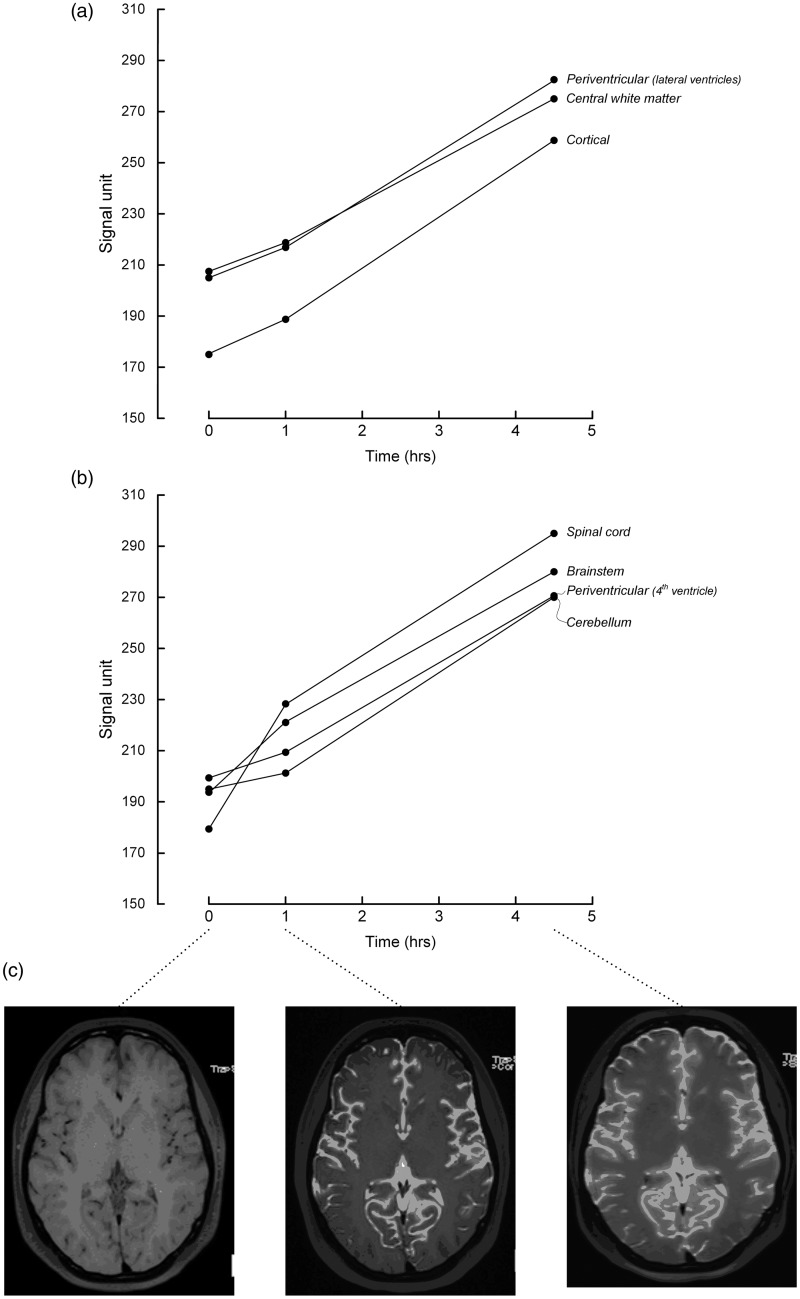
Recently, the “glymphatic system” of the brain has been discovered in rodents, which is a paravascular, transparenchymal route for clearance of excess brain metabolites and distribution of compounds in the cerebrospinal fluid. It has already been demonstrated that intrathecally administered gadolinium (Gd) contrast medium distributes along this route in rats, but so far not in humans. A 27-year-old woman underwent magnetic resonance imaging (MRI) with intrathecal administration of gadobutrol, which distributed throughout her entire brain after 1 and 4.5 h. MRI with intrathecal Gd may become a tool to study glymphatic function in the human brain.
Overview
The claims and statistics of this paper haven’t been checked yet.
Run the check — free, no account needed. It reads the open-access text and checks the sources it cites.
Integrity signals
Registry
Cites
Statistics
Figures
Verification
Claims the paper makes
The claims and statistics of this paper haven’t been checked yet.
Run the check — free, no account needed. It reads the open-access text and checks the sources it cites.
Claims other papers make about this one
Each is checked against this paper’s own text: does it say what the citing paper says it does?
The source backs part of the claim, not all of it — or backs it for a different population, condition or comparison.
In the citing paper
“However, literature reporting human in vivo CSF tracer studies is lacking, and observations made in animals have not been translated into humans.”
From Brain-wide glymphatic enhancement and clearance in humans assessed with MRI. (2018) · cites this paper as [12]
Experimental — accuracy not yet measuredThe cited source says what the claim attributes to it.
In the citing paper
“Two recent human studies demonstrated brain parenchyma enhancement subsequent to subarachnoid (intrathecal) administration of a MRI contrast agent, but observations were limited to selected ROIs (12, 13).”
From Brain-wide glymphatic enhancement and clearance in humans assessed with MRI. (2018) · cites this paper as [12]
Self-check
Percentages, sums and reported differences re-computed from the paper’s own numbers.
The self-check needs the paper’s full text.
Citations
Cites
Iliff, J. J., Wang, M., Liao, Y., Plogg, B. A., Peng, W., Gundersen, G. A., Benveniste, H., Vates, G. E., Deane, R., Goldman, S. A., et al. (2012) · Science Translational Medicine · doi:10.1126/scitranslmed.3003748 · Cited by 5,824
The glymphatic system: a beginner's guide
Jessen, N. A., Munk, A. S. F., Lundgaard, I., & Nedergaard, M. (2015) · Neurochemical Research · doi:10.1007/s11064-015-1581-6 · Cited by 2,075
Impairment of paravascular clearance pathways in the aging brain
Kress, B. T., Iliff, J. J., Xia, M., Wang, M., Wei, H. S., Zeppenfeld, D., Xie, L., Kang, H., Xu, Q., Liew, J. A., et al. (2014) · Annals of Neurology · doi:10.1002/ana.24271 · Cited by 1,521 · cited 1×
“Glymphatic function has also been shown to decrease dramatically with aging (2) and in the injured brain (3).”
BackgroundProduction of the Alzheimer Amyloid β Protein by Normal Proteolytic Processing
Shoji, M., Golde, T. E., Ghiso, J., Cheung, T. T., Estus, S., Shaffer, L. M., Cai, X.-D., McKay, D. M., Tintner, R., Frangione, B., et al. (1992) · Science · doi:10.1126/science.1439760 · Cited by 1,447
Impairment of Glymphatic Pathway Function Promotes Tau Pathology after Traumatic Brain Injury
Iliff, J. J., Chen, M. J., Plog, B. A., Zeppenfeld, D. M., Soltero, M., Yang, L., Singh, I., Deane, R., & Nedergaard, M. (2014) · The Journal of Neuroscience · doi:10.1523/jneurosci.3020-14.2014 · Cited by 1,159
Brain-wide pathway for waste clearance captured by contrast-enhanced MRI
Iliff, J. J., Lee, H., Yu, M., Feng, T., Logan, J., Nedergaard, M., & Benveniste, H. (2013) · Journal of Clinical Investigation · doi:10.1172/jci67677 · Cited by 1,148
Impaired Glymphatic Perfusion After Strokes Revealed by Contrast-Enhanced MRI
Gaberel, T., Gakuba, C., Goulay, R., de Lizarrondo, S. M., Hanouz, J., Emery, É., Touzé, E., Vivien, D., & Gauberti, M. (2014) · Stroke · doi:10.1161/strokeaha.114.006617 · Cited by 438
Akbar, J. J., Luetmer, P. H., Schwartz, K. M., et al. (2011) · American Journal of Neuroradiology · doi:10.3174/ajnr.a2815 · Cited by 119
LOCALIZATION OF CEREBROSPINAL FLUID LEAKS BY GADOLINIUM-ENHANCED MAGNETIC RESONANCE CISTERNOGRAPHY
Aydın, K., Terzibaşioğlu, E., Sencer, S., Sencer, A., Süoğlu, Y., Karasu, A., Kiris, T., & Turantan, M. I. (2008) · Neurosurgery · doi:10.1227/01.neu.0000317306.39203.24 · Cited by 68
Adam, G., Neuerburg, J. M., Spüntrup, E., et al. (1994) · Magnetic Resonance in Medicine · doi:10.1002/mrm.1910320511 · Cited by 45
Cited by
Sorted by citations
Items 101–200 of 218
Zhang, J., Liu, S., Wu, Y., Tang, Z., Wu, Y., Qi, Y., Dong, F., & Wang, Y. (2023) · Cellular and Molecular Neurobiology · doi:10.1007/s10571-023-01440-7 · Cited by 31
Taoka, T., Ito, R., Nakamichi, R., Nakane, T., Kawai, H., & Naganawa, S. (2022) · Magnetic Resonance in Medical Sciences · doi:10.2463/mrms.rev.2022-0012 · Cited by 30
Jacobsen, H. H., Sandell, T., Jørstad, Ø. K., Moe, M. C., Ringstad, G. A., & Eide, P. K. (2020) · Investigative Ophthalmology & Visual Science · doi:10.1167/iovs.61.13.24 · Cited by 28
Self-citationA SPECT-based method for dynamic imaging of the glymphatic system in rats
Sigurdsson, B., Hauglund, N. L., Lilius, T. O., Mogensen, F. L., Mortensen, K. N., Beschörner, N., Klinger, L., Bærentzen, S. L., Rosenholm, M., Shalgunov, V., et al. (2023) · Journal of Cerebral Blood Flow & Metabolism · doi:10.1177/0271678x231156982 · Cited by 30
Intrathecal Injection of Gadobutrol: A Tale of Caution
Reeves, C., Galang, E., Padalia, R. B., et al. (2017) · Journal of Pain & Palliative Care Pharmacotherapy · doi:10.1080/15360288.2017.1313353 · Cited by 27
Yin, M., Pu, T., Wang, L., Marshall, C., Wu, T., & Xiao, M. (2018) · Medical Hypotheses · doi:10.1016/j.mehy.2018.07.016 · Cited by 29
Fluid dynamics in aging-related dementias
Bah, T. M., Siler, D. A., Ibrahim, A. H., Cetas, J. S., & Alkayed, N. J. (2023) · Neurobiology of Disease · doi:10.1016/j.nbd.2022.105986 · Cited by 26
Örzsik, B., Palombo, M., Asllani, I., Dijk, D., Harrison, N. A., & Cercignani, M. (2023) · NeuroImage · doi:10.1016/j.neuroimage.2023.120124 · Cited by 26
Bao, C., Luo, H., Wang, J., Liu, X., Li, Y., Yang, J., Chen, C., Yang, R., Ba, W., Lian, X., et al. (2025) · The Journal of Prevention of Alzheimer s Disease · doi:10.1016/j.tjpad.2025.100156 · Cited by 27
Naganawa, S., Nakane, T., Kawai, H., et al. (2017) · Magnetic Resonance in Medical Sciences · doi:10.2463/mrms.ci.2016-0114 · Cited by 24
In Vivo Imaging of Neuroinflammatory Targets in Treatment-Resistant Epilepsy
Sharma, A. A., & Szaflarski, J. P. (2020) · Current Neurology and Neuroscience Reports · doi:10.1007/s11910-020-1025-9 · Cited by 24
Glymphatic Imaging in Pediatrics
Li, X., Lin, Z., Liu, C., Bai, R., Wu, D., & Yang, J. (2023) · Journal of Magnetic Resonance Imaging · doi:10.1002/jmri.29040 · Cited by 23
Neumann, K., Günther, M., Düzel, E., & Schreiber, S. (2022) · Frontiers in Aging Neuroscience · doi:10.3389/fnagi.2022.871612 · Cited by 24
Naganawa, S., Ito, R., Kato, Y., Kawai, H., Taoka, T., Yoshida, T., Maruyama, K., Murata, K., Koerzdoerfer, G., Pfeuffer, J., et al. (2020) · Magnetic Resonance in Medical Sciences · doi:10.2463/mrms.mp.2020-0030 · Cited by 23
The perivascular pathways for influx of cerebrospinal fluid are most efficient in the midbrain
Dobson, H., Sharp, M. M., Cumpsty, R., Criswell, T. P., Wellman, T., Finucane, C., Sullivan, J. M., Weller, R. O., Verma, A., & Carare, R. O. (2017) · Clinical Science · doi:10.1042/cs20171265 · Cited by 27
Öztürk, K., Nas, Ö. F., Soylu, E., & Hakyemez, B. (2018) · Investigative Radiology · doi:10.1097/rli.0000000000000472 · Cited by 22
Zedde, M., & Pascarella, R. (2024) · Brain Sciences · doi:10.3390/brainsci14100983 · Cited by 21
Smith-Cohn, M., Burley, N. B., & Grossman, S. A. (2022) · Current Neuropharmacology · doi:10.2174/1570159x20999220131163504 · Cited by 21
Dual orexin receptor antagonists as promising therapeutics for Alzheimer’s disease
Ragsdale, S. M., Radovich, J. M., Coiduras, I. I., McCall, W. V., Grant, S. C., Lee, C. S., & Wilber, A. A. (2025) · npj Biological Timing and Sleep · doi:10.1038/s44323-025-00025-5 · Cited by 20
Detection of IV-gadolinium Leakage from the Cortical Veins into the CSF Using MR Fingerprinting
Naganawa, S., Nakane, T., Kawai, H., Taoka, T., Kawaguchi, H., Maruyama, K., Murata, K., Koerzdoerfer, G., Pfeuffer, J., Nittka, M., et al. (2019) · Magnetic Resonance in Medical Sciences · doi:10.2463/mrms.mp.2019-0048 · Cited by 21
Bert, R. J., Settipalle, N., Tiwana, E., Muddasani, D., Nath, R., Wellman, B., Mihlon, F., Negahdar, M., Amini, A. A., & Boakye, M. (2019) · American Journal of Physiology-Regulatory, Integrative and Comparative Physiology · doi:10.1152/ajpregu.00101.2018 · Cited by 20
Eide, P. K., & Ringstad, G. A. (2019) · Gerontology and Geriatric Medicine · doi:10.1177/2333721419889739 · Cited by 22
Self-citationKim, M., Song, Y. S., Han, K., Bae, Y. J., Han, J. W., & Kim, K. W. (2024) · Journal of Alzheimer s Disease · doi:10.3233/jad-231131 · Cited by 20
Drenthen, G. S., Elschot, E. P., van der Knaap, N., Uher, D., Voorter, P., Backes, W. H., Jansen, J. F. A., & van der Thiel, M. M. (2023) · Journal of Magnetic Resonance Imaging · doi:10.1002/jmri.29056 · Cited by 20
Korann, V., Panganiban, K. J., Stogios, N., Remington, G., Graff‐Guerrero, A., Chintoh, A. F., Hahn, M., & Agarwal, S. M. (2024) · The Canadian Journal of Psychiatry · doi:10.1177/07067437241290193 · Cited by 17
Hu, H., Wan, S., Hu, Y., Wang, Q., Li, H., & Zhang, N. (2025) · Annals of Medicine · doi:10.1080/07853890.2024.2445194 · Cited by 20
Measuring glymphatic function: Assessing the toolkit
Ayyappan, K., Unger, L., Kitchen, P., Bill, R. M., & Salman, M. M. (2025) · Neural Regeneration Research · doi:10.4103/nrr.nrr-d-24-01013 · Cited by 27
Li, A. M., Chen, L., Liu, H., Li, Y., Duan, W., & Xu, J. (2021) · Magnetic Resonance in Medicine · doi:10.1002/mrm.29137 · Cited by 17
Glymphatic function and its influencing factors in different glucose metabolism states
Tian, B., Zhao, C., Liang, J.-L., Zhang, H., Xu, Y., Zheng, H.-L., Zhou, J., Gong, J.-N., Lu, S., & Zeng, Z. (2024) · World Journal of Diabetes · doi:10.4239/wjd.v15.i7.1537 · Cited by 20
Glymphatic MRI techniques in sleep and neurodegenerative diseases
Lee, H., Choi, S. H., & Anzai, Y. (2022) · Current Opinion in Pulmonary Medicine · doi:10.1097/mcp.0000000000000923 · Cited by 17
Xiao, M., Hou, J., Xu, M., Li, S., & Yang, B. (2023) · Advances in experimental medicine and biology · doi:10.1007/978-981-19-7415-1_7 · Cited by 17
Functional aspects of the brain lymphatic drainage system in aging and neurodegenerative diseases
Chen, Y., He, X., Cai, J., & Li, Q. (2024) · Journal of Biomedical Research · doi:10.7555/jbr.37.20230264 · Cited by 18
Association of glymphatic system dysfunction with cognitive impairment in temporal lobe epilepsy
Wang, J., Xia, X., Zhang, B., Ma, X., Shi, F., Ying, W., Li, L., & Meng, X. (2024) · Frontiers in Aging Neuroscience · doi:10.3389/fnagi.2024.1459580 · Cited by 20
Cerebrospinal fluid‐tissue exchange revealed by phase alternate labeling with null recovery MRI
Li, A. M., & Xu, J. (2021) · Magnetic Resonance in Medicine · doi:10.1002/mrm.29092 · Cited by 16
Glymphatic function decline as a mediator of core memory-related brain structures atrophy in aging
Chong, S., Wang, S., Gao, T., Yuan, K., Han, Y. K., Shi, L., Li, P., Lin, X., & Lü, L. (2025) · Journal of Translational Internal Medicine · doi:10.1515/jtim-2025-0007 · Cited by 16
The glymphatic system as a therapeutic target: TMS-induced modulation in older adults
Sundman, M., Liu, Y., Chen, N., & Chou, Y. (2025) · Frontiers in Aging Neuroscience · doi:10.3389/fnagi.2025.1597311 · Cited by 17
Imaging cerebrospinal fluid dynamics in idiopathic normal pressure hydrocephalus
Ringstad, G. A. (2018) · NORA - Norwegian Open Research Archives · Cited by 15
Self-citationBrain volume changes in spontaneous intracranial hypotension: Revisiting the Monro-Kellie doctrine
Wu, J., Wang, Y.-F., Hseu, S., Chen, S.-T., Chen, Y.-L., Wu, Y., Chen, S., Lirng, J.-F., & Wang, S. (2020) · Cephalalgia · doi:10.1177/0333102420950385 · Cited by 14
Chen, Z., Lai, J. H. C., Xu, J., Zhang, H., Huang, J., & Chan, K. W. Y. (2024) · NMR in Biomedicine · doi:10.1002/nbm.5093 · Cited by 13
Neurofluid as Assessed by Diffusion-Weighted Imaging
Taoka, T. (2021) · Magnetic Resonance Imaging Clinics of North America · doi:10.1016/j.mric.2021.01.002 · Cited by 13
Kiviniemi, V. J., Helakari, H., Raitamaa, L., Huotari, N., Rajna, Z., Järvelä, M., Elabasy, A., Tuunanen, J., Poltojainen, V., Väyrynen, T., et al. (2025) · NMR in Biomedicine · doi:10.1002/nbm.70092 · Cited by 11
Kopeć, K., Koziorowski, D., & Szlufik, S. (2025) · International Journal of Molecular Sciences · doi:10.3390/ijms26157552 · Cited by 15
Lin, W., Chen, H., Zhang, Z., Zhang, Y., Wang, X., Guo, Y., Qin, H., Liu, T., & Chen, F. (2025) · Scientific Reports · doi:10.1038/s41598-025-15923-8 · Cited by 14
Sarker, A. K., Suh, M., Choi, Y., Park, J. Y., Lee, Y., & Lee, D. S. (2023) · Scientific Reports · doi:10.1038/s41598-023-39903-y · Cited by 13
Yin, Y., Peng, Y. H., Nie, L., Li, X., Xiao, Y., Jiang, H., Gao, L., & Liu, H. (2023) · Neuroradiology · doi:10.1007/s00234-023-03269-9 · Cited by 14
Chu, M., Yue, A., Yan, S., Yu, B., He, Q., Chen, Y., Hou, J., Nan, H., Ye, H., Nie, B., et al. (2025) · Alzheimer s & Dementia · doi:10.1002/alz.70561 · Cited by 12
Guo, Y., Liu, T., Chen, H., Zhou, L., Huang, W., Zhang, K., Wang, X., wang, Y., Zhou, J. H., & Chen, F. (2025) · Brain Research Bulletin · doi:10.1016/j.brainresbull.2025.111295 · Cited by 13
Glymphatic pathway: An emerging perspective in the pathophysiology of neurodegenerative diseases
Rehman, M. U., Sehar, N., Rasool, I., Aldossari, R. M., Wani, A. B., Rashid, S. M., Wali, A. F., Ali, A., Arafah, A. M., & Khan, A. (2024) · International Journal of Geriatric Psychiatry · doi:10.1002/gps.6104 · Cited by 10
Traumatic brain injury and sleep in military and veteran populations: A literature review
Landvater, J., Kim, S. Y., Caswell, K., Kwon, C., Odafe, E., Roe, G., Tripathi, A., Vukovics, C., Wang, J., Ryan, K., et al. (2024) · Neurorehabilitation · doi:10.3233/nre-230380 · Cited by 12
Zhao, X., Cao, R., Tian, X., Liu, P., Liu, D., Yu, X., Zheng, Z., Chen, G., & Zou, L. (2025) · Neurochemical Research · doi:10.1007/s11064-025-04388-w · Cited by 10
Altered brain fluid dynamics in spontaneous intracranial hypotension
Eide, P. K., Hovd, M. H., Pripp, A. H., Gjertsen, Ø., Løvland, G., Lashkarivand, A., Lindstrøm, E. K., Valnes, L. M., & Ringstad, G. A. (2025) · Fluids and Barriers of the CNS · doi:10.1186/s12987-025-00679-0 · Cited by 10
Self-citationDoğan, S. N., Salt, V., Korkmazer, B., et al. (2020) · Neuroradiology · doi:10.1007/s00234-020-02463-3 · Cited by 10
Research on the Glial–Lymphatic System and Its Relationship With Alzheimer’s Disease
Ding, D., Wang, X., Li, Q., Li, L., & Wu, J. W. (2021) · Frontiers in Neuroscience · doi:10.3389/fnins.2021.605586 · Cited by 11
Koundal, S., Chen, X., Gursky, Z. H., Lee, H., Xu, K., Liang, F., Xie, Z., Xu, F., Lin, H., Nostrand, W. E. V., et al. (2024) · iScience · doi:10.1016/j.isci.2024.111463 · Cited by 11
Khalafi, M., Shirbandi, K., Zhou, L., Butler, T., Chen, K., Dartora, W. J., Keil, S. A., Li, Y., & Chiang, G. C.-Y. (2025) · American Journal of Neuroradiology · doi:10.3174/ajnr.a8953 · Cited by 11
Kim, H. I., Lee, I. H., Park, J. S., et al. (2020) · Journal of Clinical Medicine · doi:10.3390/jcm9093013 · Cited by 9
Riba, M., Romera, C., Alsina, R., Alsina-Scheer, G., Pelegrí, C., Vilaplana, J., & del Valle, J. (2023) · Brain Structure and Function · doi:10.1007/s00429-023-02664-5 · Cited by 11
The Glymphatic System: A Review
Oliveira, L. M., Figueiredo, E. G., & Peres, C. M. A. (2018) · Arquivos Brasileiros de Neurocirurgia Brazilian Neurosurgery · doi:10.1055/s-0038-1667052 · Cited by 9
Neurosurgery and the glymphatic system
Eide, P. K. (2024) · Acta Neurochirurgica · doi:10.1007/s00701-024-06161-4 · Cited by 9
Self-citationAdult Hydrocephalus and the Glymphatic System
Eide, P. K. (2025) · Neurosurgery Clinics of North America · doi:10.1016/j.nec.2024.11.002 · Cited by 9
Self-citationContrast Agent Clearance Dynamics and Diffusion in the Mouse Brain After Ventricular Administration
Decker, Y., Müller, A., & Proulx, S. T. (2025) · Advanced Science · doi:10.1002/advs.202501502 · Cited by 8
Berry, J. A. D., Elia, C. J., Sweiss, R., Lawandy, S., Bowen, I. E., Zampella, B., Saini, H., Brazdzionis, J., & Miulli, D. E. (2020) · Journal of Osteopathic Medicine · doi:10.7556/jaoa.2020.074 · Cited by 6
Kim, D., Huang, Y., & Liu, J. (2025) · NeuroImage · doi:10.1016/j.neuroimage.2025.121142 · Cited by 6
The Emerging Field of Perivascular Flow Dynamics: Biological Relevance and Clinical Applications
Huffman, J. N., Phillips, S. M., Taylor, G. T., & Paul, R. (2016) · Technology & Innovation · doi:10.21300/18.1.2016.63 · Cited by 7
Rare Earth Elements and Their Minerals
Aide, M. T., & Nakajima, T. (2019) · IntechOpen eBooks · doi:10.5772/intechopen.77602 · Cited by 6
Zeng, L., Yin, Z., Li, W., Wang, X., Zhang, Y., Xie, M., & Zhao, L. (2025) · Frontiers in Neurology · doi:10.3389/fneur.2025.1607723 · Cited by 6
Wu, C., Chen, S., Chung, C., Yu, K., Lin, T.-M., Luo, C., Lirng, J.-F., Lee, I., & Chang, F. (2024) · Journal of Stroke · doi:10.5853/jos.2023.04203 · Cited by 5
Current Clinical Issues: Deposition of Gadolinium Chelates
Nakajima, T., & Lamid-Ochir, O. (2020) · IntechOpen eBooks · doi:10.5772/intechopen.91260 · Cited by 4
Keser, Z., Smith, G., Cagil, E., Tufan, F., Albayram, Ö., & Albayram, S. (2022) · Journal of Neuroimaging · doi:10.1111/jon.13056 · Cited by 5
Zhou, Z., Zhao, T., Gardus, J. D., Wen, Q., Yang, F., DeLorenzo, C., Parsey, R. V., & Huang, C. (2025) · medRxiv · doi:10.1101/2025.04.07.25325241 · Cited by 5
Diffusion Tensor Imaging in Neurofluids
Levendovszky, S. R., & Meyer, B. P. (2025) · Neuroimaging Clinics of North America · doi:10.1016/j.nic.2024.11.001 · Cited by 4
Noninvasive whole-brain imaging of glymphatic dynamics
Wang, N., YU, X., Lowerison, M. R., Li, Q., Canning, A. J., He, P., Dang, L., Degan, S., Mace, B. E., Xu, Y., et al. (2026) · Science Advances · doi:10.1126/sciadv.aee4926 · Cited by 5
Ray, N. J., Cullell, N., Clark, O. J., Delgado-Sánchez, A., Caruana, G., Elias-Mas, A., Ferrer, J., Buongiorno, M., Castejón, J., Sánchez‐Benavides, G., et al. (2024) · bioRxiv (Cold Spring Harbor Laboratory) · doi:10.1101/2024.10.14.618324 · Cited by 4
Koivula, A., Perkiömäki, V., Aho, M., Rasila, A., Poltojainen, V., Korhonen, V. O., Järvelä, M., Huotari, N., Helakari, H., Tuunanen, J., et al. (2025) · NeuroImage · doi:10.1016/j.neuroimage.2025.121503 · Cited by 3
MR Imaging of Neurofluids in the Developing Brain
Agarwal, N., Klein, W. M., & Tuura, R. (2025) · Neuroimaging Clinics of North America · doi:10.1016/j.nic.2024.12.005 · Cited by 2
Simultaneous coherent–incoherent motion imaging in brain parenchyma
Jansen, I. H., Abad, N., Ajala, A., Bhushan, C., Werner, J. K., DeMarco, J. K., Morris, H. D., Pollatou, A., Kohls, G., Yalewayker, H., et al. (2025) · Interface Focus · doi:10.1098/rsfs.2024.0041 · Cited by 5
Sacchi, L., Farruggio, G., Bocca, G., Carandini, T., Riz, M. A. D., Ghezzi, L., Pintus, M., Pietroboni, A. M., Boffa, G., Scola, E., et al. (2026) · Human Brain Mapping · doi:10.1002/hbm.70603 · Cited by 3
Neurodegenerative Dementias: Improving Brain Health to Decrease Risk
Bateman, J. R., Hurley, R. A., & Taber, K. H. (2019) · Journal of Neuropsychiatry · doi:10.1176/appi.neuropsych.18110287 · Cited by 3
Woes with flows under pressure
Jansson, D., & Iliff, J. J. (2021) · Brain Communications · doi:10.1093/braincomms/fcab094 · Cited by 3
Pathobiology of the Glymphatic System in the Traumatic Brain Injury: A Narrative Review
Moscote‐Salazar, L. R., Beltran-Ruiz, J. J., Bello, J. S. R., & Lugo, C. M. R. (2022) · Panamerican Journal of Trauma Critical Care & Emergency Surgery · doi:10.5005/jp-journals-10030-1389 · Cited by 3
Deng, L., Jiang, N., Yuan, S., Jiang, D., Zhu, L., & Zhang, Y. (2025) · Journal of Affective Disorders · doi:10.1016/j.jad.2025.120427 · Cited by 3
Wu, Y., Wang, K., Ju, L., Li, A., Qin, Q., Xu, F., Lin, D. D. M., Kleinberg, L. R., & Xu, J. (2026) · Magnetic Resonance in Medicine · doi:10.1002/mrm.70298 · Cited by 4
Zhou, X. A., Man, W., Liu, X., Choi, S., Jiang, Y., Hike, D., Gomez-Cid, L., Lin, C., Nedergaard, M., & Yu, X. (2024) · Proceedings on CD-ROM - International Society for Magnetic Resonance in Medicine. Scientific Meeting and Exhibition/Proceedings of the International Society for Magnetic Resonance in Medicine, Scientific Meeting and Exhibition · doi:10.58530/2024/1126 · Cited by 2
Gadolinium-Based Imaging and the Study of Neurofluid Dynamics
Ringstad, G. A., Eide, P. K., Naganawa, S., & Agarwal, N. (2025) · Neuroimaging Clinics of North America · doi:10.1016/j.nic.2024.12.008 · Cited by 3
Self-citationLi, Y. (2025) · Frontiers in Aging Neuroscience · doi:10.3389/fnagi.2025.1581223 · Cited by 2
Human brain MRI data of CSF tracer evolution over 72h for data-integrated simulationsPreprint
Riseth, J. N., Koch, T., Lian, S. L., Storås, T. H., Zikatanov, L., Valnes, L. M., Nordengen, K., & Mardal, K. (2025) · medRxiv · doi:10.1101/2025.07.23.25331971 · Cited by 2
Gupta, S., Amyot, F., Coon, W. G., Pollatou, A., Skeiky, L., Seenivasan, S., Kenney, K., Lee, A., Lettieri, M., Penafiel, A. I., et al. (2025) · bioRxiv (Cold Spring Harbor Laboratory) · doi:10.1101/2025.10.27.684357 · Cited by 3
Pihala, T., Kiviniemi, V. J., & Martikainen, M. H. (2026) · Frontiers in Neuroscience · doi:10.3389/fnins.2025.1726054 · Cited by 1
Liu, Y., Liu, C., Liu, D., Li, Y., Li, Y., Li, S., Lu, J., Liu, P., & Gao, M. (2026) · The Journal of Sexual Medicine · doi:10.1093/jsxmed/qdag101 · Cited by 1
Asoğlu, S., Kılınçer, A., Özer, H., Topaloğlu, Ö. F., & Cebeci, H. (2024) · Clinical Imaging · doi:10.1016/j.clinimag.2024.110140 · Cited by 1
Zhou, L., Li, Y., & de Leon, M. J. (2025) · Neuroimaging Clinics of North America · doi:10.1016/j.nic.2024.12.001 · Cited by 1
Eide, P. K., & Ringstad, G. A. (2018) · Springer eBooks · doi:10.1007/978-3-319-97928-1_4 · Cited by 1
Self-citationWu, Y., Wang, K., Ju, L., Li, A., Qin, Q., Xu, F., Lin, D. D. M., Kleinberg, L. R., & Xu, J. (2025) · medRxiv · doi:10.1101/2025.08.28.25334379 · Cited by 1
Önder, H. (2018) · Turkish Journal Of Neurology · doi:10.4274/tnd.23911
Dynamics of cerebral fluids in patients suffering from hydrocephalus and pseudotumour cerebri
Lalou, A. D. (2019) · Apollo (University of Cambridge) · doi:10.17863/cam.53189
Imaging of Glymphatic Flow and Neurodegeneration
Filippi, C. G., & Watts, R. (2021) · Springer eBooks · doi:10.1007/978-3-030-82367-2_71
Imaging blood-brain barrier function in aging
Verheggen, I. C. M. (2021) · Research Publications (Maastricht University) · doi:10.26481/dis.20210601iv
Sarker, A. K., Suh, M., Choi, Y., Park, J. Y., Lee, Y., & Lee, D. S. (2023) · bioRxiv (Cold Spring Harbor Laboratory) · doi:10.1101/2023.01.18.524645
Magnetic resonance imaging-based progress in human glymphatic system research
Chen, S., Chen, Y., & Wu, R. (2023) · Radiology Science · doi:10.15212/radsci-2022-0011
Gadolinium-based imaging of the blood brain barrier and brain waste clearance pathways
(2023) · Advances in magnetic resonance technology and applications · doi:10.1016/b978-0-323-91771-1.00017-4
Metadata
- Authors
- Eide, P. K., & Ringstad, G.
- Affiliations
- Oslo University Hospital (NO)
- Corresponding author
- Eide, Per Kristian
- Venue
- Acta Radiologica Open
- Publisher
- SAGE Publications
- Published
- 2015-11-01
- Volume
- 4
- Issue
- 11
- Pages
- 205846011560963
- ISSN
- 2047-9816 (ISSN-L)
- 2058-4601
- PMC
- PMC4652208
- OpenAlex
- W2252352100
- Cited by
- 218
- References
- 10
- Citations by year
- 2026: 17 · 2025: 33 · 2024: 22 · 2023: 22 · 2022: 15 · 2021: 22 · 2020: 26 · 2019: 17 · 2018: 22 · 2017: 15 · 2016: 7
- FWCI
- 6.84
- Type
- journal article
- Language
- en
- Open access
- gold
- doi.org
- Locations
- Acta Radiologica Open (publishedVersion, cc-by-nc)
- Europe PMC (PubMed Central) (publishedVersion, cc-by-nc)
- PubMed Central (publishedVersion, cc-by-nc)
- PubMed (publishedVersion)
- DOAJ (DOAJ: Directory of Open Access Journals) (submittedVersion, cc-by-sa)
- Topics
- Health Sciences › Medicine › Clinical Neurology › Cerebrospinal fluid and hydrocephalus
- Health Sciences › Medicine › Radiology, Nuclear Medicine and Imaging › Advanced MRI Techniques and Applications
- Life Sciences › Neuroscience › Neurology › Barrier Structure and Function Studies
- Keywords
- glymphatic system, gadobutrol, cerebrospinal fluid, human brain imaging
About these checks
Registry, retraction, Cites and statistics results are computed, not judged, with one exception: a printed reference that no index lists is read once by a model before it counts as a citation error. Claims are extracted by a model and kept only when their quote appears verbatim in the text; each cited source is then checked, with a model’s help, for whether it says what the claim relies on. Those verdicts can be wrong — read the claim’s quote and the cited source before relying on one.
Statistics and arithmetic are recomputed, not judged.
Confidence bands are shown only for fields calibrated on our annotated test set. None is calibrated yet, so no band is shown.
What counts as a claim ›How we measure confidence ›How we check this ›
Are you an author, or do you see a problem with this page? Request a review
