

The DOI matches its Crossref record.
Caspase-9, caspase-3 and caspase-7 have distinct roles during intrinsic apoptosis
Abstract
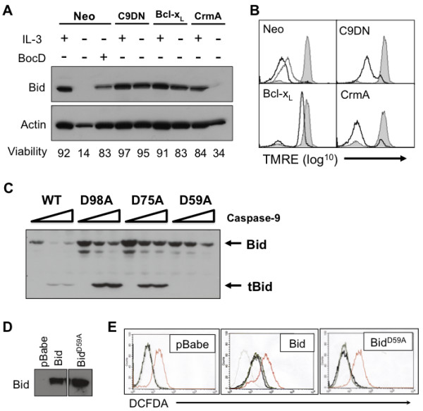
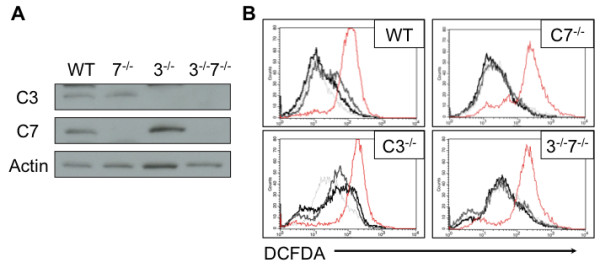
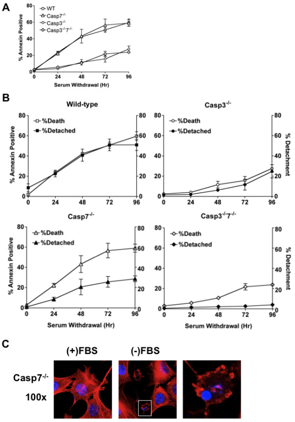
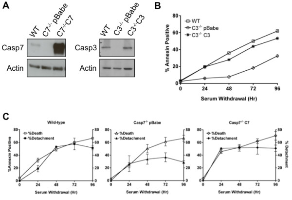
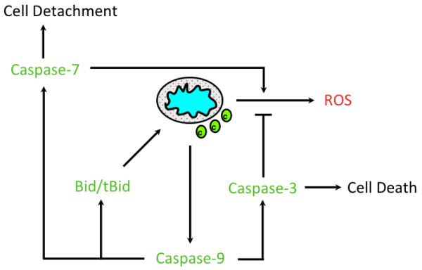
Background: Apoptosis is a form of programmed cell death that is regulated by the Bcl-2 family and caspase family of proteins. The caspase cascade responsible for executing cell death following cytochrome c release is well described; however the distinct roles of caspases-9, -3 and -7 during this process are not completely defined. Results: Here we demonstrate several unique functions for each of these caspases during cell death. Specific inhibition of caspase-9 allows for efficient release of cytochrome c , but blocks changes in mitochondrial morphology and ROS production. We show that caspase-9 can cleave Bid into tBid at amino acid 59 and that this cleavage of Bid is required for ROS production following serum withdrawal. We also demonstrate that caspase-3-deficient MEFs are less sensitive to intrinsic cell death stimulation, yet have higher ROS production. In contrast, caspase-7-deficient MEFs are not resistance to intrinsic cell death, but remain attached to the ECM. Conclusions: Taken together, these data suggest that caspase-9 is required for mitochondrial morphological changes and ROS production by cleaving and activating Bid into tBid. After activation by caspase-9, caspase-3 inhibits ROS production and is required for efficient execution of apoptosis, while effector caspase-7 is required for apoptotic cell detachment.
Overview
The claims and statistics of this paper haven’t been checked yet.
Run the check — free, no account needed. It reads the open-access text and checks the sources it cites.
Integrity signals
Registry
Cites
Statistics
Figures
Verification
Claims the paper makes
The claims and statistics of this paper haven’t been checked yet.
Run the check — free, no account needed. It reads the open-access text and checks the sources it cites.
Claims other papers make about this one
Each is checked against this paper’s own text: does it say what the citing paper says it does?
In the citing paper
“Activated by intrinsic cues like elevated reactive oxygen species (ROS) level, cytochrome c is released from the mitochondria to cytoplasm and induces apoptotic process by targeting initiator caspases.63”
Self-check
Percentages, sums and reported differences re-computed from the paper’s own numbers.
The self-check needs the paper’s full text.
Citations
Cites
No references could be resolved yet.
Research works printed but not linked: 25
- Danial NN, Korsmeyer SJ: Cell death: critical control points. Cell. 2004, 116 (2): 205-219. 10.1016/S0092-8674(04)00046-7. · DOI · Paper page
- Galluzzi L, Vitale I, Abrams JM, Alnemri ES, Baehrecke EH, Blagosklonny MV, Dawson TM, Dawson VL, El-Deiry WS, Fulda S: Molecular definitions of cell death subroutines: recommendations of the Nomenclature Committee on Cell Death 2012. Cell Death Differ. 2012, 19 (1): 107-120. 10.1038/cdd.2011.96. · DOI · Paper page
- Wei MC, Lindsten T, Mootha VK, Weiler S, Gross A, Ashiya M, Thompson CB, Korsmeyer SJ: tBID, a membrane-targeted death ligand, oligomerizes BAK to release cytochrome c. Genes Dev. 2000, 14 (16): 2060-2071. · DOI
- Eskes R, Desagher S, Antonsson B, Martinou JC: Bid induces the oligomerization and insertion of Bax into the outer mitochondrial membrane. Mol Cell Biol. 2000, 20 (3): 929-935. 10.1128/MCB.20.3.929-935.2000. · DOI
- Wei MC, Zong WX, Cheng EH, Lindsten T, Panoutsakopoulou V, Ross AJ, Roth KA, MacGregor GR, Thompson CB, Korsmeyer SJ: Proapoptotic BAX and BAK: a requisite gateway to mitochondrial dysfunction and death. Science. 2001, 292 (5517): 727-730. 10.1126/science.1059108. · DOI · Paper page
- Li P, Nijhawan D, Budihardjo I, Srinivasula SM, Ahmad M, Alnemri ES, Wang X: Cytochrome c and dATP-dependent formation of Apaf-1/caspase-9 complex initiates an apoptotic protease cascade. Cell. 1997, 91 (4): 479-489. 10.1016/S0092-8674(00)80434-1. · DOI · Paper page
- Srinivasula SM, Ahmad M, Fernandes-Alnemri T, Alnemri ES: Autoactivation of procaspase-9 by Apaf-1-mediated oligomerization. Mol Cell. 1998, 1 (7): 949-957. 10.1016/S1097-2765(00)80095-7. · DOI
- Woo M, Hakem R, Soengas MS, Duncan GS, Shahinian A, Kagi D, Hakem A, McCurrach M, Khoo W, Kaufman SA: Essential contribution of caspase 3/CPP32 to apoptosis and its associated nuclear changes. Genes Dev. 1998, 12 (6): 806-819. 10.1101/gad.12.6.806. · DOI
- Shi Y: Mechanisms of caspase activation and inhibition during apoptosis. Mol Cell. 2002, 9 (3): 459-470. 10.1016/S1097-2765(02)00482-3. · DOI
- Scorrano L, Ashiya M, Buttle K, Weiler S, Oakes SA, Mannella CA, Korsmeyer SJ: A distinct pathway remodels mitochondrial cristae and mobilizes cytochrome c during apoptosis. Dev Cell. 2002, 2 (1): 55-67. 10.1016/S1534-5807(01)00116-2. · DOI · Paper page
- Zamzami N, Susin SA, Marchetti P, Hirsch T, Gomez-Monterrey I, Castedo M, Kroemer G: Mitochondrial control of nuclear apoptosis. J Exp Med. 1996, 183 (4): 1533-1544. 10.1084/jem.183.4.1533. · DOI
- Martin SJ, Green DR: Protease activation during apoptosis: death by a thousand cuts?. Cell. 1995, 82 (3): 349-352. 10.1016/0092-8674(95)90422-0. · DOI
- Cepero E, King AM, Coffey LM, Perez RG, Boise LH: Caspase-9 and effector caspases have sequential and distinct effects on mitochondria. Oncogene. 2005, 24 (42): 6354-6366. · DOI
- Slee EA, Harte MT, Kluck RM, Wolf BB, Casiano CA, Newmeyer DD, Wang HG, Reed JC, Nicholson DW, Alnemri ES: Ordering the cytochrome c-initiated caspase cascade: hierarchical activation of caspases-2, -3, -6, -7, -8, and -10 in a caspase-9-dependent manner. J Cell Biol. 1999, 144 (2): 281-292. 10.1083/jcb.144.2.281. · DOI
- Stennicke HR, Jurgensmeier JM, Shin H, Deveraux Q, Wolf BB, Yang X, Zhou Q, Ellerby HM, Ellerby LM, Bredesen D: Pro-caspase-3 is a major physiologic target of caspase-8. J Biol Chem. 1998, 273 (42): 27084-27090. 10.1074/jbc.273.42.27084. · DOI
- Thornberry NA, Rano TA, Peterson EP, Rasper DM, Timkey T, Garcia-Calvo M, Houtzager VM, Nordstrom PA, Roy S, Vaillancourt JP: A combinatorial approach defines specificities of members of the caspase family and granzyme B. Functional relationships established for key mediators of apoptosis. J Biol Chem. 1997, 272 (29): 17907-17911. 10.1074/jbc.272.29.17907. · DOI
- Lakhani SA, Masud A, Kuida K, Porter GA, Booth CJ, Mehal WZ, Inayat I, Flavell RA: Caspases 3 and 7: key mediators of mitochondrial events of apoptosis. Science. 2006, 311 (5762): 847-851. 10.1126/science.1115035. · DOI
- Walsh JG, Cullen SP, Sheridan C, Luthi AU, Gerner C, Martin SJ: Executioner caspase-3 and caspase-7 are functionally distinct proteases. Proc Natl Acad Sci USA. 2008, 105 (35): 12815-12819. 10.1073/pnas.0707715105. · DOI
- Gregory CD, Pound JD: Microenvironmental influences of apoptosis in vivo and in vitro. Apoptosis. 2010, 15 (9): 1029-1049. 10.1007/s10495-010-0485-9. · DOI
- Johnson BW, Cepero E, Boise LH: Bcl-xL inhibits cytochrome c release but not mitochondrial depolarization during the activation of multiple death pathways by tumor necrosis factor-alpha. J Biol Chem. 2000, 275 (40): 31546-31553. · DOI
- Saraste M: Oxidative phosphorylation at the fin de siecle. Science. 1999, 283 (5407): 1488-1493. 10.1126/science.283.5407.1488. · DOI
- Cai J, Jones DP: Superoxide in apoptosis. Mitochondrial generation triggered by cytochrome c loss. J Biol Chem. 1998, 273 (19): 11401-11404. 10.1074/jbc.273.19.11401. · DOI · Paper page
- Wen LP, Fahrni JA, Troie S, Guan JL, Orth K, Rosen GD: Cleavage of focal adhesion kinase by caspases during apoptosis. J Biol Chem. 1997, 272 (41): 26056-26061. 10.1074/jbc.272.41.26056. · DOI
- Johnson BW, Boise LH: Bcl-2 and caspase inhibition cooperate to inhibit tumor necrosis factor-alpha-induced cell death in a Bcl-2 cleavage-independent fashion. J Biol Chem. 1999, 274 (26): 18552-18558. 10.1074/jbc.274.26.18552. · DOI
- Swift S, Lorens J, Achacoso P, Nolan GP: Rapid production of retroviruses for efficient gene delivery to mammalian cells using 293T cell-based systems. Curr Protoc Immunol. 2001, Chapter 10: Unit 10 17C-
Cited by
Sorted by citations
Moeinabadi-Bidgoli, K., Babajani, A., Yazdanpanah, G., Farhadihosseinabadi, B., Jamshidi, E., Bahrami, S., & Niknejad, H. (2021) · Biomedicine & Pharmacotherapy · doi:10.1016/j.biopha.2021.112026 · Cited by 70
Mishra, S., das, K., Chatterjee, S., Sahoo, P., Kundu, S., Pal, M., Bhaumik, A., & Ghosh, C. K. (2023) · ACS Omega · doi:10.1021/acsomega.2c04964 · Cited by 65
Koch, G., Di Lorenzo, F., Loizzo, S., Motta, C., Travaglione, S., Baiula, M., Rimondini, R., Ponzo, V., Bonnì, S., Toniolo, S., et al. (2017) · Scientific Reports · doi:10.1038/s41598-017-14204-3 · Cited by 64
Molecular Pathogenicity of Enteroviruses Causing Neurological Disease
Majer, A., McGreevy, A., & Booth, T. F. (2020) · Frontiers in Microbiology · doi:10.3389/fmicb.2020.00540 · Cited by 50
Amorim, R., Simões, I., Veloso, C., Carvalho, A., Simões, R., Pereira, F., Thiel, T., Normann, A., Morais, C., Jurado, A., et al. (2021) · Nutrients · doi:10.3390/nu13051723 · Cited by 21
Metadata
- Authors
- Brentnall, M., Rodriguez-Menocal, L., De Guevara, R. L., Cepero, E., & Boise, L. H.
- Venue
- BMC Cell Biology
- Published
- 2013-07-09
- Volume
- 14
- Issue
- 1
- ISSN
- 1471-2121
- PMC
- PMC3710246
- Cited by
- 1,147
- References
- 0
- Language
- en
About these checks
Registry, retraction, Cites and statistics results are computed, not judged, with one exception: a printed reference that no index lists is read once by a model before it counts as a citation error. Claims are extracted by a model and kept only when their quote appears verbatim in the text; each cited source is then checked, with a model’s help, for whether it says what the claim relies on. Those verdicts can be wrong — read the claim’s quote and the cited source before relying on one.
Statistics and arithmetic are recomputed, not judged.
Confidence bands are shown only for fields calibrated on our annotated test set. None is calibrated yet, so no band is shown.
What counts as a claim ›How we measure confidence ›How we check this ›
Are you an author, or do you see a problem with this page? Request a review
How to cite
Cite CoChat for the checks on this page, and the paper for its own findings.
- This page
- CoChat. (2026). Citation check: Caspase-9, caspase-3 and caspase-7 have distinct roles during intrinsic apoptosis. https://app.cochat.ai/papers/10.1186/1471-2121-14-32
- The paper
- Brentnall, M., Rodriguez-Menocal, L., De Guevara, R. L., Cepero, E., & Boise, L. H. (2013). Caspase-9, caspase-3 and caspase-7 have distinct roles during intrinsic apoptosis. BMC Cell Biology. https://doi.org/10.1186/1471-2121-14-32
