

The DOI matches its Crossref record; 50 of the 50 references it cites were found in a registry or an index.
A genome-wide in situhybridization map of RNA-binding proteins reveals anatomically restricted expression in the developing mouse brain
Open accessAbstract
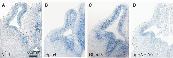
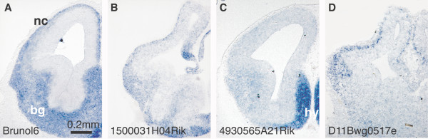
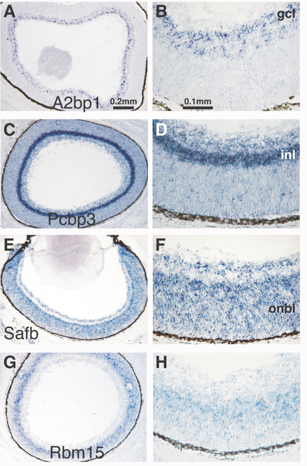
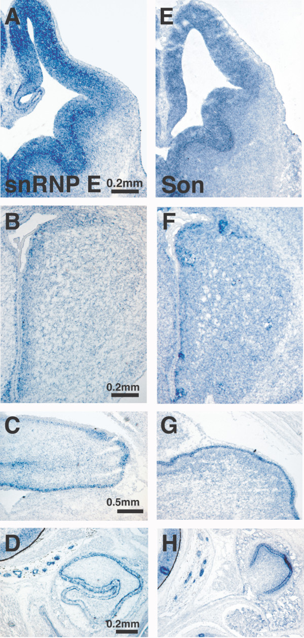
Background: In eukaryotic cells, RNA-binding proteins (RBPs) contribute to gene expression by regulating the form, abundance, and stability of both coding and non-coding RNA. In the vertebrate brain, RBPs account for many distinctive features of RNA processing such as activity-dependent transcript localization and localized protein synthesis. Several RBPs with activities that are important for the proper function of adult brain have been identified, but how many RBPs exist and where these genes are expressed in the developing brain is uncharacterized. Results: Here we describe a comprehensive catalogue of the unique RBPs encoded in the mouse genome and provide an online database of RBP expression in developing brain. We identified 380 putative RBPs in the mouse genome. Using in situ hybridization, we visualized the expression of 323 of these RBP genes in the brains of developing mice at embryonic day 13.5, when critical fate choice decisions are made and at P0, when major structural components of the adult brain are apparent. We demonstrate i) that 16 of the 323 RBPs examined show neural-specific expression at the stages we examined, and ii) that a far larger subset (221) shows regionally restricted expression in the brain. Of the regionally restricted RBPs, we describe one group that is preferentially expressed in the E13.5 ventricular areas and a second group that shows spatially restricted expression in post-mitotic regions of the embryonic brain. Additionally, we find a subset of RBPs that share the same complex pattern of expression, in proliferating regions of the embryonic and postnatal NS and peripheral tissues. Conclusion: Our data show that, in contrast to their proposed ubiquitous involvement in gene regulation, most RBPs are not uniformly expressed. Here we demonstrate the region-specific expression of RBPs in proliferating vs. post-mitotic brain regions as well as cell-type-specific RBP expression. We identify uncharacterized RBPs that exhibit neural-specific expression as well as novel RBPs that show expression in non-neural tissues. The data presented here and in an online database provide a visual filter for the functional analysis of individual RBPs.
Overview
The claims and statistics of this paper haven’t been checked yet.
Run the check — free, no account needed. It reads the open-access text and checks the sources it cites.
Integrity signals
Registry
Cites
Statistics
Figures
Verification
Claims the paper makes
The claims and statistics of this paper haven’t been checked yet.
Run the check — free, no account needed. It reads the open-access text and checks the sources it cites.
Claims other papers make about this one
Each is checked against this paper’s own text: does it say what the citing paper says it does?
The cited source says what the claim attributes to it.
In the citing paper
“whereas ELAVL2 (also known as HuB), ELAVL3 (also known as HuC) and ELAVL4 (also known as HuD) show neuronal-specific expression”
From The neurogenetics of alternative splicing (2016) · cites this paper as [51]
In the citing paper
“All three Rbfox genes are broadly expressed in the brain, with individual neuronal cell types expressing different combinations at different developmental times51–55.”
From The neurogenetics of alternative splicing (2016) · cites this paper as [51]
In the citing paper
“ELAVL1 (also known as HuR or HuA) is widely expressed in non-neuronal tissues”
From The neurogenetics of alternative splicing (2016) · cites this paper as [51]
Self-check
Percentages, sums and reported differences re-computed from the paper’s own numbers.
The self-check needs the paper’s full text.
Citations
Cites
Database resources of the National Center for Biotechnology Information: update
Wheeler, D. L. (2003) · Nucleic Acids Research · doi:10.1093/nar/gkh073 · Cited by 10,695
PANTHER: A Library of Protein Families and Subfamilies Indexed by FunctionPreprint
Thomas, P. D., Campbell, M. J., Kejariwal, A., Mi, H., Karlak, B., Daverman, R., Diemer, K., Muruganujan, A., & Narechania, A. (2003) · Genome Research · doi:10.1101/gr.772403 · Cited by 2,979
Conserved Structures and Diversity of Functions of RNA-Binding Proteins
Burd, C. G., & Dreyfuss, G. (1994) · Science · doi:10.1126/science.8036511 · Cited by 1,886
Messenger-RNA-binding proteins and the messages they carry
Dreyfuss, G., Kim, V. N., & Kataoka, N. (2002) · Nature Reviews Molecular Cell Biology · doi:10.1038/nrm760 · Cited by 1,457
CLIP Identifies Nova-Regulated RNA Networks in the Brain
Ule, J., Jensen, K. B., Ruggiu, M., Mele, A., Ule, A., & Darnell, R. B. (2003) · Science · doi:10.1126/science.1090095 · Cited by 1,120
Developmental regulation of intestinal angiogenesis by indigenous microbes via Paneth cells
Stappenbeck, T. S., Hooper, L. V., & Gordon, J. I. (2002) · Proceedings of the National Academy of Sciences · doi:10.1073/pnas.202604299 · Cited by 1,095
A Requirement for Local Protein Synthesis in Neurotrophin-Induced Hippocampal Synaptic Plasticity
Kang, H., & Schuman, E. M. (1996) · Science · doi:10.1126/science.273.5280.1402 · Cited by 996
A Unified Theory of Gene Expression
Orphanides, G., & Reinberg, D. (2002) · Cell · doi:10.1016/s0092-8674(02)00655-4 · Cited by 898
Transcription and Processing of Human microRNA Precursors
Cullen, B. R. (2004) · Molecular Cell · doi:10.1016/j.molcel.2004.12.002 · Cited by 774
Pfam: multiple sequence alignments and HMM-profiles of protein domains
Sonnhammer, E. L. L. (1998) · Nucleic Acids Research · doi:10.1093/nar/26.1.320 · Cited by 774
Basic helix-loop-helix factors in cortical development.
Ross, S. E., Greenberg, M., & Stiles, C. (2003) · Neuron · doi:10.1016/s0896-6273(03)00365-9 · Cited by 637
Self-citationGenomic Analysis of Mouse Retinal Development
Blackshaw, S., Harpavat, S., Trimarchi, J. M., Cai, L., Huang, H., Kuo, W. P., Weber, G. M., Lee, K., Fraioli, R. E., Cho, S., et al. (2004) · PLoS Biology · doi:10.1371/journal.pbio.0020247 · Cited by 586
Miller, S., Yasuda, M., Coats, J., Jones, Y., Martone, M. E., & Mayford, M. R. (2002) · Neuron · doi:10.1016/s0896-6273(02)00978-9 · Cited by 520
Early Steps in the Development of the Forebrain
Wilson, S. W., & Houart, C. (2004) · Developmental Cell · doi:10.1016/s1534-5807(04)00027-9 · Cited by 460
The staufen/pumilio Pathway Is Involved in Drosophila Long-Term Memory
Dubnau, J., Chiang, A., Grady, L., Barditch, J., Gossweiler, S., McNeil, J., Smith, P., Buldoc, F., Scott, R., Certa, U., et al. (2003) · Current Biology · doi:10.1016/s0960-9822(03)00064-2 · Cited by 449
Synexpression groups in eukaryotes
Niehrs, C., & Pollet, N. (1999) · Nature · doi:10.1038/990025 · Cited by 429
Nova-1 Regulates Neuron-Specific Alternative Splicing and Is Essential for Neuronal Viability
Jensen, K. B., Dredge, B. K., Stefani, G., Zhong, R., Buckanovich, R. J., Okano, H. J., Yang, Y. Y. L., & Darnell, R. B. (2000) · Neuron · doi:10.1016/s0896-6273(00)80900-9 · Cited by 426
Mouse Brain Organization Revealed Through Direct Genome-Scale TF Expression Analysis
Gray, P. A., Fu, H. Q., Luo, P., Zhao, Q., Yu, J., Ferrari, A., Tenzen, T., Yuk, D., Tsung, E. F., Cai, Z., et al. (2004) · Science · doi:10.1126/science.1104935 · Cited by 413
Self-citationA Hierarchy of Hu RNA Binding Proteins in Developing and Adult Neurons
Okano, H. J., & Darnell, R. B. (1997) · The Journal of Neuroscience · doi:10.1523/jneurosci.17-09-03024.1997 · Cited by 379
Cloning and characterization of PSF, a novel pre-mRNA splicing factor.Preprint
Patton, J. G., Porro, E. B., Galcerán, J., Tempst, P., & Nadal‐Ginard, B. (1993) · Genes & Development · doi:10.1101/gad.7.3.393 · Cited by 356
Sakakibara, S., Nakamura, Y., Yoshida, T., Shibata, S., Koike, M., Takano, H., Ueda, S., Uchiyama, Y., Noda, T., & Okano, H. (2002) · Proceedings of the National Academy of Sciences · doi:10.1073/pnas.232087499 · Cited by 355
The dsRNA binding protein family: critical roles, diverse cellular functions
Saunders, L. R., & Barber, G. N. (2003) · The FASEB Journal · doi:10.1096/fj.02-0958rev · Cited by 344
RNA and microRNAs in fragile X mental retardation
Jin, P., Alisch, R. S., & Warren, S. T. (2004) · Nature Cell Biology · doi:10.1038/ncb1104-1048 · Cited by 344
Alternative RNA splicing in the nervous system
Grabowski, P. J., & Black, D. L. (2001) · Progress in Neurobiology · doi:10.1016/s0301-0082(01)00007-7 · Cited by 336
Buckanovich, R. J., Posner, J. B., & Darnell, R. B. (1993) · Neuron · doi:10.1016/0896-6273(93)90077-5 · Cited by 318
Facilitation of dendritic mRNA transport by CPEBPreprint
Huang, Y., Carson, J. H., Barbarese, E., & Richter, J. D. (2003) · Genes & Development · doi:10.1101/gad.1053003 · Cited by 265
Sakakibara, S., Nakamura, Y., Satoh, H., & Okano, H. (2001) · Journal of Neuroscience · doi:10.1523/jneurosci.21-20-08091.2001 · Cited by 264
Sakakibara, S., & Okano, H. (1997) · Journal of Neuroscience · doi:10.1523/jneurosci.17-21-08300.1997 · Cited by 251
A role for a rat homolog of staufen in the transport of RNA to neuronal dendrites.
Tang, S.-J., Meulemans, D., Vazquez, L., Colaco, N., & Schuman, E. (2001) · Neuron · doi:10.1016/s0896-6273(01)00493-7 · Cited by 220
Human chromosome 21 gene expression atlas in the mouse
Reymond, A., Marigo, V., Yaylaoglu, M., Leoni, A., Ucla, C., Scamuffa, N., Caccioppoli, C., Dermitzakis, E. T., Lyle, R., Banfi, S., et al. (2002) · Nature · doi:10.1038/nature01178 · Cited by 219
Genome-wide gene expression profiles of the developing mouse hippocampus
Mody, M., Cao, Y., Cui, Z., Tay, K.-Y., Shyong, A., Shimizu, E., Pham, K., Schultz, P., Welsh, D., & Tsien, J. Z. (2001) · Proceedings of the National Academy of Sciences · doi:10.1073/pnas.141244998 · Cited by 205
Eperon, I. C., Makarova, O. V., Mayeda, A., Munroe, S. H., Cáceres, J. F., Hayward, D., & Krainer, A. R. (2000) · Molecular and Cellular Biology · doi:10.1128/mcb.20.22.8303-8318.2000 · Cited by 201
Nagai, K. (1996) · Current Opinion in Structural Biology · doi:10.1016/s0959-440x(96)80095-9 · Cited by 194
Role of HuD and other RNA‐binding proteins in neural development and plasticity
Perrone‐Bizzozero, N. I., & Bolognani, F. (2002) · Journal of Neuroscience Research · doi:10.1002/jnr.10175 · Cited by 186
Pascale, A., Gusev, P. A., Amadio, M., Dottorini, T., Govoni, S., Alkon, D. L., & Quattrone, A. (2004) · Proceedings of the National Academy of Sciences · doi:10.1073/pnas.0307674100 · Cited by 174
Protection of p27Kip1 mRNA by quaking RNA binding proteins promotes oligodendrocyte differentiation
Larocque, D., Galarneau, A., Liu, H., Scott, M. S., Almazán, G., & Richard, S. (2004) · Nature Neuroscience · doi:10.1038/nn1359 · Cited by 165
A systems view of mRNP biologyPreprint
Hieronymus, H., & Silver, P. A. (2004) · Genes & Development · doi:10.1101/gad.1256904 · Cited by 162
Self-citationLocal protein synthesis during axon guidance and synaptic plasticity
Martin, K. C. (2004) · Current Opinion in Neurobiology · doi:10.1016/j.conb.2004.05.009 · Cited by 129
A gene expression map of human chromosome 21 orthologues in the mouse
initiative, T. H. E. M., *Group 1:, Gitton, Y., Dahmane, N., Baik, S., Ruiz i Altaba, A., *Group 2:, Neidhardt, L., Scholze, M., Herrmann, B. G., et al. (2002) · Nature · doi:10.1038/nature01270 · Cited by 127
Antar, L. N., & Bassell, G. J. (2003) · Neuron · doi:10.1016/s0896-6273(03)00090-4 · Cited by 116
Harris, M. N., Özpolat, B., Abdi, F. A., Gu, S., Legler, A., Mawuenyega, K. G., Tírado-Gómez, M., Lopez‐Berestein, G., & Chen, X. (2004) · Blood · doi:10.1182/blood-2004-01-0046 · Cited by 114
Induction and patterning of neuronal development, and its connection to cell cycle control
Bally‐Cuif, L., & Hammerschmidt, M. (2003) · Current Opinion in Neurobiology · doi:10.1016/s0959-4388(03)00015-1 · Cited by 102
Molecular characterization of mouse gastric epithelial progenitor cells
Mills, J. C., Andersson, N., Hong, C. V., Stappenbeck, T. S., & Gordon, J. I. (2002) · Proceedings of the National Academy of Sciences · doi:10.1073/pnas.192574799 · Cited by 97
Zhang, W., Liu, H., Han, K., & Grabowski, P. J. (2002) · RNA · doi:10.1017/s1355838202027036 · Cited by 93
Polypyrimidine tract‐binding protein interacts with HnRNP L
Hahm, B., Cho, O. H., Kim, J.-E., Kim, Y. K., Jong Heon, K. I. M., Oh, Y. L., & Jang, S. K. (1998) · FEBS Letters · doi:10.1016/s0014-5793(98)00269-5 · Cited by 76
Regulation of local mRNA translation
Huang, Y., & Richter, J. D. (2004) · Current Opinion in Cell Biology · doi:10.1016/j.ceb.2004.03.002 · Cited by 71
Lilleväli, K., Kulla, A., & Örd, T. (2001) · Mechanisms of Development · doi:10.1016/s0925-4773(00)00566-9 · Cited by 66
Identification and expression of a mouse ortholog of A2BP1
Kiehl, T., Shibata, H., Vo, T., Huynh, D., & Pulst, S. (2001) · Mammalian Genome · doi:10.1007/s00335-001-2056-4 · Cited by 55
Gene Regulation at the RNA Layer: RNA Binding Proteins in Intercellular Signaling Networks
Lasko, P. F. (2003) · Science s STKE · doi:10.1126/stke.2003.179.re6 · Cited by 41
RNA-binding proteins and neural development: a matter of targets and complexes
Agnès, F., & Perron, M. (2004) · Neuroreport · doi:10.1097/00001756-200412030-00001 · Cited by 17
Cited by
Sorted by citations
Cahoy, J. D., Emery, B., Kaushal, A., Foo, L. C., Zamanian, J. L., Christopherson, K. S., Xing, Y., Lubischer, J. L., Krieg, P. A., Krupenko, S. A., et al. (2008) · The Journal of Neuroscience · doi:10.1523/jneurosci.4178-07.2008 · Cited by 3,129
“This gene family has been implicated in developmentally regulated alternative splicing (Ladd et al., 2001), and Brunol6 has been previously shown by ISH to be neuron-specific ( McKee et al., 2005 ).”
BackgroundTranscriptome-wide Identification of RNA-Binding Protein and MicroRNA Target Sites by PAR-CLIP
Hafner, M., Landthaler, M., Burger, L., Khorshid, M., Hausser, J., Berninger, P., Rothballer, A., Ascano, M., Jungkamp, A.-C., Munschauer, M., et al. (2010) · Cell · doi:10.1016/j.cell.2010.03.009 · Cited by 2,873
A census of human RNA-binding proteins
Gerstberger, S., Hafner, M., & Tuschl, T. (2014) · Nature Reviews Genetics · doi:10.1038/nrg3813 · Cited by 2,032
Mechanisms of alternative splicing regulation: insights from molecular and genomics approaches
Chen, M., & Manley, J. L. (2009) · Nature Reviews Molecular Cell Biology · doi:10.1038/nrm2777 · Cited by 1,284
A-to-I editing of coding and non-coding RNAs by ADARs
Nishikura, K. (2016) · Nature Reviews Molecular Cell Biology · doi:10.1038/nrm.2015.4 · Cited by 1,041
Metadata
- Authors
- McKee, A. E., Minet, E., Stern, C., Riahi, S., Stiles, C. D., & Silver, P. A.
- Affiliations
- Harvard University (US)
- University of Namur (BE)
- Dana-Farber Cancer Institute (US)
- Corresponding author
- Silver, Pamela A
- Venue
- BMC Developmental Biology
- Published
- 2005-07-20
- Volume
- 5
- Issue
- 1
- Pages
- 14-14
- ISSN
- 1471-213X (ISSN-L)
- PMC
- PMC1199591
- OpenAlex
- W2140526178
- Cited by
- 166
- References
- 50
- Citations by year
- 2026: 2 · 2025: 2 · 2024: 8 · 2023: 4 · 2022: 3 · 2021: 7 · 2020: 4 · 2019: 9 · 2018: 8 · 2017: 7 · 2016: 7 · 2015: 9 · 2014: 12 · 2013: 12 · 2012: 14
- FWCI
- 2.90
- Type
- journal article
- Language
- en
- Open access
- hybrid
- doi.org
- Licence
- cc-by
- Locations
- BMC Developmental Biology (publishedVersion, cc-by)
- PubMed Central (publishedVersion, cc-by)
- PubMed (publishedVersion)
- Digital Access to Scholarship at Harvard (DASH) (Harvard University) (submittedVersion, cc-by)
- DIAL (Catholic University of Leuven) (submittedVersion)
- DOAJ (DOAJ: Directory of Open Access Journals) (submittedVersion)
- Repository of the University of Namur (submittedVersion)
- Funders
- American Cancer Society
- Dana Foundation
- Belgian American Educational Foundation
- Fonds National de la Recherche Luxembourg
- Fonds De La Recherche Scientifique - FNRS
- National Cancer Institute (T32-CA 09361, T32CA009361)
- Topics
- Life Sciences › Biochemistry, Genetics and Molecular Biology › Molecular Biology › RNA Research and Splicing
- Life Sciences › Biochemistry, Genetics and Molecular Biology › Molecular Biology › Developmental Biology and Gene Regulation
- Life Sciences › Neuroscience › Developmental Neuroscience › Neurogenesis and neuroplasticity mechanisms
- Keywords
- RNA-binding proteins, mouse brain development, embryonic brain, postnatal brain, regional gene expression, neural stem cells, cell type-specific gene expression, transcript localization
- MeSH terms
- Animals, Brain, Embryo, Mammalian, Mice, Inbred C57BL, Tissue Distribution, RNA-Binding Proteins, Genome, In Situ Hybridization, Gene Expression Regulation, Developmental, Databases, Nucleic Acid, Cell Proliferation, Mice
About these checks
Registry, retraction, Cites and statistics results are computed, not judged, with one exception: a printed reference that no index lists is read once by a model before it counts as a citation error. Claims are extracted by a model and kept only when their quote appears verbatim in the text; each cited source is then checked, with a model’s help, for whether it says what the claim relies on. Those verdicts can be wrong — read the claim’s quote and the cited source before relying on one.
Statistics and arithmetic are recomputed, not judged.
Confidence bands are shown only for fields calibrated on our annotated test set. None is calibrated yet, so no band is shown.
What counts as a claim ›How we measure confidence ›How we check this ›
Are you an author, or do you see a problem with this page? Request a review
