

The DOI matches its Crossref record; 40 of the 40 references it cites were found in a registry or an index.
Evaluating glymphatic pathway function utilizing clinically relevant intrathecal infusion of CSF tracer
Open accessAbstract
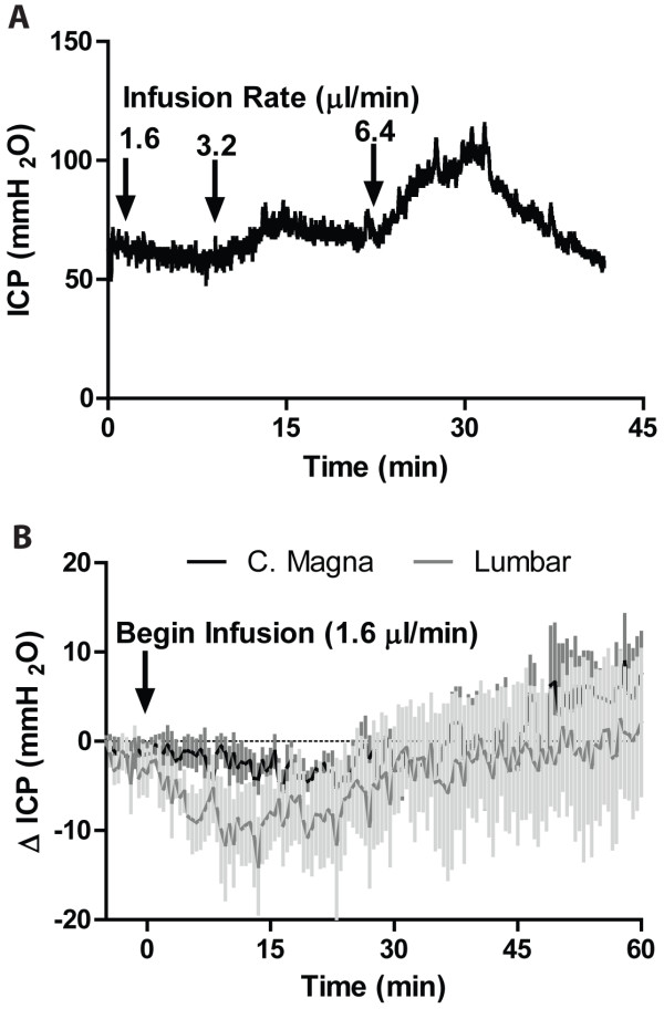
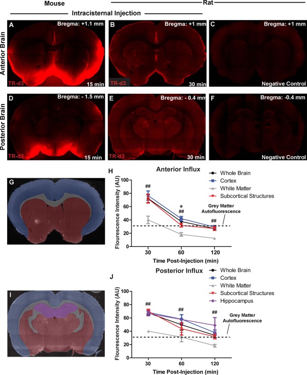
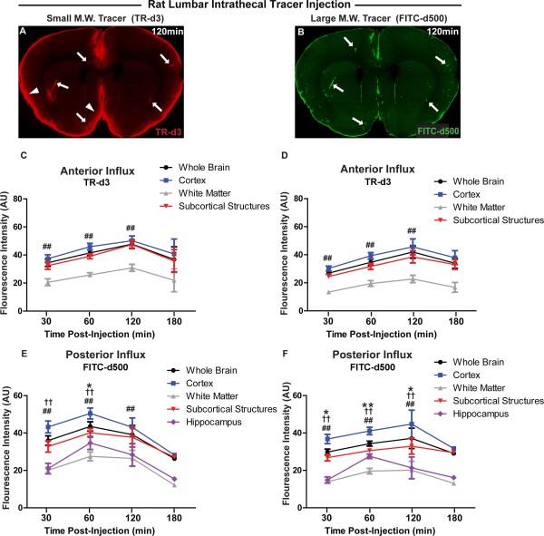
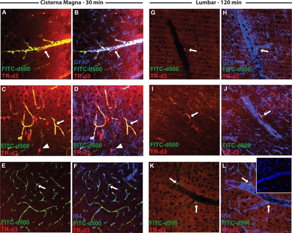
Background: Neurodegenerative diseases such as Alzheimer’s are associated with the aggregation of endogenous peptides and proteins that contribute to neuronal dysfunction and loss. The glymphatic system, a brain-wide perivascular pathway along which cerebrospinal fluid (CSF) and interstitial fluid (ISF) rapidly exchange, has recently been identified as a key contributor to the clearance of interstitial solutes from the brain, including amyloid β. These findings suggest that measuring changes in glymphatic pathway function may be an important prognostic for evaluating neurodegenerative disease susceptibility or progression. However, no clinically acceptable approach to evaluate glymphatic pathway function in humans has yet been developed. Methods: Time-sequenced ex vivo fluorescence imaging of coronal rat and mouse brain slices was performed at 30–180 min following intrathecal infusion of CSF tracer (Texas Red- dextran-3, MW 3 kD; FITC- dextran-500, MW 500 kD) into the cisterna magna or lumbar spine. Tracer influx into different brain regions (cortex, white matter, subcortical structures, and hippocampus) in rat was quantified to map the movement of CSF tracer following infusion along both routes, and to determine whether glymphatic pathway function could be evaluated after lumbar intrathecal infusion. Results: Following lumbar intrathecal infusions, small molecular weight TR-d3 entered the brain along perivascular pathways and exchanged broadly with the brain ISF, consistent with the initial characterization of the glymphatic pathway in mice. Large molecular weight FITC-d500 remained confined to the perivascular spaces. Lumbar intrathecal infusions exhibited a reduced and delayed peak parenchymal fluorescence intensity compared to intracisternal infusions. Conclusion: Lumbar intrathecal contrast delivery is a clinically useful approach that could be used in conjunction with dynamic contrast enhanced MRI nuclear imaging to assess glymphatic pathway function in humans.
Overview
The claims and statistics of this paper haven’t been checked yet.
Run the check — free, no account needed. It reads the open-access text and checks the sources it cites.
Integrity signals
Registry
Cites
Statistics
Figures
Verification
Claims the paper makes
The claims and statistics of this paper haven’t been checked yet.
Run the check — free, no account needed. It reads the open-access text and checks the sources it cites.
Claims other papers make about this one
Each is checked against this paper’s own text: does it say what the citing paper says it does?
The source backs part of the claim, not all of it — or backs it for a different population, condition or comparison.
In the citing paper
“Clearance of MRI contrast agents, such as gadobutrol, has previously been proposed to potentially resemble clearance of brain macromolecules with similar properties (14), including amyloidβ (7) and tau (21, 66).”
From Brain-wide glymphatic enhancement and clearance in humans assessed with MRI. (2018) · cites this paper as [66]
Experimental — accuracy not yet measured
Self-check
Percentages, sums and reported differences re-computed from the paper’s own numbers.
The self-check needs the paper’s full text.
Citations
Cites
The Amyloid Hypothesis of Alzheimer’s Disease: Progress and Problems on the Road to Therapeutics
Hardy, J., & Selkoe, D. J. (2002) · Science · doi:10.1126/science.1072994 · Cited by 13,864
Iliff, J. J., Wang, M., Liao, Y., Plogg, B. A., Peng, W., Gundersen, G. A., Benveniste, H., Vates, G. E., Deane, R., Goldman, S. A., et al. (2012) · Science Translational Medicine · doi:10.1126/scitranslmed.3003748 · Cited by 5,824
Self-citationProtein aggregation and neurodegenerative disease
Ross, C. A., & Poirier, M. A. (2004) · Nature Medicine · doi:10.1038/nm1066 · Cited by 3,454
Diffusion in Brain Extracellular Space
Syková, E., & Nicholson, C. (2008) · Physiological Reviews · doi:10.1152/physrev.00027.2007 · Cited by 1,481
Aggregation and Motor Neuron Toxicity of an ALS-Linked SOD1 Mutant Independent from Wild-Type SOD1
Bruijn, L. I., Houseweart, M. K., Kato, S., Anderson, K. L., Anderson, S., Ohama, E., Reaume, A. G., Scott, R. W., & Cleveland, D. W. (1998) · Science · doi:10.1126/science.281.5384.1851 · Cited by 1,250
Brain-wide pathway for waste clearance captured by contrast-enhanced MRI
Iliff, J. J., Lee, H., Yu, M., Feng, T., Logan, J., Nedergaard, M., & Benveniste, H. (2013) · Journal of Clinical Investigation · doi:10.1172/jci67677 · Cited by 1,148
Self-citationMathiisen, T. M., Lehre, K. P., Danbolt, N. C., & Ottersen, O. (2010) · Glia · doi:10.1002/glia.20990 · Cited by 893
Evidence for bulk flow of brain interstitial fluid: significance for physiology and pathology
Abbott, N. J. (2004) · Neurochemistry International · doi:10.1016/j.neuint.2003.11.006 · Cited by 786
Scherzinger, E., Sittler, A., Schweiger, K., Heiser, V., Lurz, R., Hasenbank, R., Bates, G. P., Lehrach, H. R., & Wanker, E. E. (1999) · Proceedings of the National Academy of Sciences · doi:10.1073/pnas.96.8.4604 · Cited by 707
Delivery of therapeutic agents to the central nervous system: the problems and the possibilities
Begley, D. J. (2004) · Pharmacology & Therapeutics · doi:10.1016/j.pharmthera.2004.08.001 · Cited by 679
Cited by
Sorted by citations
Items 201–300 of 318
Zhang, X., Hatoum, L., Ying, J., & Huang, C. (2025) · Psychoradiology · doi:10.1093/psyrad/kkaf031 · Cited by 11
El sistema glinfático y su implicación en las enfermedades del sistema nervioso
Toriello, M., González‐Quintanilla, V., & Pascual, J. (2021) · Medicina Clínica · doi:10.1016/j.medcli.2020.08.020 · Cited by 11
FDC:TFH Interactions within Cervical Lymph Nodes of SIV-Infected Rhesus Macaques
Dave, R. S., Sharma, R. K., Muir, R., Haddad, E. K., Gumber, S., Villinger, F. J., Nehra, A. P., Khan, Z. K., Wigdahl, B. L., Ansari, A. A., et al. (2017) · Journal of Neuroimmune Pharmacology · doi:10.1007/s11481-017-9775-0 · Cited by 11
Functional Meningeal Lymphatics and Cerebrospinal Fluid Outflow
Dave, R. S., Jain, P., & Byrareddy, S. N. (2018) · Journal of Neuroimmune Pharmacology · doi:10.1007/s11481-018-9778-5 · Cited by 10
Physiological brain clearance architecture revealed by neuronal protein tracing
Chayama, Y., Rao, N. R., Perla, D., Zhang, Z., Reid, M. M., Nelson, S., Wen, X., Ding, B., Blumenfeld, J. R., Apolonio, A., et al. (2026) · Cell · doi:10.1016/j.cell.2026.04.048 · Cited by 11
The discovery of the brain lymphatic system
Zamboni, P. (2015) · Veins and Lymphatics · doi:10.4081/vl.2015.5360 · Cited by 9
Bridging the gap between chronic cerebrospinal venous insufficiency and Ménière disease
Alpini, D. C., Bavera, P. M., Berardino, F. D., Barozzi, S., Cecconi, P., Farabola, M., Mattei, V., Mendozzi, L., Pugnetti, L., & Cesarani, A. (2016) · Veins and Lymphatics · doi:10.4081/vl.2016.5687 · Cited by 9
Thapa, A., Adhikari, R. B., Bidur, K. C., & Shakya, B. (2019) · Nepal Journal of Neuroscience · doi:10.3126/njn.v15i3.23271 · Cited by 9
Ladrón-de-Guevara, A. (2020) · bioRxiv (Cold Spring Harbor Laboratory) · doi:10.17632/t7pgxnzsxj · Cited by 9
Rey, J. A., Farid, U. M., Najjoum, C. M., Brown, A., Magdoom, K. N., Mareci, T. H., & Sarntinoranont, M. (2023) · Scientific Reports · doi:10.1038/s41598-023-34850-0 · Cited by 8
Eraky, A. M., Treffy, R., & Hedayat, H. (2022) · Cureus · doi:10.7759/cureus.31363 · Cited by 8
Xiong, R., Feng, J., Zhu, H., Li, C., Hu, P., Zou, Y., Zhou, M., Wang, Y., & Tang, X. (2025) · Sleep Medicine · doi:10.1016/j.sleep.2024.12.038 · Cited by 9
Ito, Y., Oyane, A., Yasunaga, M., Hirata, K., Hirose, M., Tsurushima, H., Ito, Y., Matsumaru, Y., & Ishikawa, E. (2022) · Journal of Biomedical Materials Research Part A · doi:10.1002/jbm.a.37434 · Cited by 8
Eraky, A. M., Treffy, R., & Hedayat, H. (2023) · Cureus · doi:10.7759/cureus.37508 · Cited by 8
Deep-learning-based segmentation of perivascular spaces on T2-Weighted 3T magnetic resonance images
Cai, D., Pan, M., Liu, C., He, W., Ge, X., Lin, J., Li, R., Liu, M., & Xia, J. (2024) · Frontiers in Aging Neuroscience · doi:10.3389/fnagi.2024.1457405 · Cited by 7
Nango, H., Takahashi, A., Suzuki, N., Kurano, T., Sakamoto, S., Nagatomo, T., Suzuki, T., Kanazawa, T., Kosuge, Y., & Miyagishi, H. (2025) · Pharmaceutics · doi:10.3390/pharmaceutics17010044 · Cited by 7
Ngono, A. E., Lepetit, M., Reindl, M., Garcia, A., Guillot, F., Genty, A., Chesneau, M., Salou, M., Michel, L., Lefrère, F., et al. (2015) · Journal of Immunology Research · doi:10.1155/2015/673503 · Cited by 7
Neuronal Impulse Theory and Alzheimer’s Disease
Barz, H. (2014) · Journal of Alzheimer’s Disease & Parkinsonism · doi:10.4172/2161-0460.1000134 · Cited by 7
Neuroinflammation in Traumatic Brain Injury
Kuo, G. Y., Tarzi, F. P., Louie, S. G., & Poblete, R. A. (2022) · IntechOpen eBooks · doi:10.5772/intechopen.105178 · Cited by 7
Persson, N. D. Å., Lohela, T. J., Anttila, J. E., Mäkelä, J., Rosenholm, M., Peltoniemi, M., Jalonen, S. C., Garcia-Horsman, A., Sarparanta, M. P., Nedergaard, M., et al. (2025) · Anesthesiology · doi:10.1097/aln.0000000000005794 · Cited by 7
Self-citationImpaired glymphatic function is associated with synaptic loss in cognitive impairment
He, K., Wang, J., Wu, J., Chen, X., Chen, H., Li, J., Guo, Q., Huang, Y. H., Guan, Y., Zhao, J., et al. (2026) · European Journal of Nuclear Medicine and Molecular Imaging · doi:10.1007/s00259-025-07716-y · Cited by 6
Kratz, S. V. (2021) · SAGE Open Medical Case Reports · doi:10.1177/2050313x20952224 · Cited by 7
The glymphatic system and its involvement in disorders of the nervous system
Toriello, M., González‐Quintanilla, V., & Pascual, J. (2021) · Medicina Clínica (English Edition) · doi:10.1016/j.medcle.2021.03.003 · Cited by 7
Sarker, A. K., Suh, M., Choi, Y., Park, J. Y., Kwon, S., Kim, H., Lee, E. J., Seo, H., Lee, Y., & Lee, D. S. (2022) · Nuclear Medicine and Molecular Imaging · doi:10.1007/s13139-022-00746-6 · Cited by 6
Dudchenko, N. G., Chimagomedova, A. S., Васенина, Е. Е., & Levin, O. S. (2022) · S S Korsakov Journal of Neurology and Psychiatry · doi:10.17116/jnevro202212207120 · Cited by 6
Cerebrospinal fluid solute transport associated with sensorimotor brain activity in rodents
Kim, E., Reet, J. V., & Yoo, S. (2023) · Scientific Reports · doi:10.1038/s41598-023-43920-2 · Cited by 6
The Emerging Field of Perivascular Flow Dynamics: Biological Relevance and Clinical Applications
Huffman, J. N., Phillips, S. M., Taylor, G. T., & Paul, R. (2016) · Technology & Innovation · doi:10.21300/18.1.2016.63 · Cited by 7
Hoz, S. S., Alramadan, A. H., Hadi, A. Q., & Moscote‐Salazar, L. R. (2018) · Journal of Neurosciences in Rural Practice · doi:10.4103/jnrp.jnrp_92_18 · Cited by 6
The involvement of perivascular spaces or tissues in the facial intradermal brain-targeted delivery
Yang, W. P., Jin, B., Chen, Y., Cao, C., Zhu, J., Zhao, Y., Yu, X., & Li, F. (2019) · Drug Delivery · doi:10.1080/10717544.2019.1587044 · Cited by 6
Levi, V., & Vetrano, I. G. (2018) · World Neurosurgery · doi:10.1016/j.wneu.2018.05.231 · Cited by 6
Lu, W., Zhu, S., Chen, R., Li, Y., Qiang, J., Ji, L., Li, C., & Zhou, D. (2025) · BMC Neurology · doi:10.1186/s12883-025-04535-4 · Cited by 5
Ciobanu-Caraus, O., Percuoco, V., Hofer, A., Sebök, M., Germans, M. R., Oertel, M. F., Regli, L., Serra, C., & Staartjes, V. E. (2024) · Neurosurgical Review · doi:10.1007/s10143-024-02954-4 · Cited by 5
Molecular and cellular mechanisms of restorative effects of sleep
Poluektov, M. G., & Spektor, E. D. (2023) · S S Korsakov Journal of Neurology and Psychiatry · doi:10.17116/jnevro202312305215 · Cited by 5
Cerebrospinal Fluid Dynamics and Intrathecal Delivery
Tangen, K. M., Mehta, A. I., & Linninger, A. A. (2018) · Elsevier eBooks · doi:10.1016/b978-0-12-805353-9.00067-x · Cited by 6
Do We Swallow the Waste From Our Brain?
Leaston, J., Kulkarni, P., Gharagouzloo, C. A., Qiao, J., Bens, N., & Ferris, C. F. (2021) · Frontiers in Neuroscience · doi:10.3389/fnins.2021.763780 · Cited by 5
Warren, K. E., Coupland, K., Hood, R. J., Kang, L., Walker, F. R., & Spratt, N. J. (2023) · Fluids and Barriers of the CNS · doi:10.1186/s12987-023-00427-2 · Cited by 5
Biomechanical forces in the aged brain: Relationship to AD
Lee, G. Y., Kim, O., Kim, E. R., & Lee, H. J. (2022) · Life Sciences · doi:10.1016/j.lfs.2022.121237 · Cited by 5
Herbert, M. R. (2014) · WORLD SCIENTIFIC eBooks · doi:10.1142/9789814602167_0021 · Cited by 5
Lazarev, V. F., Alhasan, B. A., Guzhova, I. V., & Margulis, B. A. (2025) · Frontiers in Cell and Developmental Biology · doi:10.3389/fcell.2025.1593122 · Cited by 4
Gray, S. M., & Thorner, M. O. (2017) · Endocrinology · doi:10.1210/en.2016-1884 · Cited by 4
Glymphatics, Cranium, and the Brain Homeostasis
Zibitt, M., Makhoul, V., Yu, J. C.-K., Nguyen, K. D., & Williams, J. K. (2023) · FACE · doi:10.1177/27325016231160291 · Cited by 6
Recent advances in understanding the lymphatic and glymphatic systems of the brain
Simka, M. (2015) · Phlebological Review · doi:10.5114/pr.2015.57465 · Cited by 4
Impaired cerebrospinal fluid transport due to idiopathic subdural hematoma in pig: an unusual case
Shanbhag, N. C., Bèchet, N. B., Kritsilis, M., & Lundgaard, I. (2021) · BMC Veterinary Research · doi:10.1186/s12917-021-02954-2 · Cited by 4
Vemula, R. C., Prasad, B., Banavath, H. N., Kale, P. K. G., Mohana Murali Krishna, N., & Gokanapudi, S. (2022) · Indian Journal of Neurotrauma · doi:10.1055/s-0041-1739478 · Cited by 4
Dutysheva, E. A., Викторовна, З. А., Valko, M. A., Antipina, E. O., Zimatkin, S. M., Margulis, B. A., Guzhova, I. V., & Lazarev, V. F. (2025) · International Journal of Molecular Sciences · doi:10.3390/ijms262311541 · Cited by 3
A multimodal perspective to overcome translational barriers in cerebrospinal drug delivery
Kadamus, C., Sibi, S., Bharadwaj, R., & Subramony, J. A. (2025) · Expert Opinion on Drug Delivery · doi:10.1080/17425247.2025.2578369 · Cited by 3
Jo, S. W., Park, B.-Y., Yoo, R., Choi, S. H., Ji, S., Park, T. Y., Kim, H., Park, S., Kim, M. S., Kang, S. Y., et al. (2026) · Acta Neurologica Scandinavica · doi:10.1155/ane/1538313 · Cited by 3
Piotrowska, A., Winter, K., Carare, R. O., & Bechmann, I. (2020) · Frontiers in Aging Neuroscience · doi:10.3389/fnagi.2020.00015 · Cited by 3
Activation of BDNF–TrkB Signaling in Specific Structures of the Sheep Brain by Kynurenic Acid
ROSZKOWICZ-OSTROWSKA, K. I., Młotkowska, P., Marciniak, E., Szlis, M., Barszcz, M., & Misztal, T. (2024) · Cells · doi:10.3390/cells13231928 · Cited by 3
Lowenstein, P. R., & Castro, M. G. (2016) · Advances in pharmacology · doi:10.1016/bs.apha.2016.03.002 · Cited by 3
Role of Aquaporins in Breast Cancer Progression and Metastasis
Khajah, M. A., & Luqmani, Y. A. (2016) · InTech eBooks · doi:10.5772/64446 · Cited by 3
Lomeo, A., Cacciaguerra, G., Garsia, D., & Scolaro, A. (2019) · HEGEL - HEpato-GastroEntérologie Libérale · doi:10.3917/heg.092.0114 · Cited by 3
Sontheimer, H. W. (2021) · Elsevier eBooks · doi:10.1016/b978-0-12-821228-8.00002-0 · Cited by 3
Anatomy and Physiology of the Blood-Brain Barriers*
Abbott, N. J. (2022) · AAPS advances in the pharmaceutical sciences series · doi:10.1007/978-3-030-88773-5_1 · Cited by 4
The Choroid Plexus and Cerebrospinal Fluid System: Roles in Neurodegenerative Diseases
Badaut, J., & Ghersi‐Egea, J. (2015) · Elsevier eBooks · doi:10.1016/b978-0-12-801740-1.00008-1 · Cited by 3
New Study Suggests Brain Is Connected to the Lymphatic System
Shaw, G. (2015) · Neurology Today · doi:10.1097/01.nt.0000469526.77988.9e · Cited by 3
Должиков, А. А., Shevchenko, O. A., Победа, А. С., Пересыпкина, А. А., Dolzhikova, I. N., Zhunusov, N. S., & Lugovskoy, S. S. (2020) · Research Results in Pharmacology · doi:10.3897/rrpharmacology.6.53634 · Cited by 3
Noninvasive Ultrasonic Glymphatic Induction Enhances Intrathecal Drug DeliveryPreprint
Aryal, M., Zhou, Q., Rosenthal, E. L., & Airan, R. D. (2020) · SSRN Electronic Journal · doi:10.2139/ssrn.3698892 · Cited by 3
A method for measuring first glymphatic influx of a cerebrospinal fluid tracer in the human brain
Pripp, A. H., Ringstad, G. A., Valnes, L. M., & Eide, P. K. (2025) · Frontiers in Neuroscience · doi:10.3389/fnins.2025.1703748 · Cited by 2
Molecular and Cellular Mechanisms of the Restorative Effects of Sleep
Poluektov, M. G., & Spektor, E. D. (2023) · Neuroscience and Behavioral Physiology · doi:10.1007/s11055-023-01546-x · Cited by 2
(2018) · ARC Journal of Neuroscience · doi:10.20431/2456-057x.0302005 · Cited by 2
Advancements in Brain Lymphatic System and Its Involvement in Neurological Diseases
Bari, D., Das, U., Shevalkar, G. B., Kapadia, R., Singhai, V., & Pardeshi, C. V. (2024) · Springer eBooks · doi:10.1007/978-3-031-64828-1_2 · Cited by 2
Treatment of Edema Formation in Oncology
Schnell, O., & Tonn, J. (2017) · Elsevier eBooks · doi:10.1016/b978-0-12-803196-4.00024-2 · Cited by 2
Revisiting rabies virus neutralizing antibodies through infecting BALB/c mice with live rabies virus
Qin, Y., Smith, T. G., Jackson, F. R., Gallardo-Romero, N. F., Morgan, C. N., Olson, V. A., Hutson, C. L., & Wu, X. (2018) · Virus Research · doi:10.1016/j.virusres.2018.02.012 · Cited by 2
Sachs, J. R., Zapadka, M. E., & Elster, A. D. (2018) · Journal of Computer Assisted Tomography · doi:10.1097/rct.0000000000000716 · Cited by 2
Mapping Solute Clearance From the Mouse Hippocampus Using a 3D Imaging Cryomicrotome
Naessens, D. M. P., Dobbe, J. G. G., de Vos, J., VanBavel, E., & Bakker, E. N. T. P. (2021) · Frontiers in Neuroscience · doi:10.3389/fnins.2021.631325 · Cited by 2
Evidence for the existence of a communication between the eye and the brain?
Wostyn, P., Groot, V. D., Dam, D. V., Audenaert, K., Killer, H. E., & Deyn, P. P. D. (2017) · Acta Neurochirurgica · doi:10.1007/s00701-017-3218-9 · Cited by 2
Quantifying cerebrospinal fluid tracer concentration in the brain
Zapf, B., Valnes, L. M., Mardal, K., & Zikatanov, L. (2025) · Simula SpringerBriefs on computing · doi:10.1007/978-3-032-00679-0_5 · Cited by 1
Talbot-Stetsko, H. K., Holste, K. G., Dietze-Fiedler, M. L., Hespe, G. E., Vercler, C. J., & Muraszko, K. M. (2024) · FACE · doi:10.1177/27325016231226301 · Cited by 2
Towards precision medicine for systemic lupus erythematosus and neuropsychiatric manifestations
de Aquino, B. M., Mazzola, T. N., Júlio, P. R., Silva, R. M., Sepresse, S. R., Niewold, T. B., & Appenzeller, S. (2025) · Expert Review of Precision Medicine and Drug Development · doi:10.1080/23808993.2025.2560307 · Cited by 1
Asoğlu, S., Kılınçer, A., Özer, H., Topaloğlu, Ö. F., & Cebeci, H. (2024) · Clinical Imaging · doi:10.1016/j.clinimag.2024.110140 · Cited by 1
Ito, Y., Oyane, A., Tsurushima, H., Matsumaru, Y., & Ishikawa, E. (2025) · Current Neurovascular Research · doi:10.2174/0115672026371969241224112004 · Cited by 1
Pathogenesis of Neuropsychiatric Lupus
Stock, A. D., Ben‐Zvi, A., & Putterman, C. (2019) · Elsevier eBooks · doi:10.1016/b978-0-323-47927-1.00023-2 · Cited by 1
Эргашевич, М. К., Батирбаевич, С. А., Махкамов, М. К., & Исрайилов, Д. У. (2022) · The Bulletin of Emergency Medicine · doi:10.54185/tbem/vol14_iss4/a11 · Cited by 1
Brain swelling in traumatic brain injury
Rosa, S. D., Cattin, L., Ricciardi, L., & Scerrati, A. (2022) · Elsevier eBooks · doi:10.1016/b978-0-12-823347-4.00041-5 · Cited by 1
The Mini-Spheno-Supraorbital Craniotomy for Treatment of Ruptured Anterior Circulation Aneurysms
Nerntengian, N., Tanrikulu, L., Mavromati, S., Gkasdaris, G., Kourtopoulos, H., & Birbilis, T. (2022) · MAEDICA – a Journal of Clinical Medicine · doi:10.26574/maedica.2022.17.3.583 · Cited by 1
Gruol, D. L. (2017) · MDPI eBooks · doi:10.3390/books978-3-03842-571-7 · Cited by 1
Cortical-wide impairment of “The Glymphatic system” after focal brain injuryPreprint
Azuma, M., Lee, H., Shinzaki, K., Yamane, R., & Morita, M. (2022) · bioRxiv (Cold Spring Harbor Laboratory) · doi:10.1101/2022.10.05.510560 · Cited by 1
Khan, N. R., Jones, G. M., & Klimo, P. (2014) · Stroke · doi:10.1161/strokeaha.114.007164
Open questions for Alzheimer's disease
Golde, T. E. (2014)
Önder, H. (2018) · Turkish Journal Of Neurology · doi:10.4274/tnd.23911
Sontheimer, H. W. (2015) · Elsevier eBooks · doi:10.1016/b978-0-12-800244-5.00002-1
Obrzęk mózgu u chorych po urazach czaszkowo-mózgowych
Wiśniewski, K., Posmyk, B. J., Bryl, M., Bobeff, E. J., Wójcik, R., Kowalczyk, P., & Jaskólski, D. J. (2019) · Polski Przegląd Neurologiczny · doi:10.5603/ppn.2019.0027
The Glymphatic and Meningeal Lymphatic System
Balasubramanya, V. (2019) · Materials and Methods · doi:10.13070/mm.en.9.2852
Cai, X. (2020) · Digital Repository Service (Northeastern University) · doi:10.17760/d20398331
Cisternostomy in Traumatic Brain Injury for a Novel Approach to Treatment: Review of Current Status
Agrawal, M., Mishra, K., Babal, R., & Purohit, D. K. (2021) · Indian Journal of Neurotrauma · doi:10.1055/s-0041-1726224
Alshuhri, M. S. (2021) · Enlighten: Theses (The University of Glasgow) · doi:10.5525/gla.thesis.82303
Glenfatik sistemin yapısı, fonksiyonları ve klinik açıdan önemi
Kuşat, T., & Sur, E. (2021) · Eurasian journal of veterinary sciences · doi:10.15312/eurasianjvetsci.2021.357
Hale, M., & Coles, J. A. (2022) · bioRxiv (Cold Spring Harbor Laboratory) · doi:10.1101/2021.12.30.474515
Does modulation of glymphatic system reduce delirium via waste clearance?
Naaz, S., & Asghar, A. (2022) · Journal of Anaesthesiology Clinical Pharmacology · doi:10.4103/joacp.joacp_337_19
Frontiers In Traumatic Brain Injury
Lv, X., Guo, Y., & Mao, G. (2022) · IntechOpen eBooks · doi:10.5772/intechopen.98165
Imaging blood-brain barrier function in aging
Verheggen, I. C. M. (2021) · Research Publications (Maastricht University) · doi:10.26481/dis.20210601iv
Gaillard, F. (2015) · Radiopaedia.org · doi:10.53347/rid-36699
Dissection of basal cisterns for treatment of severe traumatic brain injury
Stanishevskiy, A. V., Бабичев, К. Н., Gizatullin, S. K., Свистов, Д. В., Онницев, И. Е., & Davydov, D. V. (2022) · Russian journal of neurosurgery · doi:10.17650/1683-3295-2022-24-4-111-117
He, J., Rubini, P., Li, B.-M., Yin, H., Tang, Y., & Illés, P. (2023) · IBRO Neuroscience Reports · doi:10.1016/j.ibneur.2023.03.014
Jansson, D., & Iliff, J. J. (2023) · Springer eBooks · doi:10.1007/978-3-031-39558-1_8
Self-citationPathogenesis of neuropsychiatric lupus
Moore, E., Ben‐Zvi, A., & Putterman, C. (2024) · Elsevier eBooks · doi:10.1016/b978-0-323-93232-5.00032-0
Maynard, R. A., & Marrs, T. C. (2024) · Royal Society of Chemistry eBooks · doi:10.1039/9781839165795-00001
Çöllü, İ., & Turamanlar, O. (2024) · Farabi Tıp Dergisi · doi:10.59518/farabimedj.1420088
Metadata
- Authors
- Yang, L., Kress, B. T., Weber, H. J., Thiyagarajan, M., Wang, B., Deane, R., Benveniste, H., Iliff, J. J., & Nedergaard, M.
- Affiliations
- Hebei Medical University (CN)
- University of Rochester Medicine (US)
- Stony Brook University (US)
- Oregon Health & Science University (US)
- Corresponding author
- Iliff, Jeffrey J
- Nedergaard, Maiken
- Venue
- Journal of Translational Medicine
- Published
- 2013-05-01
- Volume
- 11
- Issue
- 1
- Pages
- 107-107
- ISSN
- 1479-5876 (ISSN-L)
- PMC
- PMC3665671
- OpenAlex
- W2166233935
- Cited by
- 317
- References
- 40
- Citations by year
- 2026: 13 · 2025: 20 · 2024: 13 · 2023: 23 · 2022: 39 · 2021: 24 · 2020: 26 · 2019: 27 · 2018: 26 · 2017: 38 · 2016: 24 · 2015: 25 · 2014: 14 · 2013: 4
- FWCI
- 10.96
- Type
- journal article
- Language
- en
- Open access
- gold
- doi.org
- Licence
- cc-by
- Locations
- Journal of Translational Medicine (publishedVersion, cc-by)
- Europe PMC (PubMed Central) (publishedVersion, cc-by)
- PubMed Central (publishedVersion, cc-by)
- PubMed (publishedVersion)
- Funders
- U.S. Department of Defense
- American Heart Association
- National Institutes of Health
- National Institute of Neurological Disorders and Stroke (R01NS078304, R01NS078167, R01NS075177)
- Topics
- Health Sciences › Medicine › Clinical Neurology › Cerebrospinal fluid and hydrocephalus
- Life Sciences › Neuroscience › Neurology › Barrier Structure and Function Studies
- Life Sciences › Neuroscience › Cellular and Molecular Neuroscience › Alzheimer's disease research and treatments
- Keywords
- glymphatic system, intrathecal infusion, interstitial fluid exchange, amyloid-beta clearance, Alzheimer's disease, neurodegenerative diseases, dynamic contrast-enhanced MRI
- MeSH terms
- Animals, Brain, Cerebrospinal Fluid, Female, Injections, Spinal, Intracranial Pressure, Lumbar Vertebrae, Male, Mice, Inbred C57BL, Molecular Weight, Molecular Probes, Rats, Sprague-Dawley, Extracellular Fluid
About these checks
Registry, retraction, Cites and statistics results are computed, not judged, with one exception: a printed reference that no index lists is read once by a model before it counts as a citation error. Claims are extracted by a model and kept only when their quote appears verbatim in the text; each cited source is then checked, with a model’s help, for whether it says what the claim relies on. Those verdicts can be wrong — read the claim’s quote and the cited source before relying on one.
Statistics and arithmetic are recomputed, not judged.
Confidence bands are shown only for fields calibrated on our annotated test set. None is calibrated yet, so no band is shown.
What counts as a claim ›How we measure confidence ›How we check this ›
Are you an author, or do you see a problem with this page? Request a review
