

The DOI matches its Crossref record; 70 of the 70 references it cites were found in a registry or an index.
Neuroprotective function for ramified microglia in hippocampal excitotoxicity
Open accessAbstract
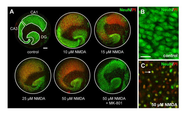
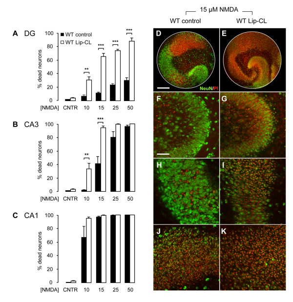
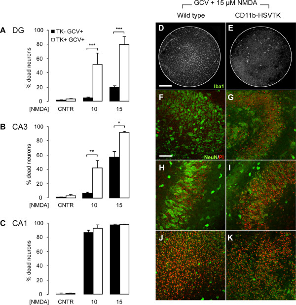
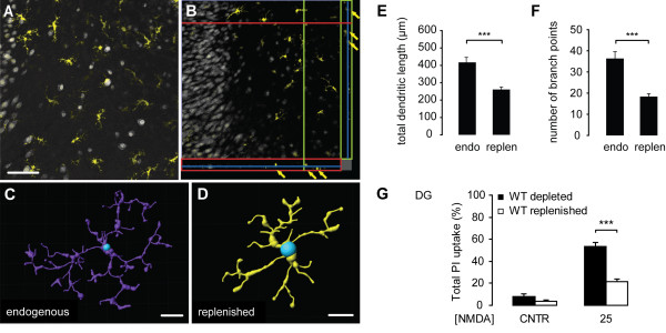
Background: Most of the known functions of microglia, including neurotoxic and neuroprotective properties, are attributed to morphologically-activated microglia. Resting, ramified microglia are suggested to primarily monitor their environment including synapses. Here, we show an active protective role of ramified microglia in excitotoxicity-induced neurodegeneration. Methods: Mouse organotypic hippocampal slice cultures were treated with N-methyl-D-aspartic acid (NMDA) to induce excitotoxic neuronal cell death. This procedure was performed in slices containing resting microglia or slices that were chemically or genetically depleted of their endogenous microglia. Results: Treatment of mouse organotypic hippocampal slice cultures with 10-50 μM N-methyl-D-aspartic acid (NMDA) induced region-specific excitotoxic neuronal cell death with CA1 neurons being most vulnerable, whereas CA3 and DG neurons were affected less. Ablation of ramified microglia severely enhanced NMDA-induced neuronal cell death in the CA3 and DG region rendering them almost as sensitive as CA1 neurons. Replenishment of microglia-free slices with microglia restored the original resistance of CA3 and DG neurons towards NMDA. Conclusions: Our data strongly suggest that ramified microglia not only screen their microenvironment but additionally protect hippocampal neurons under pathological conditions. Morphological activation of ramified microglia is thus not required to influence neuronal survival.
Overview
The claims and statistics of this paper haven’t been checked yet.
Run the check — free, no account needed. It reads the open-access text and checks the sources it cites.
Integrity signals
Registry
Cites
Statistics
Figures
Verification
Claims the paper makes
The claims and statistics of this paper haven’t been checked yet.
Run the check — free, no account needed. It reads the open-access text and checks the sources it cites.
Claims other papers make about this one
Each is checked against this paper’s own text: does it say what the citing paper says it does?
The cited source says what the claim attributes to it.
In the citing paper
“Organotypic hippocampal slice cultures are known to undergo key developmental steps similar to those observed in vivo, including synapse maturation28–31, and have been previously used to study ramified microglia function32.”
From Microglia remodel synapses by presynaptic trogocytosis and spine head filopodia induction (2018) · cites this paper as [32]
Self-check
Percentages, sums and reported differences re-computed from the paper’s own numbers.
The self-check needs the paper’s full text.
Citations
Cites
Analysis of Relative Gene Expression Data Using Real-Time Quantitative PCR and the 2−ΔΔCT Method
Livak, K. J., & Schmittgen, T. D. (2001) · Methods · doi:10.1006/meth.2001.1262 · Cited by 180,860
Resting microglial cells are highly dynamic surveillants of brain parenchyma in vivo
Nimmerjahn, A., Kirchhoff, F., & Helmchen, F. (2005) · Science · doi:10.1126/science.1110647 · Cited by 5,827 · cited 3×
“It is now clear that ramified microglia in vivo continuously scan their environment for homeostatic irregularities [6-8].”
BackgroundMicroglia: a sensor for pathological events in the CNS
Kreutzberg, G. W. (1996) · Trends in Neurosciences · doi:10.1016/0166-2236(96)10049-7 · Cited by 4,396 · cited 2×
“This microglia response has long been defined as microglia activation and based on morphological findings, microglia activation was originally described as a stereotypic and graded process [2,5].”
BackgroundATP mediates rapid microglial response to local brain injury in vivo
Davalos, D., Grutzendler, J., Yang, G., Kim, J. V., Zuo, Y., Jung, S., Littman, D. R., Dustin, M. L., & Gan, W.-B. (2005) · Nature Neuroscience · doi:10.1038/nn1472 · Cited by 3,913 · cited 3×
“It is now clear that ramified microglia in vivo continuously scan their environment for homeostatic irregularities [6-8].”
BackgroundMicroglia: Active sensor and versatile effector cells in the normal and pathologic brain
Hanisch, U.-K., & Kettenmann, H. (2007) · Nature Neuroscience · doi:10.1038/nn1997 · Cited by 3,676 · cited 3×
“This view of microglia function has been challenged in the last years [4].”
BackgroundA simple method for organotypic cultures of nervous tissue
Stoppini, L., Buchs, P., & Muller, D. (1991) · Journal of Neuroscience Methods · doi:10.1016/0165-0270(91)90128-m · Cited by 3,096 · cited 1×
“Organotypic hippocampal slice cultures were prepared as described previously [27] with minor modifications.”
van Rooijen, N., & Sanders, A. (1994) · Journal of Immunological Methods · doi:10.1016/0022-1759(94)90012-4 · Cited by 1,763 · cited 1×
“Multi-lamellar CL2MDP (clodronate)-liposomes (Lip-CL) were obtained from the Department of Molecular Cell Biology of the Free University of Amsterdam, The Netherlands (for an extensive preparation protocol see [26]).”
MethodMicroglia as a source and target of cytokines
Hanisch, U. (2002) · Glia · doi:10.1002/glia.10161 · Cited by 1,645
Onset and Progression in Inherited ALS Determined by Motor Neurons and Microglia
Boillée, S., Yamanaka, K., Lobsiger, C. S., Copeland, N. G., Jenkins, N. A., Kassiotis, G., Kollias, G., & Cleveland, D. W. (2006) · Science · doi:10.1126/science.1123511 · Cited by 1,623 · cited 2×
“However, microglial neurotoxicity can occur in case of overshooting and uncontrolled stimulation of microglia [3,17] or when microglia function is impaired [15,18,19].”
BackgroundMicroglial interactions with synapses are modulated by visual experience
Tremblay, M.-È., Lowery, R. L., & Majewska, A. K. (2010) · PLoS Biology · doi:10.1371/journal.pbio.1000527 · Cited by 1,542 · cited 1×
“The more recent findings that ramified microglia contact active synapses [20,21], suggests a cell-cell interaction between ramified microglia and neurons, which however, is not yet understood.”
Background
Cited by
Sorted by citations
Items 201–273 of 273
Vadišiūtė, A., Meijer, E. J., Therpurakal, R. N., Mueller, M., Szabó, F., Messore, F., Juršėnas, A., Bredemeyer, O., Krone, L. B., Mann, E. O., et al. (2024) · Communications Biology · doi:10.1038/s42003-024-06994-w · Cited by 10
Khiat, A. E., Hiba, O. E., Tamegart, L., Rais, H., Fdil, N., Sellami, S., Ait-El-Mokhtar, M., & Gamrani, H. (2021) · Journal of Chemical Neuroanatomy · doi:10.1016/j.jchemneu.2021.102055 · Cited by 9
A deep convolutional neural network for efficient microglia detection
Suleymanova, I., Bychkov, D., & Kopra, J. J. (2023) · Scientific Reports · doi:10.1038/s41598-023-37963-8 · Cited by 9
Establishment of mouse neuron and microglial cell co-cultured models and its action mechanism
Zhang, B., Yang, Y., Tang, J., Tao, Y., Jiang, B., Chen, Z., Feng, H., Yang, L., & Zhu, G. (2017) · Oncotarget · doi:10.18632/oncotarget.17898 · Cited by 9
“Recent studies suggest that microglial function depends on the type of stimulation and the different activation states caused by the intensity and the corresponding different immune functional phenotypes, which can simultaneously exert neuroprotective and neurotoxic effects [14, 15].”
BackgroundToshimitsu, M., Kamei, Y., Ichinose, M., Seyama, T., Imada, S., Iriyama, T., & Fujii, T. (2018) · International Journal of Developmental Neuroscience · doi:10.1016/j.ijdevneu.2018.03.011 · Cited by 9
“While ramified microglia contribute to the neuroprotection from excitotoxicity (Vinet et al., 2012), amoeboid microglia under pathological conditions are thought to be harmful to neurons (Biber et al.”
BackgroundCalderazzo, S. M., Covert, M., de Alba, D., Bowley, B. G. E., Pessina, M. A., Rosene, D. L., Buller, B., Medalla, M., & Moore, T. L. (2022) · IBRO Neuroscience Reports · doi:10.1016/j.ibneur.2022.08.001 · Cited by 8
Gellner, A., Reis, J., Fiebich, B. L., & Fritsch, B. (2023) · European Journal of Neuroscience · doi:10.1111/ejn.16206 · Cited by 7
“Of note, previous studies have shown no affection of neuron numbers, or astrocytes density and morphology after clodronate treatment in cultured slices, especially after days to weeks post-treatment (Ji et al., 2013; Vinet et al., 2012).”
ResultBalion, Z., Svirskienė, N., Svirskis, G., Inokaitis, H., Cėpla, V., Ulčinas, A., Jelinskas, T., Eimont, R., Paužienė, N., Valiokas, R., et al. (2022) · Biomedicines · doi:10.3390/biomedicines10051023 · Cited by 8
Microglia Mitigate Neuronal Activation in a Zebrafish Model of Dravet Syndrome
Brenet, A., Somkhit, J., Csaba, Z., Ciura, S., Kabashi, E., Yanicostas, C., & Soussi‐Yanicostas, N. (2024) · Cells · doi:10.3390/cells13080684 · Cited by 9
“…be harmful to hyperexcitated neuronal networks, probably owing to the synthesis and release of pro-inflammatory cytokines, which are known to promote neuron excitation [22–24], other studies suggested that microglia played a beneficial role by decreasing neuronal network hyperactivity [19,25–27].”
BackgroundSensitivity of Hypocretin System to Chronic Alcohol Exposure: A Human and Animal Study
McGregor, R., Matzeu, A., Thannickal, T. C., Wu, M.-F., Cornford, M. E., Martin‐Fardon, R., & Siegel, J. M. (2023) · Neuroscience · doi:10.1016/j.neuroscience.2023.04.018 · Cited by 7
Manickam, V., Maity, S., Murali, S. V., Gawande, D. Y., Stothert, A. R., Batalkina, L., Cardona, A. E., & Kaur, T. (2024) · Frontiers in Cellular Neuroscience · doi:10.3389/fncel.2024.1486740 · Cited by 7
“…excitotoxic injury activates and recruits microglia to the site of injury, and that those microglia protect neurons and improve synaptic recovery following excitotoxic damage (Berezovskaya et al., 1995; Vinet et al., 2012; Simard and Rivest, 2007; Eyo et al., 2014; Kato et al., 2016).”
Evidence for Neuroinflammation after Myocardial Infarction in a Mouse Model
Gouweleeuw, L., Pol, C. J., Simonides, W. S., de Kleijn, D. P. V., & Schoemaker, R. G. (2017) · Heart and Mind · doi:10.4103/hm.hm_3_18 · Cited by 7
“The hippocampus has been reported as a brain area particularly sensitive to microglia activation;[20,21] therefore, it is not surprising that microglia activation was found specifically in this region.”
BackgroundÇelik, Z. B., & Günaydın, C. (2022) · Cureus · doi:10.7759/cureus.32428 · Cited by 6
“However, the regulation of microglia in chronic conditions such as neuroinflammation has been thought of as a neuroprotective behavior [3].”
BackgroundTekko, T., Lakspere, T., Allikalt, A., End, J., Kõlvart, K. R., Jagomäe, T., Terasmaa, A., Philips, M., Visnapuu, T., Väärtnõu, F., et al. (2017) · PLoS ONE · doi:10.1371/journal.pone.0172825 · Cited by 6
vonderEmbse, A. N., Hu, Q., & DeWitt, J. C. (2019) · International Immunopharmacology · doi:10.1016/j.intimp.2019.03.039 · Cited by 6
Matuszewska, M., Wilkaniec, A., Gąssowska-Dobrowolska, M., Cieślik, M., Olech-Kochańczyk, G., Pałasz, E., Gawinek, E., Strawski, M., & Czapski, G. A. (2025) · Frontiers in Molecular Neuroscience · doi:10.3389/fnmol.2025.1619583 · Cited by 6
“This region is also more sensitive to NMDA-induced excitotoxicity than CA3 or DG, and this susceptibility is abolished in microglia-depleted preparations, indicating a microglia-dependent component of regional vulnerability (Vinet et al., 2012).”
Tan, Z., Bussies, P. L., Sarn, N., Irfan, M., DeSilva, T. M., & Eng, C. (2025) · Neuroscience · doi:10.1016/j.neuroscience.2025.02.044 · Cited by 8
“There is considerable data which show microglia play a neuro-protective role by forming specialized junctions between their processes and neuronal cell bodies (Li et al., 2012; Vinet et al., 2012; Cserep et al., 2020).”
Replenishment of Organotypic Hippocampal Slice Cultures with Neonatal or Adult Microglia
Masuch, A., & Biber, K. P. H. (2019) · Methods in molecular biology · doi:10.1007/978-1-4939-9658-2_10 · Cited by 5
Self-citationWatters, J. J., & Pocock, J. M. (2014) · Springer eBooks · doi:10.1007/978-1-4939-1429-6_3 · Cited by 5
Johnson, N. R., Yuan, P., Castillo, E., Lopez, T. P., Yue, W., Bond, A., Rivera, B. M., Sullivan, M. C., Hirouchi, M., Giles, K., et al. (2021) · bioRxiv (Cold Spring Harbor Laboratory) · doi:10.1101/2021.03.20.436288 · Cited by 5
Contribution of microglia to the epileptiform activity that results from neonatal hypoxia
Leavy, A., Phelan, J., & Jimenez-Mateos, E. M. (2024) · Neuropharmacology · doi:10.1016/j.neuropharm.2024.109968 · Cited by 4
“On the other hand, in ex vivo experiments, depletion of microglia exacerbates NMDA-induced cell death in the hippocampus (Vinet et al., 2012).”
BackgroundMalorny, N., Chausse, B., Khodaie, B., Elgez, A., Söder, L., Lewen, A., Egorov, A. V., & Kann, O. (2026) · Journal of Neuroinflammation · doi:10.1186/s12974-026-03835-x · Cited by 3
“Microglia were depleted chemically from DIV 0 on by continuously adding liposome-encapsulated clodronate (100 µg/mL) (Liposoma B.V., Amsterdam, The Netherlands) to the culture medium [26, 36, 44].”
Tamegart, L., Abbaoui, A., Oukhrib, M., Bouyatas, M. M., & Gamrani, H. (2021) · Biological Trace Element Research · doi:10.1007/s12011-021-02751-w · Cited by 3
Aderinto, N., Olatunji, G. D., Kokori, E., Ukoaka, B. M., Babalola, A. E., Ogieuhi, I. J., Moradeyo, A. A., Yusuf, I. A., Apampa, O. O., Oluwakorede, A. T., et al. (2024) · Discover Medicine · doi:10.1007/s44337-024-00161-0 · Cited by 3
“Studies have identified increased microglia ramification as a neuroprotective mechanism [37].”
Microglial TRPV1 in epilepsy: Is it druggable for new antiepileptic treatment?
HU, J., Mo, J., & Cheng, X. (2023) · Biocell · doi:10.32604/biocell.2023.029409 · Cited by 3
“It has been suggested that while brief microglial activation is advantageous (Vinet et al., 2012), the development of epilepsy is adversely affected by persistent microglial activation (Qin et al.”
BackgroundThe effects of social environment on AD-related pathology in hAPP-J20 mice and tau-P301L mice
Lanooij, S. D., Drinkenburg, W. H. I. M., Eisel, U. L. M., van der Zee, E. A., & Kas, M. J. (2023) · Neurobiology of Disease · doi:10.1016/j.nbd.2023.106309 · Cited by 4
Neuroprotection Versus Neurotoxicity
Biber, K. P. H., & Block, M. L. (2014) · Springer eBooks · doi:10.1007/978-1-4939-1429-6_6 · Cited by 3
Self-citationReyes, L. M. D. L., & Céspedes-Rubio, Á. E. (2014) · Biomédica · doi:10.7705/biomedica.v34i3.2191 · Cited by 3
Koga, M., Satoh, Y., Kashitani, M., Nakagawa, R., Sato, M., Asai, F., Ishizuka, T., Kinoshita, M., Saitoh, D., Nagamine, M., et al. (2024) · Progress in Neuro-Psychopharmacology and Biological Psychiatry · doi:10.1016/j.pnpbp.2024.111153 · Cited by 2
Jones, K., Teliska, L. H., Mirrione, M. M., & Betz, A. J. (2025) · Experimental Brain Research · doi:10.1007/s00221-025-07204-7 · Cited by 3
“Studies suggest a neuroprotective role for ramified microglia after microglial ablation enhanced NMDA-induced neuronal death in the hippocampus (Vinet et al. 2012 ).”
Kotov, V. N., Kastyro, I. V., Malakhovskiy, V. V., Korolev, A. G., & Kostyaeva, M. G. (2026) · The European Physical Journal Special Topics · doi:10.1140/epjs/s11734-026-02202-4 · Cited by 2
Skaper, S. D. (2017) · Springer eBooks · doi:10.1007/978-3-319-57371-7_12 · Cited by 2
Exercise Prevents Mental Illness
Purnomo, K. I., Doewes, M., Giri, M. K. W., Setiawan, K. H., & Wibowo, I. P. A. (2017) · IOP Conference Series Materials Science and Engineering · doi:10.1088/1757-899x/180/1/012167 · Cited by 2
“In this resting state, microglia are involved in neurogenesis [4], neuroprotective [5], and plays an important role in synaptic pruning [6].”
BackgroundA Primary Neural Cell Culture Model for NeuroinflammationPreprint
Goshi, N., Morgan, R. K., Lein, P. J., & Şeker, E. (2020) · bioRxiv (Cold Spring Harbor Laboratory) · doi:10.1101/2020.01.23.914226 · Cited by 2
“Our results suggest that microglia in the tri-culture play a significant neuroprotective role during excitotoxic events, in line with recent in vivo (Kato et al., 2016; Larochelle et al., 2015; Szalay et al., 2016) and hippocampal slice culture experiments (Masuch et al., 2015; Vinet et al., 2012).”
BackgroundResultGLIA Bilbao 2015: Abstracts Oral Presentations, Posters, Indexes
Perrier, J., Christensen, R. K., Delgado‐Lezama, R., Russo, R., Lind, B. L., Alcocer, E. L., Fabbiani, G., Schmitt, N., Lauritzen, M., Petersen, A. V., et al. (2015) · Glia · doi:10.1002/glia.22870 · Cited by 2
Parkinson's Disease Involving Glial Cell Dysfunction
Oukhrib, M., Tamegart, L., Ghachi, H. E., Haj-Khlifa, A., Hamdan, Y. A., Maloui, A. B., Gamrani, H., & Chraa, M. (2023) · Advances in bioinformatics and biomedical engineering book series · doi:10.4018/978-1-6684-9675-6.ch009 · Cited by 1
Mirzaei, S. M. M., Yousefi, M., Nasehi, M. M., Soltantooyeh, Z., Karav, S., & Sahebkar, A. (2026) · Sleep And Breathing · doi:10.1007/s11325-026-03630-1 · Cited by 1
“In epileptic brains, microglia exhibit multifaceted states: short-term activation may be beneficial, while chronic is detrimental [ 56–58].”
Microglia in Epilepsy: From Molecular Mechanism to Therapeutic Strategy
Paudel, Y. N., Angelopoulou, E., Kulkarni, S., Blair, R. E., & Deshpande, L. S. (2026) · Cells · doi:10.3390/cells15090835 · Cited by 1
“The pro-epileptic and anti-epileptic phenotypes of microglia (Figure 1) depend on their activation state, meaning that short-term microglial activation is beneficial [130,149] whereas long-term microglial activation is detrimental [150].”
Choudhary, H. H., Yoshida, J., Islam, R., Wang, Z., & Hanafy, K. A. (2026) · Journal of Cerebral Blood Flow & Metabolism · doi:10.1177/0271678x261468877 · Cited by 1
“107”
A new method allowing long‐term potentiation recordings in hippocampal organotypic slices
Paci, P., Gabriele, S., & Ris, L. (2017) · Brain and Behavior · doi:10.1002/brb3.692 · Cited by 1
“Moreover, it has been shown that ramified microglia not only monitor their microenvironment but also protect hippocampal neurons under pathological conditions (Vinet et al., 2012).”
BackgroundXu, R., Ghori, A., Schneider, U. C., & Vajkoczy, P. (2017) · Translational medicine research · doi:10.1007/978-981-10-5804-2_13 · Cited by 1
Persidsky, Y., Gofman, L., & Potula, R. (2014) · Springer eBooks · doi:10.1007/978-1-4939-1071-7_24 · Cited by 1
Microquimerismos en la enfermedad de Alzheimer y la demencia frontotemporal
Sierra-Peláez, S., & Villegas‐Lanau, C. A. (2015) · Medicina y Laboratorio · doi:10.36384/01232576.131 · Cited by 1
“Algunos estudios por imaginología y en modelos de ratones transgénicos han reportado lesiones y alteraciones desmielinizantes en la enfermedad de Alzheimer, relacionadas con la presencia de depósitos del β-amiloide, los cuales resultan tóxicos para este tipo de células [51,52].”
BackgroundInfluentialFast Single-Cell MALDI Imaging of Low-Mass Metabolites Reveals Cellular Activation MarkersPreprint
Cairns, J. L., Huber, J., Lewen, A., Jung, J., Maurer, S. J., Bausbacher, T., Schmidt, S., Levkin, P. A., Sévin, D. C., Göpfrich, K., et al. (2024) · bioRxiv (Cold Spring Harbor Laboratory) · doi:10.1101/2024.10.15.618420 · Cited by 1
“These cells engulf the liposomes that, in turn, release clodronate intracellu-379 larly, thus stopping the TCA cycle and causing apoptosis 44,45,50,51 .”
BackgroundBelhoul‐Fakir, H. (2014) · Open Access Victoria University of Wellington | Te Herenga Waka (Figshare) · doi:10.26686/wgtn.17008231.v1
“Although this strategy was shown to generate equivalent amounts of AAV particles as in HEK cell transient transfection (Urabe et al., 2004; Viraq et al., 2009), the instability of the baculovirus is very challenging as it depends on the passage number of the insect cell (Virag et al., 2009).”
BackgroundResultLee, S. L. L. (2013) · Universitätsbibliothek der FU Berlin Hochschulschriftenstelle u. Dokumentenserver · doi:10.17169/refubium-8036
Chapman, T. R. (2013) · CU Scholar (University of Colorado Boulder)
Defining microglial phenotypic diversity and the impact of ageing
Grabert, K. (2015) · ERA
Inflammasome-mediated activation of microglia : Tissue-specific features of innate immunity
Burm, S. M. (2016) · Utrecht University Repository (Utrecht University)
vonderEmbse, A. N. (2017) · The Scholarship East Carolina University's Institutional Repository (East Carolina University)
Signalisation par les récepteurs toll-like dans un contexte d'excitotoxicité
Larochelle, A. (2014) · Corpus Université Laval (Université Laval)
Hartnell, I. J. (2018) · White Rose eTheses Online (University of Leeds, The University of Sheffield, University of York)
Port’s, N. M. D. S. (2022) · Digital Library of Theses and Dissertations (Universidade de São Paulo) · doi:10.11606/d.42.2022.tde-27012023-100010
Khir, N. A. M., Noh, A. S. M., Zin, A. A. M., Long, I., Yusop, N., & Ismail, C. A. N. (2023) · Research Square · doi:10.21203/rs.3.rs-3183538/v1
Chronic paracetamol treatment induces neuroinflammation and microglia activation in rat hippocampus
Lalert, L. (2021) · Chulalongkorn Medical Journal · doi:10.58837/chula.cmj.65.3.1
“(19, 20) In response to various pathological stimuli, the microglia can transform to an ameboid shape and can produce large amounts of various cytotoxic mediators including reactive oxygen species (ROS), chemokines, and pro-inflammatory cytokines leading to promote the neuronal inflammation and…”
BackgroundTasbihgou, S. R. (2024) · doi:10.33612/diss.957310115
Glia's Role in Linking Epileptic Pathology and Neural Networks
Koyama, R., & Andoh, M. (2024) · てんかん研究 · doi:10.3805/jjes.42.11
Han, B., Jiang, W., Cui, P., Zheng, K., Dang, C., Wang, J., Li, H., Chen, L., Zhang, R., Wang, Q. M., et al. (2021) · Figshare · doi:10.6084/m9.figshare.14329336
Rodriguez, D., Church, K. A., Pietramale, A. N., Cardona, S. M., Vanegas, D., Rorex, C., Leary, M. C., Muzzio, I. A., Nash, K. R., & Cardona, A. E. (2024) · Figshare · doi:10.6084/m9.figshare.25143609
Han, B., Jiang, W., Cui, P., Zheng, K., Dang, C., Wang, J., Li, H., Chen, L., Zhang, R., Wang, Q. M., et al. (2021) · Figshare · doi:10.6084/m9.figshare.14329333
Gaigé, S., Barbouche, R., Barbot, M., Boularand, S., Dallaporta, M., Abysique, A., & Troadec, J. (2022) · Figshare · doi:10.6084/m9.figshare.21601984
Goshi, N., Morgan, R. K., Lein, P. J., & Şeker, E. (2020) · Figshare · doi:10.6084/m9.figshare.12284750
Appiah, C. B., Kunwar, K., Cunningham, R. L., & Cunningham, J. T. (2026) · Brain Behavior & Immunity - Health · doi:10.1016/j.bbih.2026.101168
“Ramified microglia have demonstrated neuroprotective capacity against excitotoxic damage in the hippocampus (Vinet et al., 2012), suggesting that maintaining ramified morphology could limit proinflammatory cytokine release and subsequent neuronal damage.”
Neuroinflammation in epilepsy: Mitochondrial connections and therapeutic targets
Olkhova, E. A., Dimitriou, A., Johnson, B. M., Hanaford, A., & Johnson, S. C. (2026) · Brain Mechanisms · doi:10.1016/j.bramec.2026.202535
In vitro and in vivo study of the effects of Levetiracetam on activated microglia during sepsis
Fleurance, R. (2021) · theses.fr (ABES) · doi:10.70675/0a82ed6ez6595z4366z832fz70cbe839c7ff
Deletion of Snap25 disrupts glial remodeling in aging mouse brain
Vadišiūtė, A., Szabó, F., Luchanskaya, S., Drevenakova, V., Messore, F., Ugwudike, A., Greene, G., Mueller, M., Morse, S. V., Hoerder‐Suabedissen, A., et al. (2026) · iScience · doi:10.1016/j.isci.2026.116478 · Cited by 1
“SNAP25 (synaptosomal-associated protein 25) is a core component of the SNARE complex 37–39 and is essential for cal-cium-evoked synaptic vesicle fusion, and global deletion re-sults in perinatal lethality, 37 demonstrating the necessity of evoked neurotransmission for survival.”
Langer, K. M., Tiemeier, E., Harmon, E., Fineberg, A., Henry, J., Veitch, I., Klopper, T., McVey, T., Dietz, R. M., Dingman, A., et al. (2026) · Pediatric Research · doi:10.1038/s41390-026-05281-0
Sentyabreva, A. V., Lyamtsev, A. S., Melnikova, E. A., Aliper, G. M., & Kosyreva, A. M. (2026) · International Journal of Molecular Sciences · doi:10.3390/ijms27188222
“According to Vinet et al. (2012), ramified microglia continuously survey the brain microenvironment and protect hippocampal neurons under both physiological and pathological conditions [34].”
Landwehr, A., Cools-Lartigue, C., Hadera, V., Bobotis, B. C., Khakpour, M., Gomes, F. V., & Tremblay, M. (2026) · bioRxiv (Cold Spring Harbor Laboratory) · doi:10.64898/2026.09.24.753926
Neuronal changes in the hippocampus of post-stroke survivors
Gemmell, E. (2014)
Metadata
- Authors
- Vinet, J., van Weering, H. R., Heinrich, A., Kälin, R. E., Wegner, A., Brouwer, N., Heppner, F. L., van Rooijen, N., Boddeke, H. W., & Biber, K.
- Affiliations
- University Medical Center Groningen (NL)
- University of Freiburg (DE)
- Charité – Universitätsmedizin Berlin (DE)
- Corresponding author
- Vinet, Jonathan
- Biber, Knut
- Venue
- Journal of Neuroinflammation
- Published
- 2012-01-31
- Volume
- 9
- Issue
- 1
- Pages
- 27-27
- ISSN
- 1742-2094 (ISSN-L)
- PMC
- PMC3292937
- OpenAlex
- W1963696719
- Cited by
- 274
- References
- 70
- Citations by year
- 2026: 11 · 2025: 6 · 2024: 18 · 2023: 16 · 2022: 14 · 2021: 20 · 2020: 22 · 2019: 20 · 2018: 18 · 2017: 28 · 2016: 24 · 2015: 18 · 2014: 24 · 2013: 26 · 2012: 9
- FWCI
- 12.38
- Type
- journal article
- Language
- en
- Open access
- gold
- doi.org
- Locations
- Journal of Neuroinflammation (publishedVersion, cc-by)
- Europe PMC (PubMed Central) (publishedVersion, cc-by)
- PubMed Central (publishedVersion, cc-by)
- PubMed (publishedVersion)
- Pure Amsterdam UMC (publishedVersion, other-oa)
- Amsterdam UMC (VU Amsterdam) - Institutional Repository (publishedVersion, other-oa)
- University of Groningen research database (University of Groningen / Centre for Information Technology) (publishedVersion, cc-by)
- Digital Academic REpository of VU University Amsterdam (Vrije Universiteit Amsterdam) (submittedVersion)
- DOAJ (DOAJ: Directory of Open Access Journals) (submittedVersion)
- UCL Discovery (University College London) (submittedVersion)
- IRIS UNIMORE (University of Modena and Reggio Emilia) (submittedVersion, cc-by)
- University of Groningen research database (University of Groningen / Centre for Information Technology) (submittedVersion, cc-by)
- Data Archiving and Networked Services (DANS) (submittedVersion, other-oa)
- Funders
- Deutsche Forschungsgemeinschaft (SFB TRR 43, NeuroCure EXC257, EXC257, FOR1336, TRR-43)
- Nederlandse Organisatie voor Wetenschappelijk Onderzoek
- Topics
- Life Sciences › Neuroscience › Neurology › Neuroinflammation and Neurodegeneration Mechanisms
- Life Sciences › Neuroscience › Cellular and Molecular Neuroscience › Neuroscience and Neuropharmacology Research
- Life Sciences › Neuroscience › Neurology › Neurological Disease Mechanisms and Treatments
- Keywords
- ramified microglia, neuroprotection, microglia depletion, organotypic hippocampal slice cultures, CA1 neurons, excitotoxic neurodegeneration
- MeSH terms
- Animals, Clodronic Acid, Dose-Response Relationship, Drug, Glial Fibrillary Acidic Protein, Hippocampus, Mice, Inbred C57BL, Mice, Transgenic, Nerve Degeneration, Neurons, Neurotoxins, Organ Culture Techniques, Phosphopyruvate Hydratase, N-Methylaspartate, Cell Death, Microglia, CD11b Antigen, Mice
About these checks
Registry, retraction, Cites and statistics results are computed, not judged, with one exception: a printed reference that no index lists is read once by a model before it counts as a citation error. Claims are extracted by a model and kept only when their quote appears verbatim in the text; each cited source is then checked, with a model’s help, for whether it says what the claim relies on. Those verdicts can be wrong — read the claim’s quote and the cited source before relying on one.
Statistics and arithmetic are recomputed, not judged.
Confidence bands are shown only for fields calibrated on our annotated test set. None is calibrated yet, so no band is shown.
What counts as a claim ›How we measure confidence ›How we check this ›
Are you an author, or do you see a problem with this page? Request a review
