

The DOI matches its Crossref record; 70 of the 70 references it cites were found in a registry or an index.
Neuroprotective function for ramified microglia in hippocampal excitotoxicity
Open accessAbstract
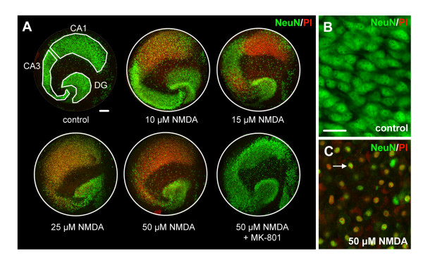
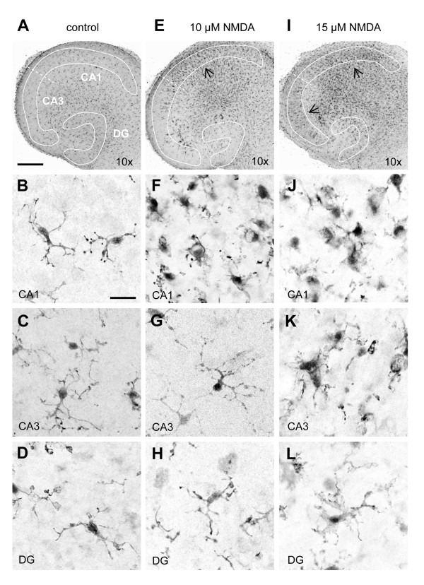
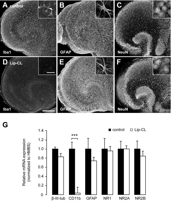
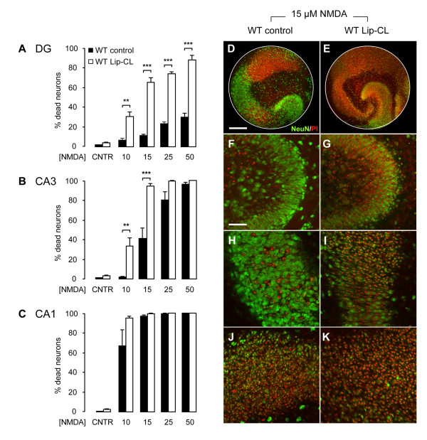
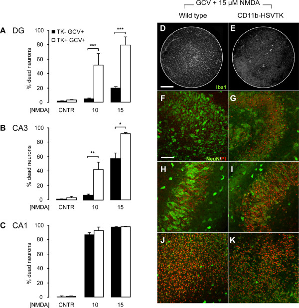
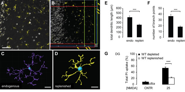
Background: Most of the known functions of microglia, including neurotoxic and neuroprotective properties, are attributed to morphologically-activated microglia. Resting, ramified microglia are suggested to primarily monitor their environment including synapses. Here, we show an active protective role of ramified microglia in excitotoxicity-induced neurodegeneration. Methods: Mouse organotypic hippocampal slice cultures were treated with N-methyl-D-aspartic acid (NMDA) to induce excitotoxic neuronal cell death. This procedure was performed in slices containing resting microglia or slices that were chemically or genetically depleted of their endogenous microglia. Results: Treatment of mouse organotypic hippocampal slice cultures with 10-50 μM N-methyl-D-aspartic acid (NMDA) induced region-specific excitotoxic neuronal cell death with CA1 neurons being most vulnerable, whereas CA3 and DG neurons were affected less. Ablation of ramified microglia severely enhanced NMDA-induced neuronal cell death in the CA3 and DG region rendering them almost as sensitive as CA1 neurons. Replenishment of microglia-free slices with microglia restored the original resistance of CA3 and DG neurons towards NMDA. Conclusions: Our data strongly suggest that ramified microglia not only screen their microenvironment but additionally protect hippocampal neurons under pathological conditions. Morphological activation of ramified microglia is thus not required to influence neuronal survival.
Overview
The claims and statistics of this paper haven’t been checked yet.
Run the check — free, no account needed. It reads the open-access text and checks the sources it cites.
Integrity signals
Registry
Cites
Statistics
Figures
Verification
Claims the paper makes
The claims and statistics of this paper haven’t been checked yet.
Run the check — free, no account needed. It reads the open-access text and checks the sources it cites.
Claims other papers make about this one
Each is checked against this paper’s own text: does it say what the citing paper says it does?
The cited source says what the claim attributes to it.
In the citing paper
“Organotypic hippocampal slice cultures are known to undergo key developmental steps similar to those observed in vivo, including synapse maturation28–31, and have been previously used to study ramified microglia function32.”
From Microglia remodel synapses by presynaptic trogocytosis and spine head filopodia induction (2018) · cites this paper as [32]
Self-check
Percentages, sums and reported differences re-computed from the paper’s own numbers.
The self-check needs the paper’s full text.
Citations
Cites
Analysis of Relative Gene Expression Data Using Real-Time Quantitative PCR and the 2−ΔΔCT Method
Livak, K. J., & Schmittgen, T. D. (2001) · Methods · doi:10.1006/meth.2001.1262 · Cited by 180,860
Resting microglial cells are highly dynamic surveillants of brain parenchyma in vivo
Nimmerjahn, A., Kirchhoff, F., & Helmchen, F. (2005) · Science · doi:10.1126/science.1110647 · Cited by 5,827 · cited 3×
“It is now clear that ramified microglia in vivo continuously scan their environment for homeostatic irregularities [6-8].”
BackgroundMicroglia: a sensor for pathological events in the CNS
Kreutzberg, G. W. (1996) · Trends in Neurosciences · doi:10.1016/0166-2236(96)10049-7 · Cited by 4,396 · cited 2×
“This microglia response has long been defined as microglia activation and based on morphological findings, microglia activation was originally described as a stereotypic and graded process [2,5].”
BackgroundATP mediates rapid microglial response to local brain injury in vivo
Davalos, D., Grutzendler, J., Yang, G., Kim, J. V., Zuo, Y., Jung, S., Littman, D. R., Dustin, M. L., & Gan, W.-B. (2005) · Nature Neuroscience · doi:10.1038/nn1472 · Cited by 3,913 · cited 3×
“It is now clear that ramified microglia in vivo continuously scan their environment for homeostatic irregularities [6-8].”
BackgroundMicroglia: Active sensor and versatile effector cells in the normal and pathologic brain
Hanisch, U.-K., & Kettenmann, H. (2007) · Nature Neuroscience · doi:10.1038/nn1997 · Cited by 3,676 · cited 3×
“This view of microglia function has been challenged in the last years [4].”
BackgroundA simple method for organotypic cultures of nervous tissue
Stoppini, L., Buchs, P., & Muller, D. (1991) · Journal of Neuroscience Methods · doi:10.1016/0165-0270(91)90128-m · Cited by 3,096 · cited 1×
“Organotypic hippocampal slice cultures were prepared as described previously [27] with minor modifications.”
van Rooijen, N., & Sanders, A. (1994) · Journal of Immunological Methods · doi:10.1016/0022-1759(94)90012-4 · Cited by 1,763 · cited 1×
“Multi-lamellar CL2MDP (clodronate)-liposomes (Lip-CL) were obtained from the Department of Molecular Cell Biology of the Free University of Amsterdam, The Netherlands (for an extensive preparation protocol see [26]).”
MethodMicroglia as a source and target of cytokines
Hanisch, U. (2002) · Glia · doi:10.1002/glia.10161 · Cited by 1,645
Onset and Progression in Inherited ALS Determined by Motor Neurons and Microglia
Boillée, S., Yamanaka, K., Lobsiger, C. S., Copeland, N. G., Jenkins, N. A., Kassiotis, G., Kollias, G., & Cleveland, D. W. (2006) · Science · doi:10.1126/science.1123511 · Cited by 1,623 · cited 2×
“However, microglial neurotoxicity can occur in case of overshooting and uncontrolled stimulation of microglia [3,17] or when microglia function is impaired [15,18,19].”
BackgroundMicroglial interactions with synapses are modulated by visual experience
Tremblay, M.-È., Lowery, R. L., & Majewska, A. K. (2010) · PLoS Biology · doi:10.1371/journal.pbio.1000527 · Cited by 1,542 · cited 1×
“The more recent findings that ramified microglia contact active synapses [20,21], suggests a cell-cell interaction between ramified microglia and neurons, which however, is not yet understood.”
Background
Cited by
Sorted by citations
Items 101–200 of 273
Microglia–neuron crosstalk through Hex–GM2–MGL2 maintains brain homeostasis
Frosch, M., Shimizu, T., Wogram, E., Amann, L., Gruber, L., Groisman, A. I., Fliegauf, M., Schwabenland, M., Chhatbar, C., Zechel, S., et al. (2025) · Nature · doi:10.1038/s41586-025-09477-y · Cited by 56
Johnson, N. R., Yuan, P., Castillo, E., Lopez, T. P., Yue, W., Bond, A., Rivera, B. M., Sullivan, M. C., Hirouchi, M., Giles, K., et al. (2023) · Nature Communications · doi:10.1038/s41467-022-35753-w · Cited by 46
Immig, K., Gericke, M. T., Menzel, F., Merz, F., Krueger, M., Schiefenhövel, F., Lösche, A., Jäger, K., Hanisch, U., Biber, K. P. H., et al. (2014) · Glia · doi:10.1002/glia.22771 · Cited by 44
Self-citationVirgili, N., Mancera, P., Wappenhans, B., Sorrosal, G., Biber, K. P. H., Pugliese, M., & Espinosa‐Parrilla, J. F. (2013) · PLoS ONE · doi:10.1371/journal.pone.0075189 · Cited by 45
“As previously described [40], neuronal cell death in microglia-depleted slice culture was enhanced in response to 10 mM NMDA compared to normal OHSC (Figure 2G).”
BackgroundMethodSelf-citationMalaguarnera, M., Llansola, M., Balzano, T., Gómez-Giménez, B., Antúnez-Muñoz, C., Martínez-Alarcón, N., Mahdinia, R., & Felipo, V. (2019) · Frontiers in Pharmacology · doi:10.3389/fphar.2019.00132 · Cited by 42
The effect of aged microglia on synaptic impairment and its relevance in neurodegenerative diseases
Triviño, J. J., & von Bernhardi, R. (2021) · Neurochemistry International · doi:10.1016/j.neuint.2021.104982 · Cited by 43
Westacott, L. J., & Wilkinson, L. S. (2022) · Frontiers in Neuroscience · doi:10.3389/fnins.2022.840266 · Cited by 48
Cabrera‐Pastor, A., Hernández‐Rabaza, V., Taoro‐González, L., Balzano, T., Llansola, M., & Felipo, V. (2016) · Brain Behavior and Immunity · doi:10.1016/j.bbi.2016.05.011 · Cited by 43
“Microglia activation was assessed by measuring the cells perimeter in 8 randomly selected areas (0.45 mm2) per section according to Vinet et al. (2012).”
InfluentialNeuroimmune System as a Driving Force for Plasticity Following CNS Injury
O’Reilly, M. L., & Tom, V. J. (2020) · Frontiers in Cellular Neuroscience · doi:10.3389/fncel.2020.00187 · Cited by 44
“Additionally, IL-1β, IL-17, and ROS have been shown to enhance post-synaptic NMDA receptor activation by increasing phosphorylation of NMDA receptor subunits NR1 and NR2A/B (Viviani et al., 2003; Gao et al., 2007; Meng et al., 2013).”
BackgroundEl-Derany, M. O., & Noureldein, M. H. (2021) · Stem Cell Research & Therapy · doi:10.1186/s13287-021-02384-9 · Cited by 41
“Therefore, IBA-1 is considered a crucial marker for microglial activation [51].”
BackgroundMevalonate Kinase Deficiency and Neuroinflammation: Balance between Apoptosis and Pyroptosis
Tricarico, P. M., Marcuzzi, A., Piscianz, E., Monasta, L., Crovella, S., & Kleiner, G. (2013) · International Journal of Molecular Sciences · doi:10.3390/ijms141223274 · Cited by 41
“physiologically patrol and monitor the CNS in a quiescent state, exerting neuroprotective and neurogenetic actions, as well as homeostasis maintenance [23–25].”
BackgroundMalaguarnera, M., Balzano, T., Castro, M. C., Llansola, M., & Felipo, V. (2021) · International Journal of Molecular Sciences · doi:10.3390/ijms22136772 · Cited by 39
“45 mm2) per section, according to [44].”
Neuro-immune interactions across development: A look at glutamate in the prefrontal cortex
Macht, V. A. (2016) · Neuroscience & Biobehavioral Reviews · doi:10.1016/j.neubiorev.2016.08.039 · Cited by 39
Beneficial effects of liraglutide (GLP1 analog) in the hippocampal inflammation
Vianna, A. R. D. C. B., Águila, M. B., & Mandarim‐de‐Lacerda, C. A. (2017) · Metabolic Brain Disease · doi:10.1007/s11011-017-0059-4 · Cited by 43
T he microglial reaction signature revealed by RNA seq from individual mice
Hirbec, H., Marmai, C., Duroux-Richard, I., Roubert, C., Esclangon, A., Croze, S., Lachuer, J., Peyroutou, R., & Rassendren, F. (2018) · Glia · doi:10.1002/glia.23295 · Cited by 39
“However, recent results indicate that microglia can become reactive without changing morphologically (Vinet et al., 2012).”
ResultUllah, F., Liang, H., Niedermayer, G., Münch, G. W., & Gyengési, E. (2020) · Frontiers in Neuroscience · doi:10.3389/fnins.2020.00170 · Cited by 39
Rodriguez, D., Church, K. A., Pietramale, A. N., Cardona, S. M., Vanegas, D., Rorex, C., Leary, M. C., Muzzio, I. A., Nash, K. R., & Cardona, A. E. (2024) · Journal of Neuroinflammation · doi:10.1186/s12974-023-02983-8 · Cited by 37
“The transformation of microglia from highly branched cells to ameboid, is captured by morphometric studies including the transformation index to detect morphological changes known to correlate with specific inflammatory phenotypes [43].”
BackgroundBhandare, A., Mohammed, S., Pilowsky, P. M., & Farnham, M. M. J. (2015) · Journal of Neuroscience · doi:10.1523/jneurosci.4058-14.2015 · Cited by 37
“DOI:10.1523/JNEUROSCI.4058-14.2015 Copyright © 2015 the authors 0270-6474/15/352191-09$15.00/0 M2 phenotype, and increase endogenous production of TGFand IL-10 (Li et al., 2007; Mosser and Edwards, 2008; Neumann et al., 2008; Loane and Byrnes, 2010; Vinet et al., 2012).”
BackgroundInfluentialNovel Methods for Microglia Segmentation, Feature Extraction, and Classification
Ding, Y., Pardon, M., Agostini, A., Faas, H., Duan, J., Ward, W. O. C., Easton, F., Auer, D. P., & Bai, L. (2016) · IEEE Transactions on Computational Biology and Bioinformatics · doi:10.1109/tcbb.2016.2591520 · Cited by 39
“They are also essential to learning andmemory [4], [5], protect neurons from damage, and mediate pain [6], [7].”
BackgroundGlial cells in Parkinson´s disease: protective or deleterious?
Domingues, A. V., Pereira, I. M., Vilaça-Faria, H., Salgado, A. J., Rodrigues, A. J., & Teixeira, F. G. (2020) · Cellular and Molecular Life Sciences · doi:10.1007/s00018-020-03584-x · Cited by 38
Kaiser, S., Selzner, L., Weber, J., & Schallner, N. (2020) · Glia · doi:10.1002/glia.23864 · Cited by 36
“The role of the neuroinflammatory response executed by microglia in neuronal injury is controversial, as increased inflammation can be detrimental by induction of neuronal apoptosis (Hanafy, 2013), but can also be neuroprotective (Vinet et al., 2012).”
BackgroundThe Role of Glial Cells in the Pathophysiology of Epilepsy
Onat, F., Andersson, M., & Çarçak, N. (2025) · Cells · doi:10.3390/cells14020094 · Cited by 35
“The Biber team [139] examined the effects of NMDA-induced excitotoxicity on neuronal degeneration and microglial activation in mouse organotypic hippocampal slice cultures.”
The Role of Stefin B in Neuro-inflammation
Kopitar‐Jerala, N. (2015) · Frontiers in Cellular Neuroscience · doi:10.3389/fncel.2015.00458 · Cited by 32
“Moreover, microglia could modulate adult neurogenesis in the brain (Vukovic et al., 2012).”
BackgroundYang, C.-H., Liang, C.-T., Jiang, S., Chen, K.-H., Yang, C.-C., Cheng, M., & Ho, H. (2019) · Journal of Virology · doi:10.1128/jvi.00183-19 · Cited by 33
“bThe sequences of the m-CD11b-F and m-CD11b-R primers were described previously (62).”
Hernández-Espinosa, D. R., Massieu, L., Montiel, T., & Morán, J. (2019) · Journal of Neuroinflammation · doi:10.1186/s12974-019-1478-4 · Cited by 32
Type I interferon‐activated microglia are critical for neuromyelitis optica pathology
Włodarczyk, A., Khorooshi, R., Marczynska, J., Holtman, I. R., Burton, M., Jensen, K. N., Blaabjerg, M., Meyer, M., Thomassen, M., Eggen, B. J. L., et al. (2020) · Glia · doi:10.1002/glia.23938 · Cited by 32
“Although clodronate liposomes (Vinet et al., 2012) achieved robust microglial depletion in an ex vivo NMO spinal cord slice culture model the outcomes were inconclusive (not shown).”
BackgroundBhandare, A., Kapoor, K., Powell, K. L., Braine, E. L., Casillas-Espinosa, P. M., O'Brien, T. J., Farnham, M. M. J., & Pilowsky, P. M. (2017) · Neuroscience · doi:10.1016/j.neuroscience.2017.03.012 · Cited by 32
Microglia‐targeted stem cell therapies for Alzheimer disease: A preclinical data review
Shen, Z., Li, X., Bao, X., & Wang, R. (2017) · Journal of Neuroscience Research · doi:10.1002/jnr.24066 · Cited by 32
“, 2017), which is associated with the ability of microglia to screen their microenvironment and protect hippocampal neurons (Vinet et al., 2012).”
Microglia replenished OHSC: A culture system to study in vivo like adult microglia
Masuch, A., van der Pijl, R., Füner, L., Wolf, Y., Eggen, B. J. L., Boddeke, E., & Biber, K. P. H. (2016) · Glia · doi:10.1002/glia.23002 · Cited by 29
Self-citationFerrer‐Martín, R. M., Martín‐Oliva, D., Sierra, A., Carrasco, M., Martín‐Estebané, M., Calvente, R., Martín‐Guerrero, S. M., Marín‐Teva, J. L., Navascués, J., & Cuadros, M. Á. (2015) · PLoS ONE · doi:10.1371/journal.pone.0135238 · Cited by 32
“Although this possibility exists, previous in vitro studies using microglial depletion with Lip-Clo have demonstrated that cells grow in number [52] or are not apparently affected [53, 64] in different model systems.”
BackgroundResultHohmann, U., Pelzer, M., Kleine, J., Hohmann, T., Ghadban, C., & Dehghani, F. (2019) · Frontiers in Neuroscience · doi:10.3389/fnins.2019.01180 · Cited by 31
Richter, M., Vidovic, N., Biber, K. P. H., Dolga, A. M., Culmsee, C., & Dodel, R. C. (2019) · Brain Pathology · doi:10.1111/bpa.12807 · Cited by 31
“Organotypic hippocampal slice cultures (OHSCs) are regarded as an excellent model system for investigating glia–neuron interplay in the context of AD-related neurodegeneration because such cultures preserve in vivo morphology and activity of all cell types present in the hippocampal region (45, 88).”
BackgroundInfluentialSelf-citationHou, B., Jiang, C. S., Wang, D., Wang, G., Wang, Z.-N., Zhu, M., Kang, Y., Su, J., Wei, P., Ren, H., et al. (2020) · Frontiers in Cellular Neuroscience · doi:10.3389/fncel.2020.00267 · Cited by 28
Gouweleeuw, L., Hovens, I. B., Liu, H., Naudé, P. J. W., & Schoemaker, R. G. (2016) · Physiology & Behavior · doi:10.1016/j.physbeh.2016.04.040 · Cited by 29
Regulation of BDNF Release by ARMS/Kidins220 through Modulation of Synaptotagmin-IV Levels
López‐Benito, S., Sánchez-Sánchez, J., Brito, V., Calvo-Enrique, L., Lisa, S., Torres-Valle, M., Palko, M. E., Vicente‐García, C., Fernández-Fernández, S., Bolaños, J. P., et al. (2018) · Journal of Neuroscience · doi:10.1523/jneurosci.1653-17.2018 · Cited by 28
“Organotypic hippocampal slice cultures were prepared from 20- to 30-week-old and 8-month-old R6/1 and KI HD mice (see below), respectively, with the corresponding controls according to the interface method previously described (Stoppini et al., 1991; Vinet et al., 2012), with minor modifications.”
MethodThe influence of sex on neuroimmune communication, pain, and physiology
Alexander, S. N., Green, A. R., Debner, E. K., Freitas, L. E. R., Abdelhadi, H. M. K., Szabo-Pardi, T. A., & Burton, M. D. (2024) · Biology of Sex Differences · doi:10.1186/s13293-024-00660-w · Cited by 30
“In contrast, in the CA1 region, microglia are not neuroprotective, adopt an amoeboid state, and NMDA-excitotoxicity induces neuronal death [146].”
BackgroundFerrer‐Martín, R. M., Martín‐Oliva, D., Sierra, A., Carrasco, M., Martín‐Estebané, M., Calvente, R., Marín‐Teva, J. L., Navascués, J., & Cuadros, M. Á. (2014) · Experimental Eye Research · doi:10.1016/j.exer.2014.02.015 · Cited by 27
“, 2004) or diminishing (Montero et al., 2009; Montero-Domínguez et al., 2009; Vinet et al., 2012) cell death in particular situations.”
BackgroundInfluentialKhiat, A. E., Tamegart, L., Draoui, A., Fari, R. E., Sellami, S., Rais, H., Hiba, O. E., & Gamrani, H. (2019) · Behavioural Brain Research · doi:10.1016/j.bbr.2019.03.046 · Cited by 27
Zhuang, H., Yang, J., Huang, Z., Liu, H., Li, X., Zhang, H., Wang, J., Yu, S., Liu, K., Liu, R., et al. (2020) · Aging · doi:10.18632/aging.103898 · Cited by 29
Quarta, A., Blon, D. L., D’aes, T., Pieters, Z., Taj, S. H., Miró‐Mur, F., Luyckx, E., Breedam, E. V., Daans, J., Goossens, H., et al. (2019) · Brain Behavior and Immunity · doi:10.1016/j.bbi.2019.09.009 · Cited by 29
Astrogliosis Is a Possible Player in Preventing Delayed Neuronal Death
Jeong, H., Ji, K., Min, K., Choi, I., Choi, D., Jou, I., & Joe, E.-H. (2014) · Molecules and Cells · doi:10.14348/molcells.2014.0046 · Cited by 26
“However, we and others have reported that brain inflammation is not neurotoxic but is instead neuroprotective and functions to repair the injured brain (Batchelor et al., 1999; Elkabes et al., 1996; Howe and Barres, 2012; Jeong et al., 2010; 2013a; 2013b; Kim et al., 2010; Koch and Szecsey, 2000; Lehrmann et al., 1998…”
BackgroundSun, Y. N., Wang, Y., Ye, F., Cui, V., Lin, D., Shi, H., Zhang, Y., Wu, A., & Wei, C. (2022) · Frontiers in Aging Neuroscience · doi:10.3389/fnagi.2022.943842 · Cited by 27
“Ramified microglia are characterized by small cell bodies and long processes and are thought to perform an active surveillance role in homeostatic neural environments (Vinet et al., 2012).”
BackgroundInflamed In Vitro Retina: Cytotoxic Neuroinflammation and Galectin-3 Expression
Bauer, P., Zalis, M. C., Abdshill, H., Deierborg, T., Johansson, F., & Englund-Johansson, U. (2016) · PLoS ONE · doi:10.1371/journal.pone.0161723 · Cited by 26
Lana, D., Traini, C., Bulli, I., Sarti, G., Magni, G., Attorre, S., Giovannini, M. G., & Vannucchi, M. G. (2024) · Frontiers in Pharmacology · doi:10.3389/fphar.2024.1451114 · Cited by 27
Neuroprotection after Hemorrhagic Stroke Depends on Cerebral Heme Oxygenase-1
Kaiser, S., Frase, S., Selzner, L., Lieberum, J.-L., Wollborn, J., Niesen, W., Foit, N. A., Heiland, D. H., & Schallner, N. (2019) · Antioxidants · doi:10.3390/antiox8100496 · Cited by 25
“This might also explain why the role of the neuroinflammatory response executed by microglia in neuronal injury remains controversial: increased inflammation can, on the one hand, be harmful when inducing neuronal apoptosis [32] but, on the other hand, also be neuroprotective [5,6].”
BackgroundXu, N., Lin, H., Lin, S., Zhang, W., Han, S., Nakajima, H., Mao, S., & Lin, J. (2021) · Analytical Chemistry · doi:10.1021/acs.analchem.0c04147 · Cited by 25
Complexity of Sex Differences and Their Impact on Alzheimer’s Disease
Kadlecová, M., Freude, K., & Haukedal, H. (2023) · Biomedicines · doi:10.3390/biomedicines11051261 · Cited by 27
“Furthermore, ramified microglia confer neuroprotection in the hippocampus under excitotoxic conditions in pathology [151], and it has been observed that there is a significantly lower amount of ramified microglia in women compared to men in response to Aβ42 [152].”
BackgroundA close look at brain dynamics: Cells and vessels seen by in vivo two-photon microscopy
Fumagalli, S., Ortolano, F., & Simoni, M. G. D. (2014) · Progress in Neurobiology · doi:10.1016/j.pneurobio.2014.06.005 · Cited by 27
Quantifying Microglial Phagocytosis of Apoptotic Cells in the Brain in Health and Disease
Beccari, S., Díaz‐Aparicio, I., & Sierra, A. (2018) · Current Protocols in Immunology · doi:10.1002/cpim.49 · Cited by 22
“Here we provide an example of treatment with the glutamate agonist NMDA (N-methyl-D-aspartate, 50 μM) as a model of excitotoxicity (Vinet et al., 2012) in which cell death is caused by excessive stimulation by neurotransmitters such as glutamate.”
Li, X., Tian, X., Lv, L., Hei, G., Huang, X., Fan, X., Zhang, J., Zhang, J., Pang, L., & Song, X. (2018) · General Psychiatry · doi:10.1136/gpsych-2018-000006 · Cited by 25
Chavez‐Valdez, R., Lechner, C., Emerson, P., Northington, F. J., & Martin, L. J. (2020) · Journal of Cerebral Blood Flow & Metabolism · doi:10.1177/0271678x20942707 · Cited by 25
“This finding may suggest that TH provides protection to the local ramified microglia, which may have a neuroprotective role.(65,66) Following the acute phase, at P18, the abundance of infiltrating activated microglia increases.”
BackgroundTripathi, S., Nathan, C. L., Tate, M. C., Horbinski, C., Templer, J. W., Rosenow, J. M., Sita, T. L., James, C. D., Deneen, B., Miller, S. D., et al. (2024) · JCI Insight · doi:10.1172/jci.insight.174753 · Cited by 27
“Neuro-genesis has been noted in preclinical MTLE models, and the role of microglia in synaptic pruning and engulfment of new granule cells might be beneficial in curbing the creation of aberrant neuronal connections that lead to abnormal electrical activity and seizures (69, 70).”
Biological agents and the aging brain: glial inflammation and neurotoxic signaling
Latham, A. S., Moreno, J. A., & Geer, C. E. (2023) · Frontiers in Aging · doi:10.3389/fragi.2023.1244149 · Cited by 27
“Excitotoxicity induced by N-methyl-D-aspartic acid (NMDA) in an environment withoutmicroglia enhances neuronal death in the hippocampus, increasing the susceptibility of neurons to damage in anatomic regions that are usually less affected (Vinet et al., 2012).”
BackgroundMao, Q., Wang, D., Li, Y., Kohler, M., Wilson, J., Parton, Z., Shmaltsuyeva, B., Gursel, D. B., Rademakers, R., Weıntraub, S., et al. (2017) · Journal of Neuropathology & Experimental Neurology · doi:10.1093/jnen/nlx085 · Cited by 24
IGF1 Gene Therapy Modifies Microglia in the Striatum of Senile Rats
Lockhart, E. F., Dolcetti, F. J. C., García‐Segura, L. M., Hereñú, C. B., & Bellini, M. J. (2019) · Frontiers in Aging Neuroscience · doi:10.3389/fnagi.2019.00048 · Cited by 23
Brain resident microglia in Alzheimer’s disease: foe or friends
Kaur, S., Malleshwari, K., Sharma, A., Giridharan, V. V., & Dandekar, M. P. (2024) · Inflammopharmacology · doi:10.1007/s10787-024-01550-8 · Cited by 23
“Moreover, activation of purinergic and glutamate receptors in microglia protects neurons from excitotoxicity (Howe and Barres 2012; Vinet et al. 2012).”
Integrity of neural extracellular matrix is required for microglia‐mediated synaptic remodeling
Cangalaya, C., Sun, W., Stoyanov, S., Dunay, I. R., & Dityatev, A. E. (2024) · Glia · doi:10.1002/glia.24588 · Cited by 27
“This hyper-ramified state of microglia is associated with a noninflammatory state (Maras et al., 2022) and neuroprotective function for microglia (Vinet et al., 2012).”
Chidambaram, S. B., Ranju, V., Sekar, S., Mani, S., Rajamani, B., & Ganapathy, R. (2017) · European Journal of Pharmacology · doi:10.1016/j.ejphar.2017.03.023 · Cited by 23
Gulino, M., Santos, S. D., & Pêgo, A. P. (2021) · Frontiers in Neuroscience · doi:10.3389/fnins.2021.787518 · Cited by 22
Repurposing of pexidartinib for microglia depletion and renewal
Weyer, M.-P., Strehle, J., Schäfer, M. K. E., & Tegeder, I. (2023) · Pharmacology & Therapeutics · doi:10.1016/j.pharmthera.2023.108565 · Cited by 23
Role of the chemokine receptors CCR2 and CX3CR1 in an experimental model of thrombotic stroke
Cisbani, G., Béhot, A. L., Plante, M.-M., Préfontaine, P., Lecordier, M., & Rivest, S. (2018) · Brain Behavior and Immunity · doi:10.1016/j.bbi.2018.03.008 · Cited by 23
Tamegart, L., Abbaoui, A., Khiat, A. E., Bouyatas, M. M., & Gamrani, H. (2021) · Journal of Trace Elements in Medicine and Biology · doi:10.1016/j.jtemb.2021.126722 · Cited by 22
The Effects of Modified Curcumin Preparations on Glial Morphology in Aging and Neuroinflammation
Ullah, F., Gamage, R. V., Sen, M. K., & Gyengési, E. (2022) · Neurochemical Research · doi:10.1007/s11064-021-03499-4 · Cited by 21
Jure, I., Lockhart, E. F., Nicola, A. F. D., Bellini, M. J., & Labombarda, F. (2021) · Molecular Neurobiology · doi:10.1007/s12035-021-02545-0 · Cited by 20
The Role of Microglial Depletion Approaches in Pathological Condition of CNS
Tahmasebi, F., & Barati, S. (2023) · Cellular and Molecular Neurobiology · doi:10.1007/s10571-023-01326-8 · Cited by 21
“Clodronate liposomes administration does not specifically deplete microglia, and also it has side effects including disorder of ATP metabolism in neurons, impairment of vascular integrity, and upregulation of proinflammatory molecules (Vinet et al. 2012).”
BackgroundNeurosteroids and Epileptogenesis
Biagini, G., Rustichelli, C., Curia, G., Vinet, J., Lucchi, C., Pugnaghi, M., & Meletti, S. (2013) · Journal of neuroendocrinology · doi:10.1111/jne.12063 · Cited by 19
Self-citationHernandez, A., Donovan, V., Grinberg, Y. Y., Obenaus, A., & Carson, M. J. (2016) · Journal of Neurochemistry · doi:10.1111/jnc.13402 · Cited by 20
“…quantification of microglial morphology, and microglial activation by flow cytometry and qPCR also supports the growing evidence that morphological and molecular activation profiles are not on a linear spectrum of microglial responses (Butovsky et al. 2014; Vinet et al. 2012; Melchior et al. 2010).”
BackgroundMicroglia attenuate the kainic acid‐induced death of hippocampal neurons in slice cultures
Araki, T., Ikegaya, Y., & Koyama, R. (2019) · Neuropsychopharmacology Reports · doi:10.1002/npr2.12086 · Cited by 20
Mass‐Guided Single‐Cell MALDI Imaging of Low‐Mass Metabolites Reveals Cellular Activation Markers
Cairns, J. L., Huber, J., Lewen, A., Jung, J., Maurer, S. J., Bausbacher, T., Schmidt, S., Levkin, P. A., Sévin, D. C., Göpfrich, K., et al. (2024) · Advanced Science · doi:10.1002/advs.202410506 · Cited by 21
Bhandare, A., Kapoor, K., Pilowsky, P. M., & Farnham, M. M. J. (2016) · Journal of Neuroscience · doi:10.1523/jneurosci.2584-15.2016 · Cited by 19
“Additionally, microglia can be pro- or anti-inflammatory in some models of diseases, such as temporal lobe epilepsy (Shapiro et al., 2008; Mirrione et al., 2010; Vinet et al., 2012).”
BackgroundPietrogrande, G., Zalewska, K., Zhao, Z., Johnson, S. J., Nilsson, M., & Walker, F. R. (2018) · Translational Stroke Research · doi:10.1007/s12975-018-0656-5 · Cited by 19
Laabbar, W., Elgot, A., Hiba, O. E., & Gamrani, H. (2019) · Journal of Chemical Neuroanatomy · doi:10.1016/j.jchemneu.2019.101654 · Cited by 19
Lewen, A., Ta, T., Cesetti, T., Hollnagel, J., Papageorgiou, I. E., Chausse, B., & Kann, O. (2020) · Journal of Neuroscience Research · doi:10.1002/jnr.24689 · Cited by 18
“…endogenous pro-inflammatory ligands, such as LPS and IFN-γ, reliably induces different microglial phenotypes in situ with varying outcomes on neuronal survival and function, including gamma oscillations (Ajmone-Cat et al., 2013; Papageorgiou et al., 2016; Ta et al., 2019; Vinet et al., 2012).”
BackgroundMethodInfluentialAbsence of Delayed Neuronal Death in ATP-Injected Brain: Possible Roles of Astrogliosis
Jeong, H., Jou, I., & Joe, E.-H. (2013) · Experimental Neurobiology · doi:10.5607/en.2013.22.4.308 · Cited by 19
“It has been reported that brain inflammation is rather neuroprotective and functions to repair the damaged sites [1, 2, 6-10].”
BackgroundPhenomic Microglia Diversity as a Druggable Target in the Hippocampus in Neurodegenerative Diseases
Lana, D., Magni, G., Landucci, E., Wenk, G. L., Pellegrini-Giampietro, D. E., & Giovannini, M. G. (2023) · International Journal of Molecular Sciences · doi:10.3390/ijms241813668 · Cited by 20
An overview on microglial origin, distribution, and phenotype in Alzheimer's disease
Islam, R., Choudhary, H. H., Rajan, R. G., Vrionis, F. D., & Hanafy, K. A. (2022) · Journal of Cellular Physiology · doi:10.1002/jcp.30829 · Cited by 18
“…is a double ‐ edged sword as these cells on one side can induce neuronal apoptosis via the triggering of the inflammatory response (Hanafy, 2013) but can also show neuroprotection; which is not unlike macrophages in other tissues (Boscia et al., 2009; Montero et al., 2009; Vinet et al., 2012).”
BackgroundHao, F., Zhang, N., Zhang, D., Bai, H., Piao, H. Z., Yuan, B., Zhu, H.-Y., Yu, H., Xiao, C.-S., & Li, A.-P. (2013) · Neurochemical Research · doi:10.1007/s11064-013-1010-7 · Cited by 17
The Dynamics of Impaired Blood-Brain Barrier Restoration in a Rat Model of Co-morbid Injury
Amtul, Z., Yang, J., Nikolova, S., Lee, T., Bartha, R., & Cechetto, D. F. (2018) · Molecular Neurobiology · doi:10.1007/s12035-018-0904-4 · Cited by 17
Kobayashi, K., Iwai, T., Sasaki‐Hamada, S., Kamanaka, G., & Oka, J. (2013) · Brain Research · doi:10.1016/j.brainres.2013.01.012 · Cited by 17
“Neurons in the CA1 region are most susceptible to excitotoxicity, which is caused by excessive stimulation by glutamate, whereas neurons in the CA3 and the DG are less vulnerable (Vinet et al., 2012).”
BackgroundSun, H., Xu, L., Wang, K., Li, Y., Bai, T., Dong, S., Wu, H., & Yao, Z. (2023) · ACS Chemical Neuroscience · doi:10.1021/acschemneuro.3c00460 · Cited by 17
Rambousek, L., Kletečková, L., Kubesova, A., Jirák, D., Valeš, K., & Fritschy, J. (2016) · Neuropharmacology · doi:10.1016/j.neuropharm.2016.02.035 · Cited by 14
Chronic methamphetamine exposure significantly decreases microglia activation in the arcuate nucleus
Lloyd, S. A., Corkill, B., Bruster, M., Roberts, R. L., & Shanks, R. A. (2017) · Journal of Chemical Neuroanatomy · doi:10.1016/j.jchemneu.2017.03.001 · Cited by 16
Tasbihgou, S. R., Netkova, M., Kalmar, A. F., Doorduin, J., Struys, M. M. R. F., Schoemaker, R. G., & Absalom, A. R. (2018) · PLoS ONE · doi:10.1371/journal.pone.0193062 · Cited by 14
“To visualize microglia, we used the ionized calcium-binding adaptor protein-1 (Iba-1), which is an actin-binding protein that is specifically expressed in microglia and is used as an immunohistochemical marker for both ramified and activated microglia [18].”
MethodA novel role for microglia in minimizing excitotoxicity
Howe, M. L., & Barres, B. A. (2012) · BMC Biology · doi:10.1186/1741-7007-10-7 · Cited by 16
Gaigé, S., Barbouche, R., Barbot, M., Boularand, S., Dallaporta, M., Abysique, A., & Troadec, J. (2022) · Journal of Neuroinflammation · doi:10.1186/s12974-022-02631-7 · Cited by 15
Neuroprotective actions of norepinephrine in neurological diseases
Ghasemi, M., & Mehranfard, N. (2024) · Pflügers Archiv - European Journal of Physiology · doi:10.1007/s00424-024-02999-w · Cited by 16
WONOEP appraisal: New genetic approaches to study epilepsy
Rossignol, E., Kobow, K., Simonato, M., Loeb, J. A., Grisar, T. M., Gilby, K. L., Vinet, J., Kadam, S. D., & Becker, A. J. (2014) · Epilepsia · doi:10.1111/epi.12692 · Cited by 16
“Treatment of slice cultures with 10–50 lM N-methyl-D-aspartate (NMDA) induced a region specific increase in neuronal cell death, with CA1 neurons being most vulnerable to NMDA exposure, followed by CA3 and DG neurons, respectively.(48)”
BackgroundSelf-citationCrosstalk between peripheral and the brain-resident immune components in epilepsy
Mu, X., Zhang, X., Gao, H., Gao, L., Li, Q., & Zhao, C. (2022) · Journal of Integrative Neuroscience · doi:10.31083/j.jin2101009 · Cited by 15
“Therefore, short-term microglial activation might be favorable [109], whereas, the”
BackgroundMitochonic acid 5 regulates mitofusin 2 to protect microglia
Tan, J., Chen, S., Lei, Q., Yi, S., Wu, N., Wang, Y., Xiao, Z., & Wu, H. (2021) · Neural Regeneration Research · doi:10.4103/1673-5374.306094 · Cited by 13
“Promotion of microglial functions may be crucial for neuronal survival under pathological conditions (Vinet et al., 2012).”
Microglia PACAP and glutamate: Friends or foes in seizure-induced autonomic dysfunction and SUDEP?
Bhandare, A., Kapoor, K., Farnham, M. M. J., & Pilowsky, P. M. (2016) · Respiratory Physiology & Neurobiology · doi:10.1016/j.resp.2016.01.003 · Cited by 14
Su, J., Dou, Z., Hong, H., Xu, F., Lu, X., Lu, Q., Ye, T., & Huang, C. (2022) · Frontiers in Pharmacology · doi:10.3389/fphar.2022.857081 · Cited by 12
“, 2019), while the elongation of microglia process produces an antineuroinflammatory phenotype (Vinet et al., 2012; Yang R. et al., 2019).”
BackgroundBanach, M., Popławska, M., & Borowicz, K. K. (2017) · Pharmacological Reports · doi:10.1016/j.pharep.2017.05.005 · Cited by 12
Don’t know what you got till it’s gone: microglial depletion and neurodegeneration
Graykowski, D. R., & Cudaback, E. (2021) · Neural Regeneration Research · doi:10.4103/1673-5374.308078 · Cited by 12
“…to in vivo studies, liposome-mediated clodronate delivery was used in vitro to eliminate contaminating microglia from primary mixed glial cultures (Kumamaru et al., 2012), as well as ex vivo to deplete microglia in an organotypic hippocampal culture model of excitotoxicity (Vinet et al., 2012).”
Jensen, S. K., & Yong, V. W. (2014) · Clinical and Experimental Neuroimmunology · doi:10.1111/cen3.12093 · Cited by 14
Virgili, N., Mancera, P., Wappenhans, B., Sorrosal, G., Biber, K. P. H., Pugliese, M., & Espinosa‐Parrilla, J. F. (2013) · PLoS ONE · doi:10.1371/annotation/0e045706-ea24-41db-be90-27d1cbcd35b1 · Cited by 12
Self-citationThe Role of Fractalkine in Diabetic Retinopathy: Pathophysiology and Clinical Implications
Lee, C.-Y., & Yang, C. (2025) · International Journal of Molecular Sciences · doi:10.3390/ijms26010378 · Cited by 11
“These mice exhibit altered microglial morphology, characterized by a transition from highly branched to amoeboid forms with truncated processes, perivascular clustering, and increased proliferation, contributing to elevated neuronal loss (Figure 2A) [99,102–104].”
Neishaboori, A. M., Eshraghi, A., Asl, A. T., Shariatpanahi, M., Yousefifard, M., & Gorji, A. (2022) · Pharmacology Research & Perspectives · doi:10.1002/prp2.977 · Cited by 10
Ex vivo analysis platforms for monitoring amyloid precursor protein cleavage
Kamikubo, Y., Jin, H., Zhou, Y., Niisato, K., Hashimoto, Y., Takasugi, N., & Sakurai, T. (2023) · Frontiers in Molecular Neuroscience · doi:10.3389/fnmol.2022.1068990 · Cited by 11
“Specific cells can be removed from cultured hippocampal slices with drug administration (Vinet et al., 2012; Richter et al., 2020).”
BackgroundMaksour, S., & Ooi, L. (2023) · Journal of Neurochemistry · doi:10.1111/jnc.15885 · Cited by 12
Rybachuk, O. A., Kopach, O., Krotov, V., Voitenko, N. V., & Pivneva, T. A. (2017) · Frontiers in Neuroscience · doi:10.3389/fnins.2017.00388 · Cited by 11
“producing anti-inflammatory factors (Vinet et al., 2012; Hu et al., 2016).”
Influential
Metadata
- Authors
- Vinet, J., van Weering, H. R., Heinrich, A., Kälin, R. E., Wegner, A., Brouwer, N., Heppner, F. L., van Rooijen, N., Boddeke, H. W., & Biber, K.
- Affiliations
- University Medical Center Groningen (NL)
- University of Freiburg (DE)
- Charité – Universitätsmedizin Berlin (DE)
- Corresponding author
- Vinet, Jonathan
- Biber, Knut
- Venue
- Journal of Neuroinflammation
- Published
- 2012-01-31
- Volume
- 9
- Issue
- 1
- Pages
- 27-27
- ISSN
- 1742-2094 (ISSN-L)
- PMC
- PMC3292937
- OpenAlex
- W1963696719
- Cited by
- 274
- References
- 70
- Citations by year
- 2026: 11 · 2025: 6 · 2024: 18 · 2023: 16 · 2022: 14 · 2021: 20 · 2020: 22 · 2019: 20 · 2018: 18 · 2017: 28 · 2016: 24 · 2015: 18 · 2014: 24 · 2013: 26 · 2012: 9
- FWCI
- 12.38
- Type
- journal article
- Language
- en
- Open access
- gold
- doi.org
- Locations
- Journal of Neuroinflammation (publishedVersion, cc-by)
- Europe PMC (PubMed Central) (publishedVersion, cc-by)
- PubMed Central (publishedVersion, cc-by)
- PubMed (publishedVersion)
- Pure Amsterdam UMC (publishedVersion, other-oa)
- Amsterdam UMC (VU Amsterdam) - Institutional Repository (publishedVersion, other-oa)
- University of Groningen research database (University of Groningen / Centre for Information Technology) (publishedVersion, cc-by)
- Digital Academic REpository of VU University Amsterdam (Vrije Universiteit Amsterdam) (submittedVersion)
- DOAJ (DOAJ: Directory of Open Access Journals) (submittedVersion)
- UCL Discovery (University College London) (submittedVersion)
- IRIS UNIMORE (University of Modena and Reggio Emilia) (submittedVersion, cc-by)
- University of Groningen research database (University of Groningen / Centre for Information Technology) (submittedVersion, cc-by)
- Data Archiving and Networked Services (DANS) (submittedVersion, other-oa)
- Funders
- Deutsche Forschungsgemeinschaft (SFB TRR 43, NeuroCure EXC257, EXC257, FOR1336, TRR-43)
- Nederlandse Organisatie voor Wetenschappelijk Onderzoek
- Topics
- Life Sciences › Neuroscience › Neurology › Neuroinflammation and Neurodegeneration Mechanisms
- Life Sciences › Neuroscience › Cellular and Molecular Neuroscience › Neuroscience and Neuropharmacology Research
- Life Sciences › Neuroscience › Neurology › Neurological Disease Mechanisms and Treatments
- Keywords
- ramified microglia, neuroprotection, microglia depletion, organotypic hippocampal slice cultures, CA1 neurons, excitotoxic neurodegeneration
- MeSH terms
- Animals, Clodronic Acid, Dose-Response Relationship, Drug, Glial Fibrillary Acidic Protein, Hippocampus, Mice, Inbred C57BL, Mice, Transgenic, Nerve Degeneration, Neurons, Neurotoxins, Organ Culture Techniques, Phosphopyruvate Hydratase, N-Methylaspartate, Cell Death, Microglia, CD11b Antigen, Mice
About these checks
Registry, retraction, Cites and statistics results are computed, not judged, with one exception: a printed reference that no index lists is read once by a model before it counts as a citation error. Claims are extracted by a model and kept only when their quote appears verbatim in the text; each cited source is then checked, with a model’s help, for whether it says what the claim relies on. Those verdicts can be wrong — read the claim’s quote and the cited source before relying on one.
Statistics and arithmetic are recomputed, not judged.
Confidence bands are shown only for fields calibrated on our annotated test set. None is calibrated yet, so no band is shown.
What counts as a claim ›How we measure confidence ›How we check this ›
Are you an author, or do you see a problem with this page? Request a review
