

The DOI matches its Crossref record; 70 of the 70 references it cites were found in a registry or an index.
Neuroprotective function for ramified microglia in hippocampal excitotoxicity
Open accessAbstract
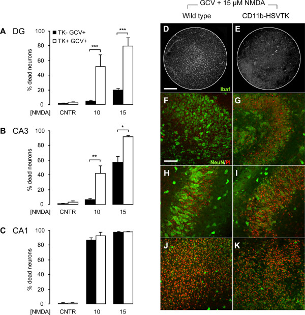
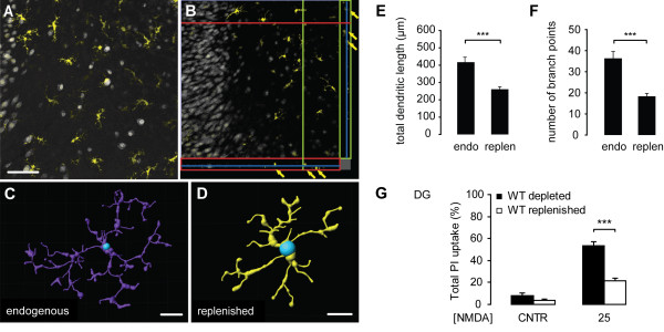
Background: Most of the known functions of microglia, including neurotoxic and neuroprotective properties, are attributed to morphologically-activated microglia. Resting, ramified microglia are suggested to primarily monitor their environment including synapses. Here, we show an active protective role of ramified microglia in excitotoxicity-induced neurodegeneration. Methods: Mouse organotypic hippocampal slice cultures were treated with N-methyl-D-aspartic acid (NMDA) to induce excitotoxic neuronal cell death. This procedure was performed in slices containing resting microglia or slices that were chemically or genetically depleted of their endogenous microglia. Results: Treatment of mouse organotypic hippocampal slice cultures with 10-50 μM N-methyl-D-aspartic acid (NMDA) induced region-specific excitotoxic neuronal cell death with CA1 neurons being most vulnerable, whereas CA3 and DG neurons were affected less. Ablation of ramified microglia severely enhanced NMDA-induced neuronal cell death in the CA3 and DG region rendering them almost as sensitive as CA1 neurons. Replenishment of microglia-free slices with microglia restored the original resistance of CA3 and DG neurons towards NMDA. Conclusions: Our data strongly suggest that ramified microglia not only screen their microenvironment but additionally protect hippocampal neurons under pathological conditions. Morphological activation of ramified microglia is thus not required to influence neuronal survival.
Overview
The claims and statistics of this paper haven’t been checked yet.
Run the check — free, no account needed. It reads the open-access text and checks the sources it cites.
Integrity signals
Registry
Cites
Statistics
Figures
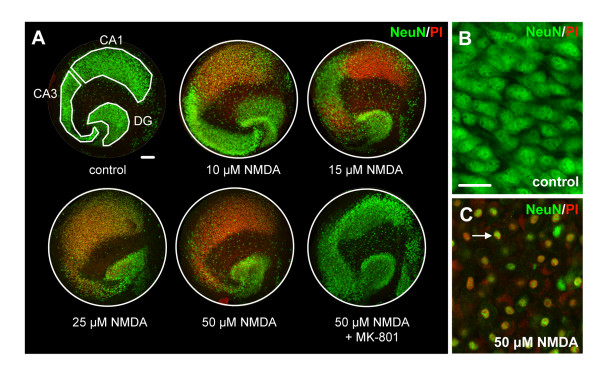
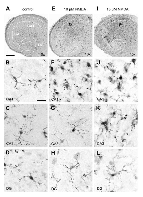
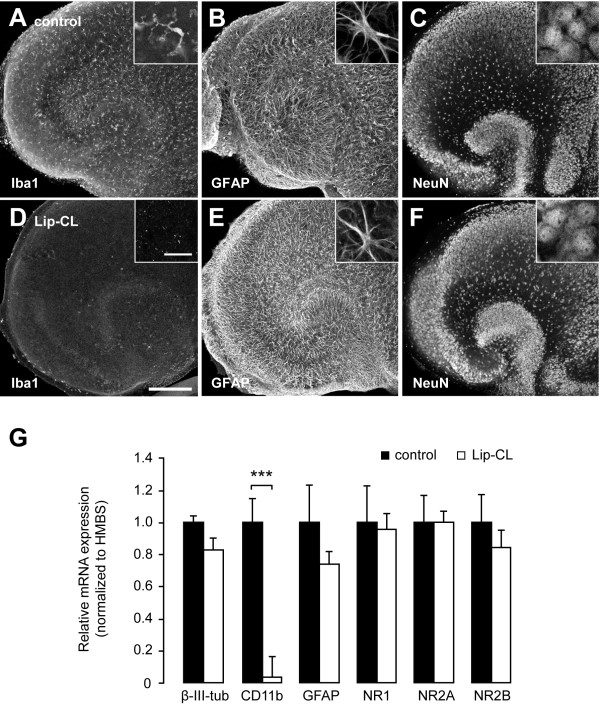
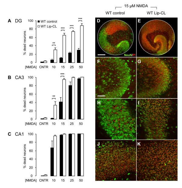
Verification
Claims the paper makes
The claims and statistics of this paper haven’t been checked yet.
Run the check — free, no account needed. It reads the open-access text and checks the sources it cites.
Claims other papers make about this one
Each is checked against this paper’s own text: does it say what the citing paper says it does?
The cited source says what the claim attributes to it.
In the citing paper
“Organotypic hippocampal slice cultures are known to undergo key developmental steps similar to those observed in vivo, including synapse maturation28–31, and have been previously used to study ramified microglia function32.”
From Microglia remodel synapses by presynaptic trogocytosis and spine head filopodia induction (2018) · cites this paper as [32]
Self-check
Percentages, sums and reported differences re-computed from the paper’s own numbers.
The self-check needs the paper’s full text.
Citations
Cites
Analysis of Relative Gene Expression Data Using Real-Time Quantitative PCR and the 2−ΔΔCT Method
Livak, K. J., & Schmittgen, T. D. (2001) · Methods · doi:10.1006/meth.2001.1262 · Cited by 180,860
Resting microglial cells are highly dynamic surveillants of brain parenchyma in vivo
Nimmerjahn, A., Kirchhoff, F., & Helmchen, F. (2005) · Science · doi:10.1126/science.1110647 · Cited by 5,827 · cited 3×
“It is now clear that ramified microglia in vivo continuously scan their environment for homeostatic irregularities [6-8].”
BackgroundMicroglia: a sensor for pathological events in the CNS
Kreutzberg, G. W. (1996) · Trends in Neurosciences · doi:10.1016/0166-2236(96)10049-7 · Cited by 4,396 · cited 2×
“This microglia response has long been defined as microglia activation and based on morphological findings, microglia activation was originally described as a stereotypic and graded process [2,5].”
BackgroundATP mediates rapid microglial response to local brain injury in vivo
Davalos, D., Grutzendler, J., Yang, G., Kim, J. V., Zuo, Y., Jung, S., Littman, D. R., Dustin, M. L., & Gan, W.-B. (2005) · Nature Neuroscience · doi:10.1038/nn1472 · Cited by 3,913 · cited 3×
“It is now clear that ramified microglia in vivo continuously scan their environment for homeostatic irregularities [6-8].”
BackgroundMicroglia: Active sensor and versatile effector cells in the normal and pathologic brain
Hanisch, U.-K., & Kettenmann, H. (2007) · Nature Neuroscience · doi:10.1038/nn1997 · Cited by 3,676 · cited 3×
“This view of microglia function has been challenged in the last years [4].”
BackgroundA simple method for organotypic cultures of nervous tissue
Stoppini, L., Buchs, P., & Muller, D. (1991) · Journal of Neuroscience Methods · doi:10.1016/0165-0270(91)90128-m · Cited by 3,096 · cited 1×
“Organotypic hippocampal slice cultures were prepared as described previously [27] with minor modifications.”
van Rooijen, N., & Sanders, A. (1994) · Journal of Immunological Methods · doi:10.1016/0022-1759(94)90012-4 · Cited by 1,763 · cited 1×
“Multi-lamellar CL2MDP (clodronate)-liposomes (Lip-CL) were obtained from the Department of Molecular Cell Biology of the Free University of Amsterdam, The Netherlands (for an extensive preparation protocol see [26]).”
MethodMicroglia as a source and target of cytokines
Hanisch, U. (2002) · Glia · doi:10.1002/glia.10161 · Cited by 1,645
Onset and Progression in Inherited ALS Determined by Motor Neurons and Microglia
Boillée, S., Yamanaka, K., Lobsiger, C. S., Copeland, N. G., Jenkins, N. A., Kassiotis, G., Kollias, G., & Cleveland, D. W. (2006) · Science · doi:10.1126/science.1123511 · Cited by 1,623 · cited 2×
“However, microglial neurotoxicity can occur in case of overshooting and uncontrolled stimulation of microglia [3,17] or when microglia function is impaired [15,18,19].”
BackgroundMicroglial interactions with synapses are modulated by visual experience
Tremblay, M.-È., Lowery, R. L., & Majewska, A. K. (2010) · PLoS Biology · doi:10.1371/journal.pbio.1000527 · Cited by 1,542 · cited 1×
“The more recent findings that ramified microglia contact active synapses [20,21], suggests a cell-cell interaction between ramified microglia and neurons, which however, is not yet understood.”
BackgroundThe P2Y12 receptor regulates microglial activation by extracellular nucleotides
Haynes, S. E., Hollopeter, G., Yang, G., Kurpius, D., Dailey, M. E., Gan, W.-B., & Julius, D. (2006) · Nature Neuroscience · doi:10.1038/nn1805 · Cited by 1,542 · cited 3×
“It is now clear that ramified microglia in vivo continuously scan their environment for homeostatic irregularities [6-8].”
BackgroundControl of microglial neurotoxicity by the fractalkine receptor
Cardona, A. E., Pioro, E. P., Sasse, M. E., Kostenko, V., Cardona, S. M., Dijkstra, I. M., Huang, D., Kidd, G., Dombrowski, S., Dutta, R., et al. (2006) · Nature Neuroscience · doi:10.1038/nn1715 · Cited by 1,531 · cited 1×
“However, microglial neurotoxicity can occur in case of overshooting and uncontrolled stimulation of microglia [3,17] or when microglia function is impaired [15,18,19].”
BackgroundMicroglia as neuroprotective, immunocompetent cells of the CNS
Streit, W. J. (2002) · Glia · doi:10.1002/glia.10154 · Cited by 919
El Khoury, J., Toft, M., Hickman, S. E., Means, T. K., Terada, K., Geula, C., & Luster, A. D. (2007) · Nature Medicine · doi:10.1038/nm1555 · Cited by 915 · cited 1×
“Moreover, protective microglia activity was described in mouse models of amyotrophic lateral sclerosis [15] and Alzheimer’s disease [16].”
BackgroundSelective Ablation of Proliferating Microglial Cells Exacerbates Ischemic Injury in the Brain
Lalancette-Hébert, M., Gowing, G., Simard, A., Weng, Y. C., & Kriz, J. (2007) · The Journal of Neuroscience · doi:10.1523/jneurosci.5360-06.2007 · Cited by 874 · cited 3×
“beneficial in a model of nitric oxide-dependent excitotoxicity [13] and in stroke [14].”
BackgroundResultNeuronal ‘On’ and ‘Off’ signals control microglia
Biber, K., Neumann, H., Inoue, K., & Boddeke, H. W. G. M. (2007) · Trends in Neurosciences · doi:10.1016/j.tins.2007.08.007 · Cited by 787 · cited 1×
“Although insensitive to NMDA, microglia most likely respond to NMDA-challenged neurons given the numerous signals that are released from these cells [67].”
BackgroundSelf-citationMicroglia Promote the Death of Developing Purkinje Cells
Marı́n-Teva, J. L., Dusart, I., Colin, C., Gervais, A., van Rooijen, N., & Mallat, M. (2004) · Neuron · doi:10.1016/s0896-6273(04)00069-8 · Cited by 757 · cited 2×
“Lip-CL has been shown to successfully deplete microglia from mouse organotypic slice cultures, without affecting other cell-types [23,24].”
BackgroundExperimental autoimmune encephalomyelitis repressed by microglial paralysis
Heppner, F. L., Greter, M., Marino, D., Falsig, J., Raivich, G., Hövelmeyer, N., Waisman, A., Rülicke, T., Prinz, M., Priller, J., et al. (2005) · Nature Medicine · doi:10.1038/nm1177 · Cited by 732 · cited 3×
“This mouse strain expresses the herpes simplex virus thymidine kinase (HSV-TK) under control of the CD11b promoter [25].”
BackgroundSelf-citationElkabes, S., Dicicco-Bloom, E., & Black, I. (1996) · Journal of Neuroscience · doi:10.1523/jneurosci.16-08-02508.1996 · Cited by 697 · cited 1×
“Whether or not ramified microglia in response release some of their neurotrophic factors [68,69] should, however, be further investigated.”
BackgroundSelective vulnerability in the gerbil hippocampus following transient ischemia
Kirino, T., & Sano, K. (1984) · Acta Neuropathologica · doi:10.1007/bf00691853 · Cited by 561 · cited 1×
“Similar patterns towards excitotoxicity or (hypoxic-) ischemic insults in the hippocampus have been observed previously both in vivo [34-38] and in vitro in organotypic slice cultures [32,39-44], corroborating our findings.”
ResultWhat is the blood–brain barrier (not)?
Bechmann, I., Galea, I., & Perry, V. H. (2007) · Trends in Immunology · doi:10.1016/j.it.2006.11.007 · Cited by 520 · cited 1×
“From the outside, the brain is protected by the skull and the blood-brain barrier [1].”
BackgroundMicroglial phenotype: is the commitment reversible?
Schwartz, M., Butovsky, O., Brück, W., & Hanisch, U.-K. (2006) · Trends in Neurosciences · doi:10.1016/j.tins.2005.12.005 · Cited by 434
Formation and maintenance of Alzheimer's disease β-amyloid plaques in the absence of microglia
Grathwohl, S. A., Kälin, R. E., Bolmont, T., Prokop, S., Winkelmann, G., Kaeser, S. A., Odenthal, J., Radde, R., Eldh, T., Gandy, S., et al. (2009) · Nature Neuroscience · doi:10.1038/nn.2432 · Cited by 401 · cited 3×
“In two mouse models of Alzheimer’s disease (AD) no change in amyloid-beta plaque load was seen in the absence of microglia [52].”
BackgroundSelf-citationMicroglial senescence: does the brain's immune system have an expiration date?
Streit, W. J. (2006) · Trends in Neurosciences · doi:10.1016/j.tins.2006.07.001 · Cited by 397 · cited 1×
“However, microglial neurotoxicity can occur in case of overshooting and uncontrolled stimulation of microglia [3,17] or when microglia function is impaired [15,18,19].”
BackgroundMicroglial CB2 cannabinoid receptors are neuroprotective in Huntington's disease excitotoxicity
Palazuelos, J., Aguado, T., Pazos, M. R., Julien, B., Carrasco, C., Resel, E., Sagredo, O., Benito, C., Romero, J., Azcoitia, I., et al. (2009) · Brain · doi:10.1093/brain/awp239 · Cited by 382 · cited 1×
“There is also evidence for the involvement of cannabinoid receptor 2 activation in microglia in neuroprotection against excitotoxicity in Huntington’s disease [60].”
BackgroundMicroglia Protect Neurons against Ischemia by Synthesis of Tumor Necrosis Factor
Lambertsen, K. L., Clausen, B. H., Babcock, A. A., Gregersen, R., Fenger, C., Nielsen, H. H., Haugaard, L. S., Wirenfeldt, M., Nielsen, M., Dagnæs‐Hansen, F., et al. (2009) · Journal of Neuroscience · doi:10.1523/jneurosci.5505-08.2009 · Cited by 374 · cited 1×
“Moreover, microglia in the hippocampus of rats subjected to stroke were found to specifically express neuroprotective TNFa [61].”
BackgroundIdentification of a microglia phenotype supportive of remyelination
Olah, M., Amor, S., Brouwer, N., Vinet, J., Eggen, B., Biber, K., & Boddeke, H. W. G. M. (2012) · Glia · doi:10.1002/glia.21266 · Cited by 358 · cited 1×
“Moreover, we just published a mRNA expression analysis of ramified white matter microglia from the adult mouse and did not find significant expression of NMDA receptor mRNA in these cells [65].”
BackgroundSelf-citationFontainhas, A. M., Wang, M., Liang, K. J., Chen, S., Mettu, P., Damani, M., Fariss, R. N., Li, W., & Wong, W. T. (2011) · PLoS ONE · doi:10.1371/journal.pone.0015973 · Cited by 334 · cited 1×
“The more recent findings that ramified microglia contact active synapses [20,21], suggests a cell-cell interaction between ramified microglia and neurons, which however, is not yet understood.”
BackgroundMicroglia Stimulate the Invasiveness of Glioma Cells by Increasing the Activity of Metalloprotease-2
Markovic, D. S., Glaß, R., Synowitz, M., van Rooijen, N., & Kettenmann, H. (2005) · Journal of Neuropathology & Experimental Neurology · doi:10.1097/01.jnen.0000178445.33972.a9 · Cited by 318 · cited 2×
“Lip-CL has been shown to successfully deplete microglia from mouse organotypic slice cultures, without affecting other cell-types [23,24].”
BackgroundMicroglia provide neuroprotection after ischemia
Neumann, J. H. L., Gunzer, M., Gutzeit, H. O., Ullrich, O., Reymann, K. G., & Dinkel, K. (2006) · The FASEB Journal · doi:10.1096/fj.05-4882fje · Cited by 284 · cited 1×
“In line with these results, it has been shown that application of the microglia cell line BV-2 to slice cultures reduced oxygen-glucose deprivation-induced neuronal damage [63].”
ResultOlah, M., Biber, K. P. H., Vinet, J., & Boddeke, H. W. G. M. (2011) · CNS & Neurological Disorders - Drug Targets · doi:10.2174/187152711794488575 · Cited by 260 · cited 1×
“It therefore is suggested that the NMDA-receptor expressing amoeboid microglia in the developing white matter recently described by Murugan and colleagues [64] is a specialized phenotype of microglia and these data add up to the concept that there are subtypes of microglia cells with different functions [66].”
BackgroundSelf-citationNeumann, H., & Takahashi, K. (2007) · Journal of Neuroimmunology · doi:10.1016/j.jneuroim.2006.11.032 · Cited by 249 · cited 1×
“However, microglial neurotoxicity can occur in case of overshooting and uncontrolled stimulation of microglia [3,17] or when microglia function is impaired [15,18,19].”
BackgroundDelayed neuronal death and delayed neuronal recovery in the human brain following global ischemia
Horn, M., & Schlote, W. (1992) · Acta Neuropathologica · doi:10.1007/bf00304636 · Cited by 241 · cited 1×
“Similar patterns towards excitotoxicity or (hypoxic-) ischemic insults in the hippocampus have been observed previously both in vivo [34-38] and in vitro in organotypic slice cultures [32,39-44], corroborating our findings.”
Resultvan Rossum, D., & Hanisch, U. (2004) · Metabolic Brain Disease · doi:10.1023/b:mebr.0000043984.73063.d8 · Cited by 239
Bernardino, L. I., Xapelli, S., Silva, A. P., Jakobsen, B., Poulsen, F. R., Oliveira, C. R., Vezzani, A., Malva, J. O., & Zimmer, J. (2005) · Journal of Neuroscience · doi:10.1523/jneurosci.1510-05.2005 · Cited by 224 · cited 1×
“This morphological activation, induced by neuronal stress or cell death signals, is a well-known feature of microglia that has already been reported for by del Rio-Hortega about a century ago [56] and ever since numerous times both in vivo [36] and in vitro in slice cultures [57].”
BackgroundNeuroprotective Effect of Exogenous Microglia in Global Brain Ischemia
Imai, F., Suzuki, H., Oda, J., Ninomiya, T., Ono, K., Sano, H., & Sawada, M. (2006) · Journal of Cerebral Blood Flow & Metabolism · doi:10.1038/sj.jcbfm.9600362 · Cited by 207 · cited 1×
“Thus, not the ablation technique but the absence of microglia enhanced the neuronal sensitivity, which is in agreement with earlier findings by us and others [13,14,33,53-55].”
ResultCytology and Cellular Pathology of the Nervous System.
(1932) · Archives of Neurology And Psychiatry · doi:10.1001/archneurpsyc.1932.02240030272017 · Cited by 198
Vornov, J. J., Tasker, R. C., & Coyle, J. T. (1991) · Experimental Neurology · doi:10.1016/0014-4886(91)90079-r · Cited by 172 · cited 6×
“Although insensitive to NMDA, microglia most likely respond to NMDA-challenged neurons given the numerous signals that are released from these cells [67].”
BackgroundResultInfluentialIGF‐I and microglia/macrophage proliferation in the ischemic mouse brain
O'Donnell, S. L., Frederick, T. J., Krady, J. K., Vannucci, S. J., & Wood, T. L. (2002) · Glia · doi:10.1002/glia.10081 · Cited by 139
Biber, K. P. H., Klotz, K., Berger, T., Gebicke‐Haerter, P. J., & van Calker, D. (1997) · Journal of Neuroscience · doi:10.1523/jneurosci.17-13-04956.1997 · Cited by 134 · cited 1×
“Total RNA was transcribed into cDNA as described previously [29] and gene expression for b-III-tubulin, GFAP, CD11b and the NMDA receptor subunits NR1, NR2A and NR2B (see Table 1 for primers) were analyzed using the ABI Prism 7900 HT real time PCR instrument and the iTAQTM SYBR Green Supermix with ROX (Bio-Rad 172-585…”
MethodSelf-citationTurrin, N. P., & Rivest, S. (2006) · Journal of Neuroscience · doi:10.1523/jneurosci.4032-05.2006 · Cited by 129 · cited 2×
“by us and others [13,14,33,53-55].”
BackgroundKristensen, B. W., Noraberg, J., & Zimmer, J. (2001) · Brain Research · doi:10.1016/s0006-8993(01)02900-6 · Cited by 129 · cited 2×
“Accordingly, it has been demonstrated that CA1 neurons are most vulnerable to glutamate- and NMDA-induced insults, whereas CA3 (and DG) neurons are most sensitive to the excitotoxin kainic acid [32,37,43,48].”
BackgroundResultStrasser, U., & Fischer, G. (1995) · Journal of Neuroscience Methods · doi:10.1016/0165-0270(94)00147-9 · Cited by 112 · cited 1×
“Similar patterns towards excitotoxicity or (hypoxic-) ischemic insults in the hippocampus have been observed previously both in vivo [34-38] and in vitro in organotypic slice cultures [32,39-44], corroborating our findings.”
ResultHailer, N. P., Grampp, A., & Nitsch, R. (1999) · European Journal of Neuroscience · doi:10.1046/j.1460-9568.1999.00808.x · Cited by 107 · cited 1×
“The microglia response to injury can also be accompanied by proliferation [9,10].”
BackgroundNarantuya, D., Nagai, A., Sheikh, A. M., Masuda, J., Kobayashi, S., Yamaguchi, S., & Kim, S. U. (2010) · PLoS ONE · doi:10.1371/journal.pone.0011746 · Cited by 106 · cited 1×
“Furthermore, it was recently described that an intravenous injection of the human microglial cell line HMO6 in ischemic rats leads to reduced infarct size and improved behavioral outcome, suggesting a neuroprotective function of these cells by the upregulation of several inflammatory mediators and neurotrophic factors…”
BackgroundLauro, C., Cipriani, R., Catalano, M., Trettel, F., Chece, G., Brusadin, V., Antonilli, L., van Rooijen, N., Eusebi, F., Fredholm, B. B., et al. (2010) · Neuropsychopharmacology · doi:10.1038/npp.2010.26 · Cited by 105 · cited 1×
“For instance, it was shown that CXC3CL1 expression on neurons leads to secretion of adenosine by microglia, which in turn leads to neuronal increase of adenosine A1 receptors and neuroprotection [58].”
BackgroundIntracerebroventricular injection of microglia protects against focal brain ischemia
Kitamura, Y., Takata, K., Inden, M., Tsuchiya, D., Yanagisawa, D., Nakata, J., & Taniguchi, T. (2004) · Journal of Pharmacological Sciences · doi:10.1254/jphs.94.203 · Cited by 104 · cited 1×
“Thus, not the ablation technique but the absence of microglia enhanced the neuronal sensitivity, which is in agreement with earlier findings by us and others [13,14,33,53-55].”
ResultMirrione, M. M., Konomos, D. K., Gravanis, I., Dewey, S. L., Aguzzi, A., Heppner, F. L., & Tsirka, S. E. (2010) · Neurobiology of Disease · doi:10.1016/j.nbd.2010.04.001 · Cited by 102 · cited 4×
“In kainate-induced neurotoxicity it recently was found that the ablation of morphologically activated microglia was correlated to reduced neuronal loss, showing that morphologically activated microglia can also promote the death of neurons [48].”
BackgroundInfluentialSelf-citationMurugan, M., Viswanathan, S., Lu, J., Ling, E., & Kaur, C. (2011) · Glia · doi:10.1002/glia.21121 · Cited by 94
Pozzo‐Miller, L. D., Mahanty, N. K., Connor, J. A., & Landis, D. M. D. (1994) · Neuroscience · doi:10.1016/0306-4522(94)90544-4 · Cited by 91 · cited 4×
“Although insensitive to NMDA, microglia most likely respond to NMDA-challenged neurons given the numerous signals that are released from these cells [67].”
InfluentialCoultrap, S. J., Nixon, K. M., Alvestad, R. M., Valenzuela, C. F., & Browning, M. D. (2005) · Molecular Brain Research · doi:10.1016/j.molbrainres.2004.12.005 · Cited by 86 · cited 1×
“It has been shown that CA1 neurons express relatively high levels of AMPA- and NMDA-receptor (-subtypes), while neurons in the CA3 region express relatively high levels of kainate-receptors [46,47].”
BackgroundNMDA receptors and the differential ischemic vulnerability of hippocampal neurons
Gee, C. E., Benquet, P., Raineteau, O., Rietschin, L., Kirbach, S. W., & Gerber, U. R. (2006) · European Journal of Neuroscience · doi:10.1111/j.1460-9568.2006.04786.x · Cited by 83 · cited 1×
“Similar patterns towards excitotoxicity or (hypoxic-) ischemic insults in the hippocampus have been observed previously both in vivo [34-38] and in vitro in organotypic slice cultures [32,39-44], corroborating our findings.”
ResultCytology and Cellular Pathology of the Nervous System.
(1932) · Annals of Internal Medicine · doi:10.7326/0003-4819-6-3-452_2 · Cited by 80
Retigabine and flupirtine exert neuroprotective actions in organotypic hippocampal cultures
Boscia, F., Annunziato, L., & Taglialatela, M. (2006) · Neuropharmacology · doi:10.1016/j.neuropharm.2006.03.024 · Cited by 79 · cited 1×
“Similar patterns towards excitotoxicity or (hypoxic-) ischemic insults in the hippocampus have been observed previously both in vivo [34-38] and in vitro in organotypic slice cultures [32,39-44], corroborating our findings.”
ResultSchauwecker, P. (2002) · Experimental Neurology · doi:10.1006/exnr.2002.8038 · Cited by 69 · cited 2×
“Accordingly, it has been demonstrated that CA1 neurons are most vulnerable to glutamate- and NMDA-induced insults, whereas CA3 (and DG) neurons are most sensitive to the excitotoxin kainic acid [32,37,43,48].”
BackgroundResultExpression, transport, and axonal sorting of neuronal CCL21 in large dense‐core vesicles
de Jong, E. K., Vinet, J., Stanulovic, V., Meijer, M., Wesseling, E. M., Sjollema, K. A., Boddeke, H. W. G. M., & Biber, K. P. H. (2008) · The FASEB Journal · doi:10.1096/fj.07-101907 · Cited by 61
Self-citationvan Weering, H. R. J., Boddeke, H. W. G. M., Vinet, J., Brouwer, N., Haas, A. H. D., van Rooijen, N., Thomsen, A. R., & Biber, K. P. H. (2010) · Hippocampus · doi:10.1002/hipo.20742 · Cited by 59 · cited 2×
“Thus, not the ablation technique but the absence of microglia enhanced the neuronal sensitivity, which is in agreement with earlier findings by us and others [13,14,33,53-55].”
MethodResultSelf-citationXapelli, S., Bernardino, L. I., Ferreira, R., Grade, S., Silva, A. P., Rosmaninho‐Salgado, J., Cavadas, C., Grouzmann, E., Poulsen, F. R., Jakobsen, B., et al. (2008) · European Journal of Neuroscience · doi:10.1111/j.1460-9568.2008.06172.x · Cited by 59 · cited 1×
“Whether or not ramified microglia in response release some of their neurotrophic factors [68,69] should, however, be further investigated.”
BackgroundKohl, A., Dehghani, F., Korf, H., & Hailer, N. P. (2003) · Experimental Neurology · doi:10.1016/s0014-4886(02)00049-3 · Cited by 59 · cited 1×
“Since microglia can be specifically eliminated from these slice cultures [22] without affecting other cell types [23-25], this model provides an ideal system to analyze the function of microglia in their ramified state.”
BackgroundBoscia, F., Esposito, C. L., Crisci, A. D., de Franciscis, V., Annunziato, L., & Cerchia, L. (2009) · PLoS ONE · doi:10.1371/journal.pone.0006486 · Cited by 57 · cited 1×
“Exposure of hippocampal slice cultures to GDNF has been shown to activate microglia, leading to increased neuronal survival [59].”
BackgroundMontero, M., González, B., & Zimmer, J. (2009) · Brain Research · doi:10.1016/j.brainres.2009.06.097 · Cited by 54 · cited 1×
“Thus, not the ablation technique but the absence of microglia enhanced the neuronal sensitivity, which is in agreement with earlier findings by us and others [13,14,33,53-55].”
ResultSakaguchi, T., Okada, M., Kuno, M., & Kawasaki, K. (1997) · Neuroscience · doi:10.1016/s0306-4522(96)00403-4 · Cited by 53 · cited 1×
“Thus, variability in glutamate receptor (-subtype) expression and/or endogenous properties of the distinct neuronal populations in the CA1, CA3 and DG regions [41,49-51] could (in part) explain their selective vulnerability towards excitotoxicity.”
BackgroundRegional distribution of neurotrophin receptors in the developing auditory brainstem
Hafidi, A., Moore, T., & Sanes, D. H. (1996) · The Journal of Comparative Neurology · doi:10.1002/(sici)1096-9861(19960408)367:3 · Cited by 51
Grishin, A. A., Gee, C. E., Gerber, U. R., & Benquet, P. (2004) · Journal of Neuroscience · doi:10.1523/jneurosci.4933-03.2004 · Cited by 50 · cited 1×
“Thus, variability in glutamate receptor (-subtype) expression and/or endogenous properties of the distinct neuronal populations in the CA1, CA3 and DG regions [41,49-51] could (in part) explain their selective vulnerability towards excitotoxicity.”
BackgroundKeynes, R. G., Duport, S., & Garthwaite, J. (2004) · European Journal of Neuroscience · doi:10.1111/j.1460-9568.2004.03217.x · Cited by 41 · cited 1×
“Similar patterns towards excitotoxicity or (hypoxic-) ischemic insults in the hippocampus have been observed previously both in vivo [34-38] and in vitro in organotypic slice cultures [32,39-44], corroborating our findings.”
ResultMartens, U., Capito, B., & Wree, A. (1998) · Anatomy and Embryology · doi:10.1007/s004290050177 · Cited by 38 · cited 1×
“It has been shown that CA1 neurons express relatively high levels of AMPA- and NMDA-receptor (-subtypes), while neurons in the CA3 region express relatively high levels of kainate-receptors [46,47].”
BackgroundNuclear factor kappa B-mediated kainate neurotoxicity in the rat and hamster hippocampus
Won, S. J., Ko, H. W., Kim, E. Y., Park, E. C., Huh, K. H., Jung, N. P., Choi, I., Oh, Y. K., Shin, H.-S., & Gwag, B. J. (1999) · Neuroscience · doi:10.1016/s0306-4522(99)00196-7 · Cited by 32 · cited 1×
“Similar patterns towards excitotoxicity or (hypoxic-) ischemic insults in the hippocampus have been observed previously both in vivo [34-38] and in vitro in organotypic slice cultures [32,39-44], corroborating our findings.”
ResultIkegaya, Y., & Matsuki, N. (2002) · Neuroscience · doi:10.1016/s0306-4522(02)00217-8 · Cited by 24 · cited 1×
“Interestingly, selectivity towards NMDA has been shown to be independent of an intact hippocampal neuronal circuitry as isolated CA3, CA1 and DG slice cultures still respond with a selective vulnerability towards NMDA, with the CA1 and CA3 regions being more susceptible to NMDA than the DG region [45].”
BackgroundCronberg, T., Jensen, K., Rytter, A., & Wieloch, T. W. (2005) · European Journal of Neuroscience · doi:10.1111/j.1460-9568.2005.04235.x · Cited by 21 · cited 2×
“Thus, variability in glutamate receptor (-subtype) expression and/or endogenous properties of the distinct neuronal populations in the CA1, CA3 and DG regions [41,49-51] could (in part) explain their selective vulnerability towards excitotoxicity.”
BackgroundResultChen, Y., Chad, J. E., Cannon, R. C., & Wheal, H. V. (1999) · Neuroscience · doi:10.1016/s0306-4522(98)00253-x · Cited by 20
Cited by
Sorted by citations
Microglia emerge from erythromyeloid precursors via Pu.1- and Irf8-dependent pathways
Kierdorf, K., Erny, D., Goldmann, T., Sander, V., Schulz, C., Perdiguero, E. G., Wieghofer, P., Heinrich, A., Riemke, P., Hölscher, C., et al. (2013) · Nature Neuroscience · doi:10.1038/nn.3318 · Cited by 1,457
Self-citationMicroglia remodel synapses by presynaptic trogocytosis and spine head filopodia induction
Weinhard, L., di Bartolomei, G., Bolasco, G., Machado, P., Schieber, N. L., Neniskyte, U., Exiga, M., Vadisiute, A., Raggioli, A., Schertel, A., et al. (2018) · Nature Communications · doi:10.1038/s41467-018-03566-5 · Cited by 790
Neuroinflammation: The role and consequences
Lyman, M., Lloyd, D. G., Ji, X., Vizcaychipi, M. P., & Ma, D. (2013) · Neuroscience Research · doi:10.1016/j.neures.2013.10.004 · Cited by 776
Filipello, F., Morini, R., Corradini, I., Zerbi, V., Canzi, A., Michalski, B., Erreni, M., Markicevic, M., Starvaggi-Cucuzza, C., Otero, K., et al. (2018) · Immunity · doi:10.1016/j.immuni.2018.04.016 · Cited by 695
Valdearcos, M., Robblee, M. M., Benjamin, D. I., Nomura, D. K., Xu, A. W., & Koliwad, S. K. (2014) · Cell Reports · doi:10.1016/j.celrep.2014.11.018 · Cited by 629
“Depleting Microglia from Hypothalamic Slice Cultures Microglia were depleted from hypothalamic slice cultures without altering other cell types by modifying prior approaches (Okamura et al., 2012; Vinet et al., 2012).”
Metadata
- Authors
- Vinet, J., van Weering, H. R., Heinrich, A., Kälin, R. E., Wegner, A., Brouwer, N., Heppner, F. L., van Rooijen, N., Boddeke, H. W., & Biber, K.
- Affiliations
- University Medical Center Groningen (NL)
- University of Freiburg (DE)
- Charité – Universitätsmedizin Berlin (DE)
- Corresponding author
- Vinet, Jonathan
- Biber, Knut
- Venue
- Journal of Neuroinflammation
- Published
- 2012-01-31
- Volume
- 9
- Issue
- 1
- Pages
- 27-27
- ISSN
- 1742-2094 (ISSN-L)
- PMC
- PMC3292937
- OpenAlex
- W1963696719
- Cited by
- 274
- References
- 70
- Citations by year
- 2026: 11 · 2025: 6 · 2024: 18 · 2023: 16 · 2022: 14 · 2021: 20 · 2020: 22 · 2019: 20 · 2018: 18 · 2017: 28 · 2016: 24 · 2015: 18 · 2014: 24 · 2013: 26 · 2012: 9
- FWCI
- 12.38
- Type
- journal article
- Language
- en
- Open access
- gold
- doi.org
- Locations
- Journal of Neuroinflammation (publishedVersion, cc-by)
- Europe PMC (PubMed Central) (publishedVersion, cc-by)
- PubMed Central (publishedVersion, cc-by)
- PubMed (publishedVersion)
- Pure Amsterdam UMC (publishedVersion, other-oa)
- Amsterdam UMC (VU Amsterdam) - Institutional Repository (publishedVersion, other-oa)
- University of Groningen research database (University of Groningen / Centre for Information Technology) (publishedVersion, cc-by)
- Digital Academic REpository of VU University Amsterdam (Vrije Universiteit Amsterdam) (submittedVersion)
- DOAJ (DOAJ: Directory of Open Access Journals) (submittedVersion)
- UCL Discovery (University College London) (submittedVersion)
- IRIS UNIMORE (University of Modena and Reggio Emilia) (submittedVersion, cc-by)
- University of Groningen research database (University of Groningen / Centre for Information Technology) (submittedVersion, cc-by)
- Data Archiving and Networked Services (DANS) (submittedVersion, other-oa)
- Funders
- Deutsche Forschungsgemeinschaft (SFB TRR 43, NeuroCure EXC257, EXC257, FOR1336, TRR-43)
- Nederlandse Organisatie voor Wetenschappelijk Onderzoek
- Topics
- Life Sciences › Neuroscience › Neurology › Neuroinflammation and Neurodegeneration Mechanisms
- Life Sciences › Neuroscience › Cellular and Molecular Neuroscience › Neuroscience and Neuropharmacology Research
- Life Sciences › Neuroscience › Neurology › Neurological Disease Mechanisms and Treatments
- Keywords
- ramified microglia, neuroprotection, microglia depletion, organotypic hippocampal slice cultures, CA1 neurons, excitotoxic neurodegeneration
- MeSH terms
- Animals, Clodronic Acid, Dose-Response Relationship, Drug, Glial Fibrillary Acidic Protein, Hippocampus, Mice, Inbred C57BL, Mice, Transgenic, Nerve Degeneration, Neurons, Neurotoxins, Organ Culture Techniques, Phosphopyruvate Hydratase, N-Methylaspartate, Cell Death, Microglia, CD11b Antigen, Mice
About these checks
Registry, retraction, Cites and statistics results are computed, not judged, with one exception: a printed reference that no index lists is read once by a model before it counts as a citation error. Claims are extracted by a model and kept only when their quote appears verbatim in the text; each cited source is then checked, with a model’s help, for whether it says what the claim relies on. Those verdicts can be wrong — read the claim’s quote and the cited source before relying on one.
Statistics and arithmetic are recomputed, not judged.
Confidence bands are shown only for fields calibrated on our annotated test set. None is calibrated yet, so no band is shown.
What counts as a claim ›How we measure confidence ›How we check this ›
Are you an author, or do you see a problem with this page? Request a review
