

Altered monocyte and fibrocyte phenotype and function in scleroderma interstitial lung disease: reversal by caveolin-1 scaffolding domain peptide
Open accessThe DOI matches its Crossref record; 30 of the 32 references it cites were found in a registry or an index.
Abstract
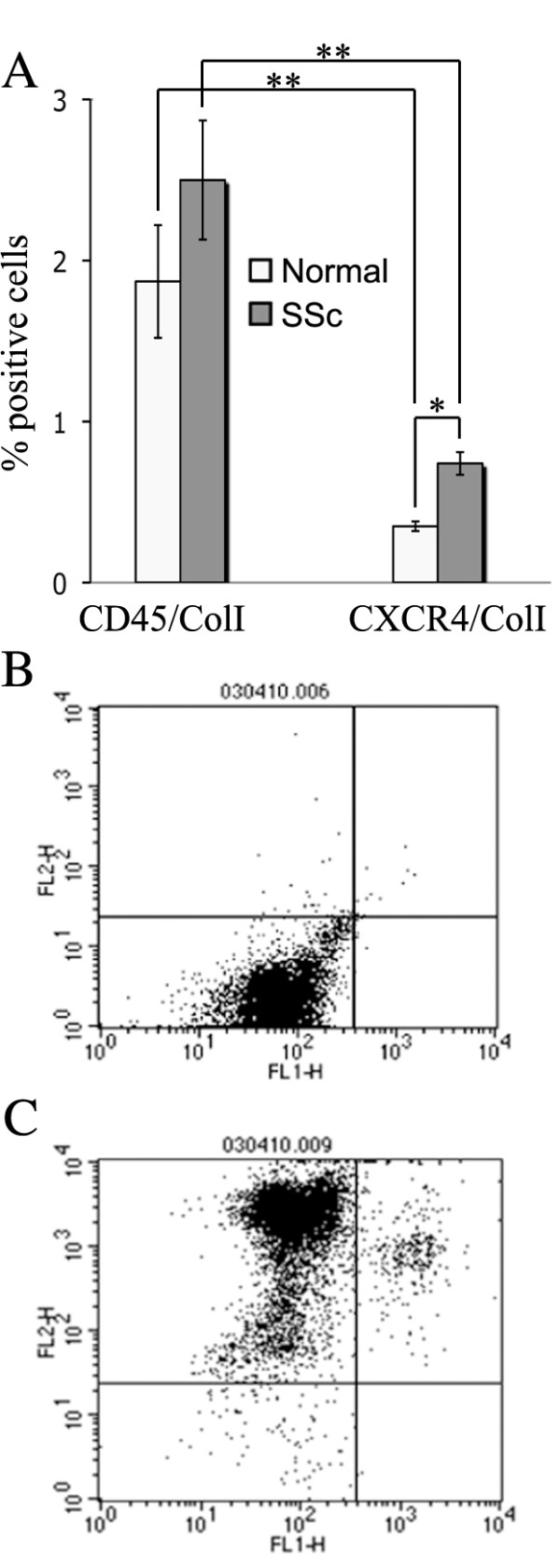
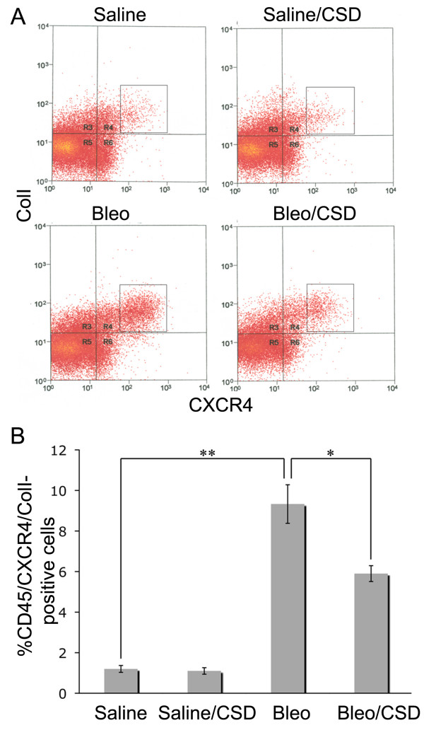
Interstitial lung disease (ILD) is a major cause of morbidity and mortality in scleroderma (systemic sclerosis, or SSc). Fibrocytes are a monocyte-derived cell population implicated in the pathogenesis of fibrosing disorders. Given the recently recognized importance of caveolin-1 in regulating function and signaling in SSc monocytes, in the present study we examined the role of caveolin-1 in the migration and/or trafficking and phenotype of monocytes and fibrocytes in fibrotic lung disease in human patients and an animal model. These studies fill a gap in our understanding of how monocytes and fibrocytes contribute to SSc-ILD pathology. We found that C-X-C chemokine receptor type 4-positive (CXCR4+)/collagen I-positive (ColI+), CD34+/ColI+ and CD45+/ColI+ cells are present in SSc-ILD lungs, but not in control lungs, with CXCR4+ cells being most prevalent. Expression of CXCR4 and its ligand, stromal cell-derived factor 1 (CXCL12), are also highly upregulated in SSc-ILD lung tissue. SSc monocytes, which lack caveolin-1 and therefore overexpress CXCR4, exhibit almost sevenfold increased migration toward CXCL12 compared to control monocytes. Restoration of caveolin-1 function by administering the caveolin scaffolding domain (CSD) peptide reverses this hypermigration. Similarly, transforming growth factor β-treated normal monocytes lose caveolin-1, overexpress CXCR4 and exhibit 15-fold increased monocyte migration that is CSD peptide-sensitive. SSc monocytes exhibit a different phenotype than normal monocytes, expressing high levels of ColI, CD14 and CD34. Because ColI+/CD14+ cells are prevalent in SSc blood, we looked for such cells in lung tissue and confirmed their presence in SSc-ILD lungs but not in normal lungs. Finally, in the bleomycin model of lung fibrosis, we show that CSD peptide diminishes fibrocyte accumulation in the lungs. Our results suggest that low caveolin-1 in SSc monocytes contributes to ILD via effects on cell migration and phenotype and that the hyperaccumulation of fibrocytes in SSc-ILD may result from the altered phenotype and migratory activity of their monocyte precursors.
Overview
The claims and statistics of this paper haven’t been checked yet.
Free: it reads the open-access text and checks the first 5 claims that cite a source we can check. Create a free account to check every claim.
Integrity signals
Registry
Cites
Statistics
Figures





Verification
Claims the paper makes
The claims and statistics of this paper haven’t been checked yet.
Free: it reads the open-access text and checks the first 5 claims that cite a source we can check. Create a free account to check every claim.
Claims other papers make about this one
Each is checked against this paper’s own text: does it say what the citing paper says it does?
In the citing paper
“Of note, fibrocytes can originate from monocytes, and, importantly, SSc monocytes display increased maturation toward myofibroblasts as indicated by αSMA expression when compared to monocytes from healthy controls (87).”
From Unraveling SSc Pathophysiology; The Myofibroblast (2018) · cites this paper as [87]
In the citing paper
“Paradoxically, fibrocyte numbers in blood are lower in SSc patients than in healthy controls.”
From Unraveling SSc Pathophysiology; The Myofibroblast (2018) · cites this paper as [87]
In the citing paper
“Furthermore, fibrocyte presence and involvement in pulmonary fibrosis can readily be detected in SSc (87).”
From Unraveling SSc Pathophysiology; The Myofibroblast (2018) · cites this paper as [87]
Self-check
Percentages, sums and reported differences re-computed from the paper’s own numbers.
The self-check needs the paper’s full text.
Citations
Cites
Preliminary criteria for the classification of systemic sclerosis (scleroderma)
Masi, A. T., & Subcommittee For Scleroderma Criteria of the American Rheumatism Association Diagnostic and Therapeutic Criteria Committee (1980) · Arthritis & Rheumatism · doi:10.1002/art.1780230510 · Cited by 4,898
Klinge, U., Park, J.-K., & Klosterhalfen, B. (2013) · Pathobiology · doi:10.1159/000348446 · Cited by 1,783
Circulating Fibrocytes Define a New Leukocyte Subpopulation That Mediates Tissue Repair
Bucala, R., Spiegel, L. A., Chesney, J., Hogan, M., & Cerami, A. (1994) · Molecular Medicine · doi:10.1007/bf03403533 · Cited by 1,308
Circulating fibrocytes traffic to the lungs in response to CXCL12 and mediate fibrosis
Phillips, R. J., Burdick, M. D., Hong, K., Lutz, M. A., Murray, L. A., Xue, Y. Y., Belperio, J. A., Keane, M. P., & Strieter, R. M. (2004) · Journal of Clinical Investigation · doi:10.1172/jci200420997 · Cited by 1,079
Peripheral Blood Fibrocytes: Differentiation Pathway and Migration to Wound Sites
Abe, R., Donnelly, S. C., Peng, T., Bucala, R., & Metz, C. N. (2001) · The Journal of Immunology · doi:10.4049/jimmunol.166.12.7556 · Cited by 967 · cited 2×
“In addition, fibrocytes contribute to tissue remodeling by producing high levels of cytokines, fibrogenic growth factors, extracellular matrix proteins and matrix metalloproteinase [1,8-12].”
BackgroundMethodCirculating Fibrocytes Are an Indicator of Poor Prognosis in Idiopathic Pulmonary Fibrosis
Moeller, A., Gilpin, S. E., Ask, K., Cox, G., Cook, D., Gauldie, J., Margetts, P. J., Farkas, L., Dobranowski, J., Boylan, C., et al. (2009) · American Journal of Respiratory and Critical Care Medicine · doi:10.1164/rccm.200810-1534oc · Cited by 513
CCR2-Mediated Recruitment of Fibrocytes to the Alveolar Space after Fibrotic Injury
Moore, B. B., Kolodsick, J. E., Thannickal, V. J., Cooke, K., Moore, T. A., Hogaboam, C., Wilke, C. A., & Toews, G. B. (2005) · The American Journal of Pathology · doi:10.1016/s0002-9440(10)62289-4 · Cited by 441
Pilling, D., Fan, T., Huang, D., Kaul, B., & Gomer, R. H. (2009) · PLoS ONE · doi:10.1371/journal.pone.0007475 · Cited by 433
Caveolin-1: a critical regulator of lung fibrosis in idiopathic pulmonary fibrosis
Wang, X. M., Zhang, Y., Kim, H. P., Zhou, Z., Feghali-Bostwick, C. A., Liu, F., Ifedigbo, E., Xu, X., Oury, T. D., Kaminski, N., et al. (2006) · The Journal of Experimental Medicine · doi:10.1084/jem.20061536 · Cited by 397 · cited 1×
“Our data, together with data from other groups, strongly suggest that caveolin-1 is a key signaling molecule in the monocyte-fibrocyte-fibroblast lineage and is responsible for functional differences observed among cells isolated from SSc-ILD and idiopathic pulmonary fibrosis (IPF) patients compared to control subject…”
ResultFibrocytes are a potential source of lung fibroblasts in idiopathic pulmonary fibrosis
Andersson-Sjöland, A., de Alba, C. G., Nihlberg, K., Becerril, C., Ramírez, R., Pardo, A., Westergren-Thorsson, G., & Selman, M. (2008) · The International Journal of Biochemistry & Cell Biology · doi:10.1016/j.biocel.2008.02.012 · Cited by 360
Research works printed but not linked: 3
- Metchnikov II: Immunity of Infectious Diseases. 1901, Paris, France. Masson
- Maximow AA: Cultures of blood leucocytes: from lymphocyte and monocyte to connective tissue. Arch Exp Zellforsch. 1928, 5: 169-268.
- Fischer AC: Transformation outside of organism of mononuclears into fibroblasts. Compt Rend Soc Biol. 1925, 92: 109-112.
Cited by
Fibroblasts in fibrosis: novel roles and mediators
Kendall, R. T., & Feghali-Bostwick, C. A. (2014) · Frontiers in Pharmacology · doi:10.3389/fphar.2014.00123 · Cited by 1,082
Scleroderma pathogenesis: a pivotal role for fibroblasts as effector cells
Gilbane, A. J., Denton, C. P., & Holmes, A. M. (2013) · Arthritis Research & Therapy · doi:10.1186/ar4230 · Cited by 196
The Living Scar – Cardiac Fibroblasts and the Injured Heart
Rog-Zielinska, E. A., Norris, R. A., Kohl, P., & Markwald, R. (2016) · Trends in Molecular Medicine · doi:10.1016/j.molmed.2015.12.006 · Cited by 171
Unraveling SSc Pathophysiology; The Myofibroblast
van Caam, A., Vonk, M., van den Hoogen, F., van Lent, P., & van der Kraan, P. (2018) · Frontiers in Immunology · doi:10.3389/fimmu.2018.02452 · Cited by 153
“Furthermore, fibrocyte presence and involvement in pulmonary fibrosis can readily be detected in SSc (87).”
BackgroundWermuth, P. J., & Jimenez, S. A. (2015) · Clinical and Translational Medicine · doi:10.1186/s40169-015-0047-4 · Cited by 141
Metadata
- Authors
- Tourkina, E., Bonner, M., Oates, J., Hofbauer, A., Richard, M., Znoyko, S., Visconti, R. P., Zhang, J., Hatfield, C. M., Silver, R. M., & Hoffman, S.
- Affiliations
- Medical University of South Carolina (US)
- Corresponding author
- Tourkina, Elena
- Venue
- Fibrogenesis & Tissue Repair
- Published
- 2011-07-01
- Volume
- 4
- Issue
- 1
- Pages
- 15-15
- ISSN
- 1755-1536 (ISSN-L)
- PMC
- PMC3155832
- OpenAlex
- W2016100345
- Cited by
- 104
- References
- 32
- Citations by year
- 2026: 1 · 2025: 3 · 2024: 4 · 2023: 3 · 2022: 5 · 2021: 2 · 2020: 7 · 2019: 10 · 2018: 8 · 2017: 11 · 2016: 8 · 2015: 4 · 2014: 9 · 2013: 12 · 2012: 15
- FWCI
- 8.53
- Type
- journal article
- Language
- en
- Open access
- hybrid
- doi.org
- Licence
- cc-by
- creativecommons.org/licenses/by/2.0 (tdm)
- Locations
- Fibrogenesis & Tissue Repair (publishedVersion, cc-by)
- Europe PMC (PubMed Central) (publishedVersion, cc-by)
- PubMed Central (publishedVersion, cc-by)
- PubMed (publishedVersion)
- Funders
- Yale University
- Medical University of South Carolina (C06 RR015455)
- University of South Carolina
- Fondation Leducq
- National Institutes of Health (1c06rr015455-01a1, RR-016434, AR049459)
- National Center for Complementary and Alternative Medicine
- National Institute of Arthritis and Musculoskeletal and Skin Diseases (R01AR062078, P60AR049459, P60AR062755)
- National Center for Research Resources (UL1RR029882, P20RR021949, C06 RR-015455)
- National Center for Complementary and Integrative Health (R21AT004450)
- Topics
- Health Sciences › Medicine › Rheumatology › Systemic Sclerosis and Related Diseases
- Health Sciences › Medicine › Pulmonary and Respiratory Medicine › Interstitial Lung Diseases and Idiopathic Pulmonary Fibrosis
- Health Sciences › Medicine › Pulmonary and Respiratory Medicine › Pulmonary Hypertension Research and Treatments
- Keywords
- systemic sclerosis, interstitial lung disease, caveolin-1, monocytes, fibrocytes, CXCR4, CXCL12, monocyte migration, pulmonary fibrosis, bleomycin-induced lung fibrosis, type I collagen, TGF-beta, CD14, CD34
About these checks
Registry, retraction, Cites and statistics results are computed, not judged, with one exception: a printed reference that no index lists is read once by a model before it counts as a citation error. Claims are extracted by a model and kept only when their quote appears verbatim in the text; each cited source is then checked, with a model’s help, for whether it says what the claim relies on. Those verdicts can be wrong — read the claim’s quote and the cited source before relying on one.
Statistics and arithmetic are recomputed, not judged.
Confidence bands are shown only for fields calibrated on our annotated test set. None is calibrated yet, so no band is shown.
What counts as a claim ›How we measure confidence ›How we check this ›
Are you an author, or do you see a problem with this page? Request a review
How to cite
Cite CoChat for the checks on this page, and the paper for its own findings.
- This page
- CoChat. (2026). Citation check: Altered monocyte and fibrocyte phenotype and function in scleroderma interstitial lung disease: reversal by caveolin-1 scaffolding domain peptide. https://app.cochat.ai/papers/10.1186/1755-1536-4-15
- The paper
- Tourkina, E., Bonner, M., Oates, J., Hofbauer, A., Richard, M., Znoyko, S., Visconti, R. P., Zhang, J., Hatfield, C. M., Silver, R. M., & Hoffman, S. (2011). Altered monocyte and fibrocyte phenotype and function in scleroderma interstitial lung disease: reversal by caveolin-1 scaffolding domain peptide. Fibrogenesis & Tissue Repair. https://doi.org/10.1186/1755-1536-4-15
Data sources
Bibliographic data: Semantic Scholar, OpenAlex, Crossref.
Contains information from the Semantic Scholar Academic Graph Datasets, made available under the ODC Attribution License (ODC-BY 1.0).
Semantic Scholar’s data is described in: Kinney, R., et al. (2023). The Semantic Scholar Open Data Platform. arXiv:2301.10140.
