

The DOI matches its Crossref record; 148 of the 150 references it cites were found in a registry or an index.
Regulation of skeletal muscle growth by the IGF1-Akt/PKB pathway: insights from genetic models
Open accessAbstract
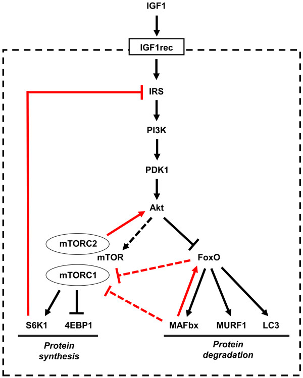
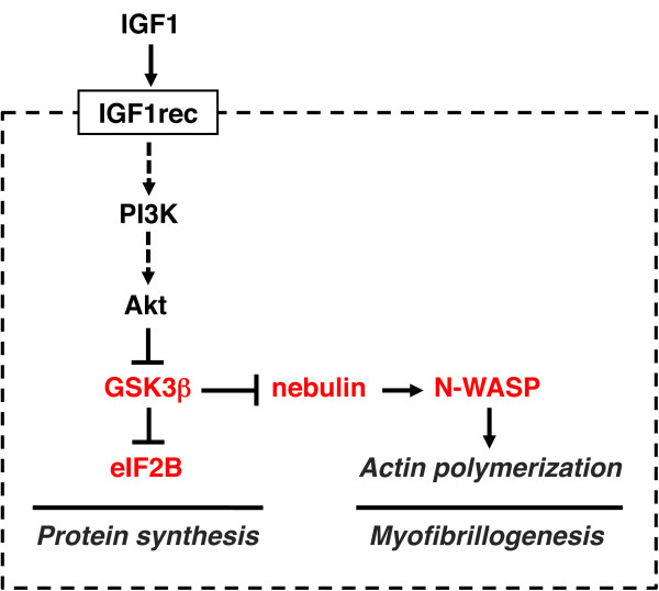
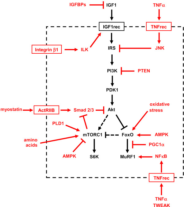
A highly conserved signaling pathway involving insulin-like growth factor 1 (IGF1), and a cascade of intracellular components that mediate its effects, plays a major role in the regulation of skeletal muscle growth. A central component in this cascade is the kinase Akt, also called protein kinase B (PKB), which controls both protein synthesis, via the kinases mammalian target of rapamycin (mTOR) and glycogen synthase kinase 3β (GSK3β), and protein degradation, via the transcription factors of the FoxO family. In this paper, we review the composition and function of this pathway in skeletal muscle fibers, focusing on evidence obtained in vivo by transgenic and knockout models and by muscle transient transfection experiments. Although this pathway is essential for muscle growth during development and regeneration, its role in adult muscle response to mechanical load is less clear. A full understanding of the operation of this pathway could help to design molecularly targeted therapeutics aimed at preventing muscle wasting, which occurs in a variety of pathologic contexts and in the course of aging.
Overview
The claims and statistics of this paper haven’t been checked yet.
Run the check — free, no account needed. It reads the open-access text and checks the sources it cites.
Integrity signals
Registry
Cites
Statistics
Figures
Verification
Claims the paper makes
The claims and statistics of this paper haven’t been checked yet.
Run the check — free, no account needed. It reads the open-access text and checks the sources it cites.
Claims other papers make about this one
Each is checked against this paper’s own text: does it say what the citing paper says it does?
The cited source says what the claim attributes to it.
In the citing paper
“The Akt‐mTOR pathway is central to the regulation of muscle mass because it promotes protein synthesis and inhibits protein degradation (Schiaffino & Mammucari, 2011).”
From Parkin overexpression protects from ageing‐related loss of muscle mass and strength (2019) · cites this paper as Schiaffino & Mammucari, 2011
In the citing paper
“Additionally, it has been discovered that PEs present in cosmetic preparation protect skin from pigmentation and aging163.”
From The Phytochemical, Biological, and Medicinal Attributes of Phytoecdysteroids: An Updated Review (2021) · cites this paper as [163]
Self-check
Percentages, sums and reported differences re-computed from the paper’s own numbers.
The self-check needs the paper’s full text.
Citations
Cites
Phosphorylation and regulation of Akt/PKB by the rictor-mTOR complex
Sarbassov, D. D., Guertin, D. A., Ali, S. M., & Sabatini, D. M. (2005) · Science · doi:10.1126/science.1106148 · Cited by 6,216
AKT/PKB signaling: navigating downstream
Manning, B. D., & Cantley, L. C. (2007) · Cell · doi:10.1016/j.cell.2007.06.009 · Cited by 6,115 · cited 1×
“transcription factors of the FoxO family, and stimulates protein synthesis via the mammalian target of rapamycin (mTOR) and glycogen synthase kinase 3b (GSK3b) [4].”
BackgroundRegulation of skeletal muscle mass in mice by a new TGF-p superfamily member
McPherron, A. C., Lawler, A. M., & Lee, S.-J. (1997) · Nature · doi:10.1038/387083a0 · Cited by 4,013
mTOR: from growth signal integration to cancer, diabetes and ageing
Zoncu, R., Efeyan, A., & Sabatini, D. M. (2011) · Nature Reviews Molecular Cell Biology · doi:10.1038/nrm3025 · Cited by 3,941 · cited 15×
“Deletion of S6K1 is sufficient to reproduce this atrophic phenotype.”
BackgroundInfluentialAMPK Phosphorylation of Raptor Mediates a Metabolic Checkpoint
Gwinn, D. M., Shackelford, D. B., Egan, D. F., Mihaylova, M. M., Mery, A., Vasquez, D. S., Turk, B. E., & Shaw, R. J. (2008) · Molecular Cell · doi:10.1016/j.molcel.2008.03.003 · Cited by 3,849 · cited 13×
“(AMPK), an energy status sensor activated by ATP depletion, inhibits mTORC1 by phosphorylating and inactivating Raptor [23], and by phosphorylating and activating TSC2 [24].”
BackgroundInfluentialIdentification of ubiquitin ligases required for skeletal muscle atrophy.
Bodine, S. C., Latres, E., Baumhueter, S., Lai, V. K.-M., Nunez, L., Clarke, B. A., Poueymirou, W. T., Panaro, F. J., Na, E., Dharmarajan, K., et al. (2001) · Science · doi:10.1126/science.1065874 · Cited by 3,521 · cited 5×
“Their role in muscle-mass regulation is supported by the finding that muscle atrophy induced by denervation is partially prevented in both MuRF1 null and atrogin1/MAFbx null mice [ 92 ].”
BackgroundInfluentialTSC2 is phosphorylated and inhibited by Akt and suppresses mTOR signalling
Inoki, K., Li, Y., Zhu, T., Wu, J., & Guan, K.-L. (2002) · Nature Cell Biology · doi:10.1038/ncb839 · Cited by 3,083 · cited 3×
“Rictor-dependent phosphorylation of Akt at serine 473 is required for Akt-mediated phosphorylation of FoxO but not of TSC2, thus it does not affect activation of S6K [5].”
BackgroundInfluentialSandri, M., Sandri, C., Gilbert, A., Skurk, C., Calabria, E., Picard, A., Walsh, K., Schiaffino, S., Lecker, S. H., & Goldberg, A. L. (2004) · Cell · doi:10.1016/s0092-8674(04)00400-3 · Cited by 3,012 · cited 5×
“Small interfering RNA to FoxO1 and FoxO3 Knockdown of FoxO1 and FoxO3 by RNA interference Prevention of atrogin-1 reporter upregulation induced by starvation [ 91 ]”
BackgroundInfluentialSelf-citationLiu, J.-P., Baker, J. C., Perkins, A. S., Robertson, E. J., & Efstratiadis, A. (1993) · Cell · doi:10.1016/s0092-8674(05)80084-4 · Cited by 2,854
The Rag GTPases Bind Raptor and Mediate Amino Acid Signaling to mTORC1
Sancak, Y., Peterson, T. R., Shaul, Y. D., Lindquist, R. A., Thoreen, C. C., Bar-Peled, L., & Sabatini, D. M. (2008) · Science · doi:10.1126/science.1157535 · Cited by 2,773 · cited 13×
“(AMPK), an energy status sensor activated by ATP depletion, inhibits mTORC1 by phosphorylating and inactivating Raptor [23], and by phosphorylating and activating TSC2 [24].”
BackgroundInfluential
Cited by
Sorted by citations
Mechanisms regulating skeletal muscle growth and atrophy
Schiaffino, S., Dyar, K. A., Ciciliot, S., Blaauw, B., & Sandri, M. (2013) · The FEBS Journal · doi:10.1111/febs.12253 · Cited by 1,492
Self-citationCellular and molecular mechanisms of muscle atrophy
Bonaldo, P., & Sandri, M. (2013) · Disease Models & Mechanisms · doi:10.1242/dmm.010389 · Cited by 1,284
“Insulin-like growth factor 1 (IGF1), a circulating growth factor, is also produced locally by many tissues, including skeletal muscle (reviewed in Schiaffino and Mammucari, 2011).”
Muscle wasting in disease: molecular mechanisms and promising therapies
Cohen, S., Nathan, J. A., & Goldberg, A. L. (2015) · Nature Reviews Drug Discovery · doi:10.1038/nrd4467 · Cited by 1,089
A PGC-1α isoform induced by resistance training regulates skeletal muscle hypertrophy
Ruas, J. L., White, J. P., Rao, R. R., Kleiner, S., Brannan, K. T., Harrison, B. C., Greene, N. P., Wu, J., Estall, J. L., Irving, B. A., et al. (2012) · Cell · doi:10.1016/j.cell.2012.10.050 · Cited by 677
Skeletal muscle: A review of molecular structure and function, in health and disease
Mukund, K., & Subramaniam, S. (2019) · WIREs Systems Biology and Medicine · doi:10.1002/wsbm.1462 · Cited by 789
“The major signaling pathway that regulates muscle mass and protein synthesis is the IGF1-Akt/PKB-mTOR signaling pathway (Schiaffino & Mammucari, 2011; Figure 12).”
Background
Metadata
- Authors
- Schiaffino, S., & Mammucari, C.
- Affiliations
- Veneto Institute of Molecular Medicine (IT)
- University of Padua (IT)
- Corresponding author
- Schiaffino, Stefano
- Venue
- Skeletal Muscle
- Published
- 2011
- Volume
- 1
- Issue
- 1
- Pages
- 4
- ISSN
- 2044-5040 (ISSN-L)
- PMC
- PMC3143906
- OpenAlex
- W2150059080
- Cited by
- 816
- References
- 150
- Citations by year
- 2026: 42 · 2025: 70 · 2024: 68 · 2023: 63 · 2022: 55 · 2021: 59 · 2020: 56 · 2019: 69 · 2018: 68 · 2017: 57 · 2016: 42 · 2015: 46 · 2014: 48 · 2013: 44 · 2012: 23
- FWCI
- 15.73
- Type
- journal article
- Language
- en
- Open access
- gold
- doi.org
- Licence
- cc-by
- Locations
- Skeletal Muscle (publishedVersion, cc-by)
- Europe PMC (PubMed Central) (publishedVersion, cc-by)
- PubMed Central (publishedVersion, cc-by)
- PubMed (publishedVersion)
- Padua Research Archive (University of Padova) (submittedVersion, cc-by)
- DOAJ (DOAJ: Directory of Open Access Journals) (submittedVersion)
- Funders
- European Commission
- Università degli Studi di Padova
- Topics
- Life Sciences › Biochemistry, Genetics and Molecular Biology › Molecular Biology › Muscle Physiology and Disorders
- Health Sciences › Nursing › Nutrition and Dietetics › Muscle metabolism and nutrition
- Life Sciences › Biochemistry, Genetics and Molecular Biology › Molecular Biology › Metabolism, Diabetes, and Cancer
- Keywords
- skeletal muscle growth, IGF-1 signaling, mTOR, GSK3β, FOXO transcription factors, muscle regeneration, myogenesis, mechanical loading, muscle atrophy, muscle protein synthesis, protein degradation
About these checks
Registry, retraction, Cites and statistics results are computed, not judged, with one exception: a printed reference that no index lists is read once by a model before it counts as a citation error. Claims are extracted by a model and kept only when their quote appears verbatim in the text; each cited source is then checked, with a model’s help, for whether it says what the claim relies on. Those verdicts can be wrong — read the claim’s quote and the cited source before relying on one.
Statistics and arithmetic are recomputed, not judged.
Confidence bands are shown only for fields calibrated on our annotated test set. None is calibrated yet, so no band is shown.
What counts as a claim ›How we measure confidence ›How we check this ›
Are you an author, or do you see a problem with this page? Request a review
