

The DOI matches its Crossref record; 148 of the 150 references it cites were found in a registry or an index.
Regulation of skeletal muscle growth by the IGF1-Akt/PKB pathway: insights from genetic models
Open accessAbstract
A highly conserved signaling pathway involving insulin-like growth factor 1 (IGF1), and a cascade of intracellular components that mediate its effects, plays a major role in the regulation of skeletal muscle growth. A central component in this cascade is the kinase Akt, also called protein kinase B (PKB), which controls both protein synthesis, via the kinases mammalian target of rapamycin (mTOR) and glycogen synthase kinase 3β (GSK3β), and protein degradation, via the transcription factors of the FoxO family. In this paper, we review the composition and function of this pathway in skeletal muscle fibers, focusing on evidence obtained in vivo by transgenic and knockout models and by muscle transient transfection experiments. Although this pathway is essential for muscle growth during development and regeneration, its role in adult muscle response to mechanical load is less clear. A full understanding of the operation of this pathway could help to design molecularly targeted therapeutics aimed at preventing muscle wasting, which occurs in a variety of pathologic contexts and in the course of aging.
Overview
The claims and statistics of this paper haven’t been checked yet.
Run the check — free, no account needed. It reads the open-access text and checks the sources it cites.
Integrity signals
Registry
Cites
Statistics
Figures
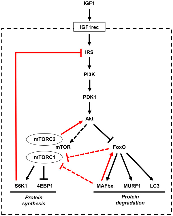
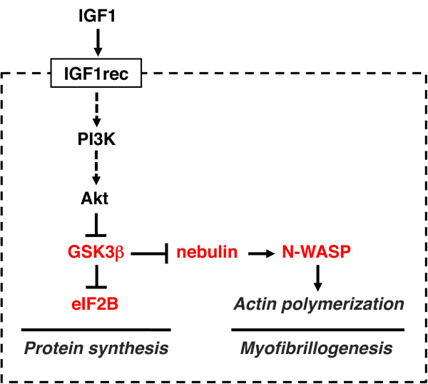
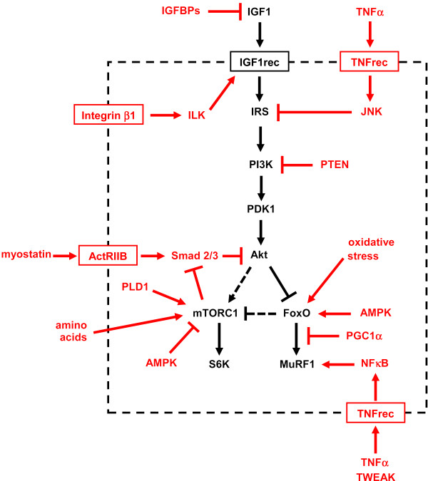
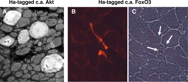
Verification
Claims the paper makes
The claims and statistics of this paper haven’t been checked yet.
Run the check — free, no account needed. It reads the open-access text and checks the sources it cites.
Claims other papers make about this one
Each is checked against this paper’s own text: does it say what the citing paper says it does?
The cited source says what the claim attributes to it.
In the citing paper
“The Akt‐mTOR pathway is central to the regulation of muscle mass because it promotes protein synthesis and inhibits protein degradation (Schiaffino & Mammucari, 2011).”
From Parkin overexpression protects from ageing‐related loss of muscle mass and strength (2019) · cites this paper as Schiaffino & Mammucari, 2011
In the citing paper
“Additionally, it has been discovered that PEs present in cosmetic preparation protect skin from pigmentation and aging163.”
From The Phytochemical, Biological, and Medicinal Attributes of Phytoecdysteroids: An Updated Review (2021) · cites this paper as [163]
Self-check
Percentages, sums and reported differences re-computed from the paper’s own numbers.
The self-check needs the paper’s full text.
Citations
Cites
Phosphorylation and regulation of Akt/PKB by the rictor-mTOR complex
Sarbassov, D. D., Guertin, D. A., Ali, S. M., & Sabatini, D. M. (2005) · Science · doi:10.1126/science.1106148 · Cited by 6,216
AKT/PKB signaling: navigating downstream
Manning, B. D., & Cantley, L. C. (2007) · Cell · doi:10.1016/j.cell.2007.06.009 · Cited by 6,115 · cited 1×
“transcription factors of the FoxO family, and stimulates protein synthesis via the mammalian target of rapamycin (mTOR) and glycogen synthase kinase 3b (GSK3b) [4].”
BackgroundRegulation of skeletal muscle mass in mice by a new TGF-p superfamily member
McPherron, A. C., Lawler, A. M., & Lee, S.-J. (1997) · Nature · doi:10.1038/387083a0 · Cited by 4,013
mTOR: from growth signal integration to cancer, diabetes and ageing
Zoncu, R., Efeyan, A., & Sabatini, D. M. (2011) · Nature Reviews Molecular Cell Biology · doi:10.1038/nrm3025 · Cited by 3,941 · cited 15×
“Deletion of S6K1 is sufficient to reproduce this atrophic phenotype.”
BackgroundInfluentialAMPK Phosphorylation of Raptor Mediates a Metabolic Checkpoint
Gwinn, D. M., Shackelford, D. B., Egan, D. F., Mihaylova, M. M., Mery, A., Vasquez, D. S., Turk, B. E., & Shaw, R. J. (2008) · Molecular Cell · doi:10.1016/j.molcel.2008.03.003 · Cited by 3,849 · cited 13×
“(AMPK), an energy status sensor activated by ATP depletion, inhibits mTORC1 by phosphorylating and inactivating Raptor [23], and by phosphorylating and activating TSC2 [24].”
BackgroundInfluentialIdentification of ubiquitin ligases required for skeletal muscle atrophy.
Bodine, S. C., Latres, E., Baumhueter, S., Lai, V. K.-M., Nunez, L., Clarke, B. A., Poueymirou, W. T., Panaro, F. J., Na, E., Dharmarajan, K., et al. (2001) · Science · doi:10.1126/science.1065874 · Cited by 3,521 · cited 5×
“Their role in muscle-mass regulation is supported by the finding that muscle atrophy induced by denervation is partially prevented in both MuRF1 null and atrogin1/MAFbx null mice [ 92 ].”
BackgroundInfluentialTSC2 is phosphorylated and inhibited by Akt and suppresses mTOR signalling
Inoki, K., Li, Y., Zhu, T., Wu, J., & Guan, K.-L. (2002) · Nature Cell Biology · doi:10.1038/ncb839 · Cited by 3,083 · cited 3×
“Rictor-dependent phosphorylation of Akt at serine 473 is required for Akt-mediated phosphorylation of FoxO but not of TSC2, thus it does not affect activation of S6K [5].”
BackgroundInfluentialSandri, M., Sandri, C., Gilbert, A., Skurk, C., Calabria, E., Picard, A., Walsh, K., Schiaffino, S., Lecker, S. H., & Goldberg, A. L. (2004) · Cell · doi:10.1016/s0092-8674(04)00400-3 · Cited by 3,012 · cited 5×
“Small interfering RNA to FoxO1 and FoxO3 Knockdown of FoxO1 and FoxO3 by RNA interference Prevention of atrogin-1 reporter upregulation induced by starvation [ 91 ]”
BackgroundInfluentialSelf-citationLiu, J.-P., Baker, J. C., Perkins, A. S., Robertson, E. J., & Efstratiadis, A. (1993) · Cell · doi:10.1016/s0092-8674(05)80084-4 · Cited by 2,854
The Rag GTPases Bind Raptor and Mediate Amino Acid Signaling to mTORC1
Sancak, Y., Peterson, T. R., Shaul, Y. D., Lindquist, R. A., Thoreen, C. C., Bar-Peled, L., & Sabatini, D. M. (2008) · Science · doi:10.1126/science.1157535 · Cited by 2,773 · cited 13×
“(AMPK), an energy status sensor activated by ATP depletion, inhibits mTORC1 by phosphorylating and inactivating Raptor [23], and by phosphorylating and activating TSC2 [24].”
BackgroundInfluential
Cited by
Sorted by citations
Mechanisms regulating skeletal muscle growth and atrophy
Schiaffino, S., Dyar, K. A., Ciciliot, S., Blaauw, B., & Sandri, M. (2013) · The FEBS Journal · doi:10.1111/febs.12253 · Cited by 1,492
Self-citationCellular and molecular mechanisms of muscle atrophy
Bonaldo, P., & Sandri, M. (2013) · Disease Models & Mechanisms · doi:10.1242/dmm.010389 · Cited by 1,284
“Insulin-like growth factor 1 (IGF1), a circulating growth factor, is also produced locally by many tissues, including skeletal muscle (reviewed in Schiaffino and Mammucari, 2011).”
Muscle wasting in disease: molecular mechanisms and promising therapies
Cohen, S., Nathan, J. A., & Goldberg, A. L. (2015) · Nature Reviews Drug Discovery · doi:10.1038/nrd4467 · Cited by 1,089
A PGC-1α isoform induced by resistance training regulates skeletal muscle hypertrophy
Ruas, J. L., White, J. P., Rao, R. R., Kleiner, S., Brannan, K. T., Harrison, B. C., Greene, N. P., Wu, J., Estall, J. L., Irving, B. A., et al. (2012) · Cell · doi:10.1016/j.cell.2012.10.050 · Cited by 677
Skeletal muscle: A review of molecular structure and function, in health and disease
Mukund, K., & Subramaniam, S. (2019) · WIREs Systems Biology and Medicine · doi:10.1002/wsbm.1462 · Cited by 789
“The major signaling pathway that regulates muscle mass and protein synthesis is the IGF1-Akt/PKB-mTOR signaling pathway (Schiaffino & Mammucari, 2011; Figure 12).”
BackgroundFrailty and sarcopenia: The potential role of an aged immune system
Wilson, D., Jackson, T., Sapey, E., & Lord, J. M. (2017) · Ageing Research Reviews · doi:10.1016/j.arr.2017.01.006 · Cited by 573
“Studies in rodents suggest that muscle synthesis is intrinsically linked with muscle atrophy via the PI3k-Akt pathway which is central to both processes (Schiaffino and Mammucari, 2011).”
mTOR as a key regulator in maintaining skeletal muscle mass
Yoon, M.-S. (2017) · Frontiers in Physiology · doi:10.3389/fphys.2017.00788 · Cited by 495
“In addition, Akt regulates muscle mass by phosphorylating and deactivating glycogen synthase kinase (GSK) β1, followed by the GSK β1-dependent inhibition of the eukaryotic translation initiation factor 2B (eIF2B) (Manning and Cantley, 2007; Schiaffino and Mammucari, 2011).”
BackgroundMolecular Choreography of Acute Exercise
Contrepois, K., Wu, S., Moneghetti, K. J., Hornburg, D., Ahadi, S., Tsai, M.-S., Metwally, A. A., Wei, E., Lee-McMullen, B., Quijada, J. V., et al. (2020) · Cell · doi:10.1016/j.cell.2020.04.043 · Cited by 502
Muscle wasting and cachexia in heart failure: mechanisms and therapies
von Haehling, S., Ebner, N., dos Santos, M. R., Springer, J., & Anker, S. D. (2017) · Nature Reviews Cardiology · doi:10.1038/nrcardio.2017.51 · Cited by 434
Muscle and bone, two interconnected tissues
Tagliaferri, C., Wittrant, Y., Davicco, M.-J., Walrand, S., & Coxam, V. (2015) · Ageing Research Reviews · doi:10.1016/j.arr.2015.03.002 · Cited by 406
Atrogin-1, MuRF-1, and sarcopenia
Gumucio, J. P., & Mendias, C. L. (2012) · Endocrine · doi:10.1007/s12020-012-9751-7 · Cited by 378
Dzik, K. P., & Kaczor, J. J. (2019) · European Journal of Applied Physiology · doi:10.1007/s00421-019-04104-x · Cited by 357
Sandri, M., Barberi, L., Bijlsma, A. Y., Blaauw, B., Dyar, K. A., Milan, G., Mammucari, C., Meskers, C. G. M., Pallafacchina, G., Paoli, A., et al. (2013) · Biogerontology · doi:10.1007/s10522-013-9432-9 · Cited by 340
Self-citationHuman skeletal muscle possesses an epigenetic memory of hypertrophy
Seaborne, R. A., Strauss, J., Cocks, M., Shepherd, S., O’Brien, T. D., van Someren, K. A., Bell, P. G., Murgatroyd, C., Morton, J. P., Stewart, C. E., et al. (2018) · Scientific Reports · doi:10.1038/s41598-018-20287-3 · Cited by 334
“Importantly, following reloading induced muscle growth we observed an increase in the number of epigenetically modified sites (27,155) and an enhanced number of hypomethylated DNA sites (18,816, Fig.”
BackgroundMolecular circuitry of stem cell fate in skeletal muscle regeneration, ageing and disease
Almada, A. E., & Wagers, A. J. (2016) · Nature Reviews Molecular Cell Biology · doi:10.1038/nrm.2016.7 · Cited by 322
Myostatin/activin pathway antagonism: molecular basis and therapeutic potential
Han, H. Q., Zhou, X., Mitch, W. E., & Goldberg, A. L. (2013) · The International Journal of Biochemistry & Cell Biology · doi:10.1016/j.biocel.2013.05.019 · Cited by 314
Vanweert, F., Schrauwen, P., & Phielix, E. (2022) · Nutrition and Diabetes · doi:10.1038/s41387-022-00213-3 · Cited by 331
Recessive mutations in EPG5 cause Vici syndrome, a multisystem disorder with defective autophagy
Cullup, T., Kho, A. L., Dionisi‐Vici, C., Brandmeier, B., Smith, F., Urry, Z., Simpson, M. A., Yau, S., Bertini, E., McClelland, V. M., et al. (2012) · Nature Genetics · doi:10.1038/ng.2497 · Cited by 287
“To see if these changes also involve upstream regulatory mechanisms of autophagy, we analysed the AKT-mTOR protein kinase pathway, a major pathway controlling the expression of autophagy proteins by inhibiting the transcriptional activity of FoxO transcription factors 35 .”
Muscle injuries and strategies for improving their repair
Laumonier, T., & Ménétrey, J. (2016) · Journal of Experimental Orthopaedics · doi:10.1186/s40634-016-0051-7 · Cited by 297
“IGF-I stimulates myoblasts proliferation and differentiation (Engert et al. 1996) and is implicated in the regulation of muscle growth (Schiaffino and Mammucari 2011).”
BackgroundMuscle Atrophy Induced by Mechanical Unloading: Mechanisms and Potential Countermeasures
Gao, Y., Arfat, Y., Wang, H., & Goswami, N. (2018) · Frontiers in Physiology · doi:10.3389/fphys.2018.00235 · Cited by 289
“Under normal physiological condition, the IGF-1-Akt-mTOR pathway acts as a key regulator in the translation initiation step of protein synthesis in skeletal muscle (Erbay et al., 2003; Han et al., 2008; Schiaffino and Mammucari, 2011).”
BackgroundEgan, B., & Sharples, A. P. (2023) · Physiological Reviews · doi:10.1152/physrev.00054.2021 · Cited by 278
Caloric restriction mimetics: towards a molecular definition
Madeo, F., Pietrocola, F., Eisenberg, T., & Kroemer, G. (2014) · Nature Reviews Drug Discovery · doi:10.1038/nrd4391 · Cited by 245
Mechanisms Modulating Skeletal Muscle Phenotype
Blaauw, B., Schiaffino, S., & Reggiani, C. (2013) · Comprehensive physiology · doi:10.1002/cphy.c130009 · Cited by 298
Self-citationStress-induced Skeletal Muscle Gadd45a Expression Reprograms Myonuclei and Causes Muscle Atrophy
Ebert, S. M., Dyle, M. C., Kunkel, S. D., Bullard, S. A., Bongers, K. S., Fox, D. K., Dierdorff, J. M., Foster, E. D., & Adams, C. M. (2012) · Journal of Biological Chemistry · doi:10.1074/jbc.m112.374777 · Cited by 221
Roberts, M. D., McCarthy, J. J., Hornberger, T. A., Phillips, S. M., Mackey, A. L., Nader, G. A., Boppart, M. D., Kavazis, A. N., Reidy, P. T., Ogasawara, R., et al. (2023) · Physiological Reviews · doi:10.1152/physrev.00039.2022 · Cited by 214
Genomic basis for the convergent evolution of electric organs
Gallant, J. R., Traeger, L. L., Volkening, J. D., Moffett, H. F., Chen, P.-H., Novina, C. D., Phillips, G. N., Anand, R., Wells, G. B., Pinch, M., et al. (2014) · Science · doi:10.1126/science.1254432 · Cited by 207
Fibro-adipogenic progenitors in skeletal muscle homeostasis, regeneration and diseases
Molina, T., Fabre, P., & Dumont, N. A. (2021) · Open Biology · doi:10.1098/rsob.210110 · Cited by 205
Luo, L., Lu, A.-M., Wang, Y., Hong, A., Chen, Y., Hu, J., Li, X., & Qin, Z.-H. (2013) · Experimental Gerontology · doi:10.1016/j.exger.2013.02.009 · Cited by 192
Gehlert, S., Bloch, W., & Suhr, F. (2015) · International Journal of Molecular Sciences · doi:10.3390/ijms16011066 · Cited by 200
The Mitochondrial Calcium Uniporter Controls Skeletal Muscle Trophism In Vivo
Mammucari, C., Gherardi, G., Zamparo, I., Raffaello, A., Boncompagni, S., Chemello, F., Cagnin, S., Braga, A., Zanin, S., Pallafacchina, G., et al. (2015) · Cell Reports · doi:10.1016/j.celrep.2015.01.056 · Cited by 190
“Specific AKT downstream targets were phosphorylated: in detail, 4E-BP1 and GSK3β, two inhibitors of protein translation (Schiaffino and Mammucari, 2011), were phosphorylated, and thus inhibited, in AAV-MCU muscles (Figure 3M).”
BackgroundSelf-citationLncIRS1 controls muscle atrophy via sponging miR‐15 family to activate IGF1‐PI3K/AKT pathway
Li, Z., Cai, B., Abdalla, B. A., Zhu, X., Zheng, M., Han, P., Nie, Q., & Zhang, X. (2019) · Journal of Cachexia Sarcopenia and Muscle · doi:10.1002/jcsm.12374 · Cited by 190
“IGF1-PI3K/AKT pathway is a key intracellular signalling mechanism controlling muscle hypertrophy.(7) In addition, evidence suggests that in complex organism development, RNA contains a hidden layer of regulatory information.”
BackgroundInfluentialUpdate on the effects of microgravity on the musculoskeletal system
Juhl, O. J., Buettmann, E. G., Friedman, M. A., DeNapoli, R. C., Hoppock, G. A., & Donahue, H. J. (2021) · npj Microgravity · doi:10.1038/s41526-021-00158-4 · Cited by 204
Regulation of Skeletal Muscle Glucose Transport and Glucose Metabolism by Exercise Training
Evans, P. L., McMillin, S. L., Weyrauch, L. A., & Witczak, C. A. (2019) · Nutrients · doi:10.3390/nu11102432 · Cited by 190
“Akt1 is a kinase found in skeletal muscle that plays a critical role in mediating resistance exercise training-induced muscle hypertrophic growth and protein synthesis (reviewed in [208,209]).”
BackgroundDifferential role of insulin/IGF-1 receptor signaling in muscle growth and glucose homeostasis
O’Neill, B. T., Lauritzen, H. P. M. M., Hirshman, M. F., Smyth, G., Goodyear, L. J., & Kahn, C. R. (2015) · Cell Reports · doi:10.1016/j.celrep.2015.04.037 · Cited by 171
“It has been known for some time that IGF-1 treatment can induce muscle hypertrophy via the Akt-mTOR pathways, yet previous studies have indicated that deletion of IGF1Rs alone in muscle only modestly changes myocyte size and morphology (Schiaffino and Mammucari, 2011; Mavalli et al., 2010).”
Pala, F., Di Girolamo, D., Mella, S., Yennek, S., Chatre, L., Ricchetti, M., & Tajbakhsh, S. (2018) · Journal of Cell Science · doi:10.1242/jcs.212977 · Cited by 184
“Depending on their fibre composition (fast type II and slow type I fibres), different muscles exhibit more glycolytic or oxidative activity (Schiaffino and Mammucari, 2011).”
BackgroundInsulin and IGF-1 receptors regulate FoxO-mediated signaling in muscle proteostasis
O'Neill, B. T., Lee, K. Y., Klaus, K. A., Softic, S., Krumpoch, M. T., Fentz, J., Stanford, K. I., Robinson, M. M., Cai, W., Kleinridders, A., et al. (2016) · Journal of Clinical Investigation · doi:10.1172/jci86522 · Cited by 185
Role of satellite cells in muscle growth and maintenance of muscle mass
Pallafacchina, G., Blaauw, B., & Schiaffino, S. (2012) · Nutrition Metabolism and Cardiovascular Diseases · doi:10.1016/j.numecd.2012.02.002 · Cited by 189
Self-citationThe Role of Reactive Oxygen Species in the Life Cycle of the Mitochondrion
Venditti, P., & Meo, S. D. (2020) · International Journal of Molecular Sciences · doi:10.3390/ijms21062173 · Cited by 176
Schwalm, C., Jamart, C., Benoit, N., Naslain, D., Prémont, C., Prévet, J., Thienen, R. V., Deldicque, L., & Francaux, M. (2015) · The FASEB Journal · doi:10.1096/fj.14-267187 · Cited by 174
“Indeed, beyond muscle damage, autophagy can be activated while the energetic status of the cell is impaired or in response to changes in hormonal environment (21, 22).”
BackgroundMolecular Mechanisms of Skeletal Muscle Hypertrophy
Schiaffino, S., Reggiani, C., Akimoto, T., & Blaauw, B. (2020) · Journal of Neuromuscular Diseases · doi:10.3233/jnd-200568 · Cited by 192
Self-citationSarcopenia and Cognitive Decline in Older Adults: Targeting the Muscle–Brain Axis
Arosio, B., Calvani, R., Ferri, E., Coelho-Junior, H. J., Carandina, A., Campanelli, F., Ghiglieri, V., Marzetti, E., & Picca, A. (2023) · Nutrients · doi:10.3390/nu15081853 · Cited by 165
“The PI3K–AKT–mTOR pathway is activated by this protein’s interaction with the insulin receptor substrate 1 (IRS1), which enhances muscle protein synthesis and prevents protein degradation [73].”
BackgroundInsulin-Like Growth Factor-1 Receptor Is Regulated by microRNA-133 during Skeletal Myogenesis
Huang, M., Xu, H. M., Xie, S., Zhou, H., & Qu, L. (2011) · PLoS ONE · doi:10.1371/journal.pone.0029173 · Cited by 166
“phosphorylation of Akt, which plays a key role in multiple cellular processes such as glucose metabolism, cell proliferation, differentiation, growth and apoptosis [25,26].”
BackgroundOno, Y., Maejima, Y., Saito, M., Sakamoto, K., Horita, S., Shimomura, K., Inoue, S., & Kotani, J. (2020) · Scientific Reports · doi:10.1038/s41598-020-57714-3 · Cited by 169
CURRENT NUTRITIONAL RECOMMENDATIONS AND NOVEL DIETARY STRATEGIES TO MANAGE SARCOPENIA
Calvani, R., Miccheli, A., Landi, F., Bossola, M., Cesari, M., Leeuwenburgh, C., Sieber, C. C., Bernabei, R., & Marzetti, E. (2013) · The Journal of Frailty & Aging · doi:10.14283/jfa.2013.7 · Cited by 164
Antioxidant and Adaptative Response Mediated by Nrf2 during Physical Exercise
Vargas-Mendoza, N., Morales‐González, Á., Madrigal‐Santillán, E., Madrigal‐Bujaidar, E., Álvarez‐González, I., García-Melo, L. F., Anguiano‐Robledo, L., Fregoso-Aguilar, T. A., & Morales‐González, J. A. (2019) · Antioxidants · doi:10.3390/antiox8060196 · Cited by 170
“Simultaneously, the activation of PGC-1α4 also promotes the synthesis of IGF-1, intervening in the formation of muscle mass [107].”
BackgroundInfluentialCell therapy to improve regeneration of skeletal muscle injuries
Qazi, T. H., Duda, G. N., Ort, M., Perka, C., Geißler, S., & Winkler, T. (2019) · Journal of Cachexia Sarcopenia and Muscle · doi:10.1002/jcsm.12416 · Cited by 160
Sestrin prevents atrophy of disused and aging muscles by integrating anabolic and catabolic signals
Segalés, J., Perdiguero, E., Serrano, A. L., Sousa-Victor, P., Ortet, L., Jardí, M., Budanov, A. V., Garcia-Prat, L., Sandri, M., Thomson, D. M., et al. (2020) · Nature Communications · doi:10.1038/s41467-019-13832-9 · Cited by 153
Bone and skeletal muscle: Key players in mechanotransduction and potential overlapping mechanisms
Goodman, C. A., Hornberger, T. A., & Robling, A. G. (2015) · Bone · doi:10.1016/j.bone.2015.04.014 · Cited by 163
“stages of growth and development and under various conditions that may include increased mechanical loading (for recent reviews on these topics, see [32–41]).”
BackgroundBarclay, R. D., Burd, N. A., Tyler, C. J., Tillin, N. A., & Mackenzie, R. W. (2019) · Frontiers in Nutrition · doi:10.3389/fnut.2019.00146 · Cited by 161
Myogenin is an essential regulator of adult myofibre growth and muscle stem cell homeostasis
Ganassi, M., Badodi, S., Wanders, K., Zammit, P. S., & Hughes, S. M. (2020) · eLife · doi:10.7554/elife.60445 · Cited by 159
Glucocorticoid Signaling: An Update from a Genomic Perspective
Sacta, M. A., Chinenov, Y. V., & Rogatsky, I. (2015) · Annual Review of Physiology · doi:10.1146/annurev-physiol-021115-105323 · Cited by 155
Gharahdaghi, N., Phillips, B. E., Szewczyk, N. J., Smith, K., Wilkinson, D. J., & Atherton, P. J. (2021) · Frontiers in Physiology · doi:10.3389/fphys.2020.621226 · Cited by 161
“…et al., 2016), neurogenesis (Trejo et al., 2001), and improved muscle strength with RET (Bjersing et al., 2017) by improving translational efficiency (Schiaffino and Mammucari, 2011) and satellite cell proliferation (Velloso, 2008) in muscle and in the central nervous system (CNS) (Mainardi et…”
BackgroundTang, H., Inoki, K., Lee, M. C., Wright, E., Khuong, A., Khuong, A., Sugiarto, S., Garner, M. S., Paik, J., DePinho, R. A., et al. (2014) · Science Signaling · doi:10.1126/scisignal.2004809 · Cited by 145
Functionally heterogeneous human satellite cells identified by single cell RNA sequencing
Barruet, E., Garcia, S. M., Striedinger, K., Wu, J., Lee, S., Byrnes, L. E., Wong, A., Sun, X., Tamaki, S. J., Brack, A. S., et al. (2020) · eLife · doi:10.7554/elife.51576 · Cited by 142
Experimental Models of Sarcopenia: Bridging Molecular Mechanism and Therapeutic Strategy
Mankhong, S., Kim, S., Moon, S., Kwak, H., Park, D., & Kang, J. (2020) · Cells · doi:10.3390/cells9061385 · Cited by 150
Regulation of ribosome biogenesis in skeletal muscle hypertrophy
Figueiredo, V. C., & McCarthy, J. J. (2019) · Physiology · doi:10.1152/physiol.00034.2018 · Cited by 140
Exosome-Mediated Benefits of Cell Therapy in Mouse and Human Models of Duchenne Muscular Dystrophy
Aminzadeh, M. A., Rogers, R. G., Fournier, M., Tobin, R. E., Guan, X., Childers, M. K., Andres, A. M., TAYLOR, D. J., Ibrahim, A. G.-E., Ding, X., et al. (2018) · Stem Cell Reports · doi:10.1016/j.stemcr.2018.01.023 · Cited by 142
“In physiologic muscle growth, insulin growth factor (IGF)-1 is commonly implicated as an upstream signal (Schiaffino and Mammucari, 2011), but the effects of CDC exosomes onmdx soleus muscle were independent of IGF-1 receptors (Figure 5E).”
BackgroundGoodman, C. A. (2013) · Reviews of physiology, biochemistry and pharmacology · doi:10.1007/112_2013_17 · Cited by 145
“, (Dardevet et al. 1996; Rommel et al. 2001; Glass 2010; Schiaffino and Mammucari 2011)].”
BackgroundRole of mTORC1 in mechanically induced increases in translation and skeletal muscle mass
Goodman, C. A. (2019) · Journal of Applied Physiology · doi:10.1152/japplphysiol.01011.2018 · Cited by 133
“the direct activation of mTORC1, further upstream activation of the phosphatidylinositol 3-kinase (PI3K)/Akt pathway by IGF-1 is also sufficient to activate rapamycin-sensitive mTORC1 signaling, increase protein synthesis, and induce skeletal muscle hypertrophy (28, 81).”
BackgroundInteraction Between Bone and Muscle in Older Persons with Mobility Limitations
Ferrucci, L., Baroni, M., Ranchelli, A., Lauretani, F., Maggio, M. G., Mecocci, P., & Ruggiero, C. (2014) · Current Pharmaceutical Design · doi:10.2174/13816128113196660690 · Cited by 136
Muscle wasting in heart failure: An overview
von Haehling, S., Steinbeck, L., Doehner, W., Springer, J., & Anker, S. D. (2013) · The International Journal of Biochemistry & Cell Biology · doi:10.1016/j.biocel.2013.04.025 · Cited by 129
Skeletal muscle glycoprotein 130's role in Lewis lung carcinoma–induced cachexia
Puppa, M. J., Gao, S., Narsale, A. A., & Carson, J. A. (2014) · The FASEB Journal · doi:10.1096/fj.13-240580 · Cited by 143
Role of Pro-inflammatory Cytokines in Regulation of Skeletal Muscle Metabolism: A Systematic Review
Sharma, B., & Dabur, R. (2018) · Current Medicinal Chemistry · doi:10.2174/0929867326666181129095309 · Cited by 129
Mitochondrial dysfunction and skeletal muscle atrophy: Causes, mechanisms, and treatment strategies
Kubat, G. B., Bouhamida, E., Ülger, Ö., Türkel, İ., Pedriali, G., Ramaccini, D., Ekinci, Ö., Özerkliğ, B., Atalay, Ö., Patergnani, S., et al. (2023) · Mitochondrion · doi:10.1016/j.mito.2023.07.003 · Cited by 134
Judge, S. M., Howard, T. M., Ahn, B., Ferreira, L. F., & Judge, A. R. (2013) · PLoS ONE · doi:10.1371/journal.pone.0062687 · Cited by 127
Myostatin signaling regulates Akt activity via the regulation of miR-486 expression
Hitachi, K., Nakatani, M., & Tsuchida, K. (2013) · The International Journal of Biochemistry & Cell Biology · doi:10.1016/j.biocel.2013.12.003 · Cited by 127
Parkin overexpression protects from ageing‐related loss of muscle mass and strength
Leduc‐Gaudet, J., Reynaud, O., Hussain, S. N., & Gouspillou, G. (2019) · The Journal of Physiology · doi:10.1113/jp277157 · Cited by 119
“The Akt-mTOR pathway is central to the regulation of muscle mass because it promotes protein synthesis and inhibits protein degradation (Schiaffino & Mammucari, 2011).”
BackgroundSubversion of Systemic Glucose Metabolism as a Mechanism to Support the Growth of Leukemia Cells
Ye, H., Adane, B., Khan, N., Alexeev, E. E., Nusbacher, N. M., Minhajuddin, M., Stevens, B. M., Winters, A. C., Lin, X., Ashton, J. M., et al. (2018) · Cancer Cell · doi:10.1016/j.ccell.2018.08.016 · Cited by 130
“IGF1 plays a similar role in glucose metabolism as insulin (Schiaffino and Mammucari, 2011).”
BackgroundDiet, exercise, and pharmacotherapy for sarcopenia in people with diabetes
Hashimoto, Y., Takahashi, F., Okamura, T., Hamaguchi, M., & Fukui, M. (2023) · Metabolism · doi:10.1016/j.metabol.2023.155585 · Cited by 126
Strasser, B., & Pesta, D. H. (2013) · BioMed Research International · doi:10.1155/2013/805217 · Cited by 135
“which results in increased protein synthesis [74].”
Molecular Mechanisms of Obesity-Induced Osteoporosis and Muscle Atrophy
Roy, B., Curtis, M. E., Fears, L., Nahashon, S. N., & Fentress, H. M. (2016) · Frontiers in Physiology · doi:10.3389/fphys.2016.00439 · Cited by 122
“This finding suggests the role of obesity as a positive modulator of IL-6R and IL-6 expression in adipose tissue (Sindhu et al., 2015).”
BackgroundEnhanced Skeletal Muscle for Effective Glucose Homeostasis
Yang, J. (2014) · Progress in molecular biology and translational science · doi:10.1016/b978-0-12-800101-1.00005-3 · Cited by 122
Epigenetics and developmental programming of welfare and production traits in farm animals
Sinclair, K. D., Rutherford, K., Wallace, J. M., Brameld, J. M., Stöger, R. J., Alberio, R., Sweetman, D., Gardner, D. S., Perry, V. E. A., Adam, C. L., et al. (2016) · Reproduction Fertility and Development · doi:10.1071/rd16102 · Cited by 124
“However, the exact mechanisms that lead to muscle growth, even for very well-characterised signals with long-established roles, such as the insulin-like growth factor (IGF) family (Schiaffino and Mammucari 2011), remain to be established.”
BackgroundSestrins are evolutionarily conserved mediators of exercise benefits
Kim, M., Sujkowski, A. L., Namkoong, S., Gu, B., Cobb, T., Kim, B. Y., Kowalsky, A. H., Cho, C., Semple, I. A., Ro, S., et al. (2020) · Nature Communications · doi:10.1038/s41467-019-13442-5 · Cited by 119
The Phytochemical, Biological, and Medicinal Attributes of Phytoecdysteroids: An Updated Review
Das, N., Mishra, S. K., Bishayee, A., Ali, E. S., & Bishayee, A. (2021) · Acta Pharmaceutica Sinica B · doi:10.1016/j.apsb.2020.10.012 · Cited by 115
“breviflora(117), decumbesterone A (157)(118) and ajugacetalsterone E (158)(119) isolated from Ajuga decumbens, 22-dehydrocyasterone-2glucoside (159), ajugacetalsterone A (160) and ajugacetalsterone B (161), isolated from Ajuga nipponensis(120), 25-hydroxy-atrotosterone A (162), 11-hydroxy-cyasterone (163), 11-hydroxys…”
BackgroundYang, Y. J., & Kim, D. J. (2021) · International Journal of Molecular Sciences · doi:10.3390/ijms22052604 · Cited by 119
Ohashi, K., Nagata, Y., Wada, E., Zammit, P. S., Shiozuka, M., & Matsuda, R. (2015) · Experimental Cell Research · doi:10.1016/j.yexcr.2015.03.003 · Cited by 114
“The phospho-tyrosine of IR allows activation of insulin receptor substrate-1 (IRS-1), which operates through several signaling pathways including the phosphoinositide 3-kinase (PI3K)/Akt cascade [4,5].”
Understanding the Role of Exercise in Cancer Cachexia Therapy
Hardee, J. P., Counts, B. R., & Carson, J. A. (2017) · American Journal of Lifestyle Medicine · doi:10.1177/1559827617725283 · Cited by 111
“nutrients, and mechanical loading to regulate protein synthesis.(91,92,97) Growth”
InfluentialVandoorne, T., Smet, S. D., Ramaekers, M., Thienen, R. V., Bock, K. D., Clarke, K., & Hespel, P. (2017) · Frontiers in Physiology · doi:10.3389/fphys.2017.00310 · Cited by 116
“Muscle protein synthesis following exercise is mainly controlled by mTORC1 (Bodine et al., 2001; Schiaffino and Mammucari, 2011).”
BackgroundInfluentialOut of Control: The Role of the Ubiquitin Proteasome System in Skeletal Muscle during Inflammation
Haberecht-Müller, S., Krüger, E., & Fielitz, J. (2021) · Biomolecules · doi:10.3390/biom11091327 · Cited by 108
Autophagy as a Potential Target for Sarcopenia: AUTOPHAGY REGULATION IN SARCOPENIA
Fan, J., Kou, X., Jia, S., Yang, X., Yang, Y., & Chen, N. (2016) · Journal of Cellular Physiology · doi:10.1002/jcp.25260 · Cited by 101
“For instance, the activation of Akt signaling by insulin or IGF-1 inactivates FoxO-dependent transcription (Schiaffino and Mammucari, 2011), and mice with the musclespecific loss of IGF-1 receptor have reduced mass and fiber number of skeletal muscle (Mavalli et al., 2010), suggesting that…”
BackgroundRoberts, M. D., Haun, C. T., Mobley, C. B., Mumford, P. W., Romero, M. A., Roberson, P. A., Vann, C. G., & McCarthy, J. J. (2018) · Frontiers in Physiology · doi:10.3389/fphys.2018.00834 · Cited by 107
Damiano, S., Muscariello, E., Rosa, G. L., Maro, M. D., Mondola, P., & Santillo, M. (2019) · International Journal of Molecular Sciences · doi:10.3390/ijms20153815 · Cited by 116
Kuttappan, V. A., Bottje, W. G., Ramnathan, R., Hartson, S. D., Coon, C. N., Kong, B. C., Owens, C. M., Vazquez-Anon, M., & Hargis, B. M. (2017) · Poultry Science · doi:10.3382/ps/pex069 · Cited by 103
Gheller, B. J. F., Riddle, E. S., Lem, M. R., & Thalacker-Mercer, A. E. (2016) · Annual Review of Nutrition · doi:10.1146/annurev-nutr-071715-050901 · Cited by 98
The clinical impact and biological mechanisms of skeletal muscle aging
Aversa, Z., Zhang, X., Fielding, R. A., Lanza, I. R., & LeBrasseur, N. K. (2019) · Bone · doi:10.1016/j.bone.2019.05.021 · Cited by 113
Molecular Mechanisms of Bone Metastasis and Associated Muscle Weakness
Waning, D. L., & Guise, T. A. (2014) · Clinical Cancer Research · doi:10.1158/1078-0432.ccr-13-1590 · Cited by 101
MicroRNA Profile and Adaptive Response to Exercise Training: A Review
Domańska-Senderowska, D., Laguette, M.-J. N., Jegier, A., Cięszczyk, P., September, A. V., & Brzeziańska‐Lasota, E. (2019) · International Journal of Sports Medicine · doi:10.1055/a-0824-4813 · Cited by 104
“The IGF1/PI3K/AKT/mTOR signalling pathway is activated by several factors under various conditions such as growth, glucose homeostasis regulators and/or muscle atrophy/hypertrophy factors that are especially secreted during resistance training [45].”
BackgroundInfluentialde Sire, R., Rizzatti, G., Ingravalle, F., Pizzoferrato, M., Petito, V., Lopetuso, L. R., Graziani, C., Alessandro de, S. I. R. E., Mentella, M. C., Mele, M. C., et al. (2018) · Minerva Gastroenterologica e Dietologica · doi:10.23736/s1121-421x.18.02511-4 · Cited by 93
The AKT/mTOR signaling pathway plays a key role in statin-induced myotoxicity
Bonifacio, A., Sanvee, G. M., Bouitbir, J., & Krähenbühl, S. (2015) · Biochimica et Biophysica Acta (BBA) - Molecular Cell Research · doi:10.1016/j.bbamcr.2015.04.010 · Cited by 93
“AKT functions as a key regulator of both protein synthesis and degradation by activating the protein kinase mammalian target of rapamycin (mTOR) [15] and by inhibiting forkhead box O (FoxO) transcription factors [16].”
BackgroundLeucine as a treatment for muscle wasting: a critical review.
Ham, D. J., Caldow, M., Lynch, G., & Koopman, R. (2014) · Clinical Nutrition · doi:10.1016/j.clnu.2014.09.016 · Cited by 90
“This fact, especially in the light of robust increases in protein synthesis and activation of mTORC1 with leucine treatment, raises a more pertinent question: can chronic activation of mTORC1 increase muscle mass? Genetic overexpression of Akt in mouse skeletal muscle has been shown repeatedly to induce marked skeleta…”
BackgroundMurine models of atrophy, cachexia, and sarcopenia in skeletal muscle
Romanick, M. A., Thompson, L. V., & Brown‐Borg, H. M. (2013) · Biochimica et Biophysica Acta (BBA) - Molecular Basis of Disease · doi:10.1016/j.bbadis.2013.03.011 · Cited by 104
Nair, V. D., Ge, Y., Li, S., Pinças, H., Jain, N., Seenarine, N., Amper, M. A. S., Goodpaster, B. H., Walsh, M. J., Coen, P. M., et al. (2020) · Frontiers in Physiology · doi:10.3389/fphys.2020.00605 · Cited by 92
Oxidative Stress in Brain Function
Trofin, D. M., Sardaru, D.-P., Trofin, D., Onu, I., Tutu, A., Onu, A., Onita, C. A., Galaction, A., & Matei, D. V. (2025) · Antioxidants · doi:10.3390/antiox14030297 · Cited by 105
Quantitative measures of healthy aging and biological age
Kim, S., & Jazwinski, S. M. (2015) · Healthy Aging Research · doi:10.12715/har.2015.4.26 · Cited by 93
“Of these, IGF1 has the potential to affect RMR by inducing skeletal muscle growth through activation of the Akt-mTOR pathway [50, 51].”
BackgroundRavenscroft, G., Laing, N. G., & Bönnemann, C. G. (2014) · Brain · doi:10.1093/brain/awu368 · Cited by 94
“Skeletal muscle bulk is determined by the net effect of hypertrophy and atrophy processes: protein synthesis and degradation (comprising atrophy and autophagy) (reviewed in Schiaffino and Mammucari, 2011).”
BackgroundMolinari, F., Pin, F., Gorini, S., Chiandotto, S., Pontecorvo, L., Penna, F., Rizzuto, E., Pisu, S., Musarò, A., Costelli, P., et al. (2017) · Journal of Cachexia Sarcopenia and Muscle · doi:10.1002/jcsm.12226 · Cited by 88
Cunha, P. M., Nunes, J. P., Tomeleri, C. M., do Nascimento, M. A., Schoenfeld, B. J., Antunes, M., Gobbo, L. A., Teixeira, D. D. C., & Cyrino, E. S. (2018) · The Journal of Strength and Conditioning Research · doi:10.1519/jsc.0000000000002847 · Cited by 87
“However, although the IGF-1/ AKT/PI3K pathway is obligatory for hypertrophy during human maturation, the extent of its involvement in muscular adaptations to regimented RT remains unclear (33).”
BackgroundSarcopenia, cachexia, and muscle performance in heart failure: Review update 2016
Saitoh, M., Ishida, J., Doehner, W., von Haehling, S., Anker, M. S., Coats, A. J. S., Anker, S. D., & Springer, J. (2017) · International Journal of Cardiology · doi:10.1016/j.ijcard.2017.03.155 · Cited by 85
Bekfani, T., Bekhite, M. M., Derlien, S., Nisser, J., Westermann, M., Nietzsche, S., Hamadanchi, A., Fröb, E., Westphal, J. G., Haase, D., et al. (2020) · Circulation Heart Failure · doi:10.1161/circheartfailure.120.007198 · Cited by 88
Metadata
- Authors
- Schiaffino, S., & Mammucari, C.
- Affiliations
- Veneto Institute of Molecular Medicine (IT)
- University of Padua (IT)
- Corresponding author
- Schiaffino, Stefano
- Venue
- Skeletal Muscle
- Published
- 2011
- Volume
- 1
- Issue
- 1
- Pages
- 4
- ISSN
- 2044-5040 (ISSN-L)
- PMC
- PMC3143906
- OpenAlex
- W2150059080
- Cited by
- 816
- References
- 150
- Citations by year
- 2026: 42 · 2025: 70 · 2024: 68 · 2023: 63 · 2022: 55 · 2021: 59 · 2020: 56 · 2019: 69 · 2018: 68 · 2017: 57 · 2016: 42 · 2015: 46 · 2014: 48 · 2013: 44 · 2012: 23
- FWCI
- 15.73
- Type
- journal article
- Language
- en
- Open access
- gold
- doi.org
- Licence
- cc-by
- Locations
- Skeletal Muscle (publishedVersion, cc-by)
- Europe PMC (PubMed Central) (publishedVersion, cc-by)
- PubMed Central (publishedVersion, cc-by)
- PubMed (publishedVersion)
- Padua Research Archive (University of Padova) (submittedVersion, cc-by)
- DOAJ (DOAJ: Directory of Open Access Journals) (submittedVersion)
- Funders
- European Commission
- Università degli Studi di Padova
- Topics
- Life Sciences › Biochemistry, Genetics and Molecular Biology › Molecular Biology › Muscle Physiology and Disorders
- Health Sciences › Nursing › Nutrition and Dietetics › Muscle metabolism and nutrition
- Life Sciences › Biochemistry, Genetics and Molecular Biology › Molecular Biology › Metabolism, Diabetes, and Cancer
- Keywords
- skeletal muscle growth, IGF-1 signaling, mTOR, GSK3β, FOXO transcription factors, muscle regeneration, myogenesis, mechanical loading, muscle atrophy, muscle protein synthesis, protein degradation
About these checks
Registry, retraction, Cites and statistics results are computed, not judged, with one exception: a printed reference that no index lists is read once by a model before it counts as a citation error. Claims are extracted by a model and kept only when their quote appears verbatim in the text; each cited source is then checked, with a model’s help, for whether it says what the claim relies on. Those verdicts can be wrong — read the claim’s quote and the cited source before relying on one.
Statistics and arithmetic are recomputed, not judged.
Confidence bands are shown only for fields calibrated on our annotated test set. None is calibrated yet, so no band is shown.
What counts as a claim ›How we measure confidence ›How we check this ›
Are you an author, or do you see a problem with this page? Request a review
