

The DOI matches its Crossref record; 148 of the 150 references it cites were found in a registry or an index.
Regulation of skeletal muscle growth by the IGF1-Akt/PKB pathway: insights from genetic models
Open accessAbstract
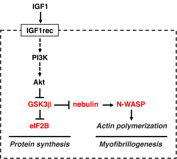
A highly conserved signaling pathway involving insulin-like growth factor 1 (IGF1), and a cascade of intracellular components that mediate its effects, plays a major role in the regulation of skeletal muscle growth. A central component in this cascade is the kinase Akt, also called protein kinase B (PKB), which controls both protein synthesis, via the kinases mammalian target of rapamycin (mTOR) and glycogen synthase kinase 3β (GSK3β), and protein degradation, via the transcription factors of the FoxO family. In this paper, we review the composition and function of this pathway in skeletal muscle fibers, focusing on evidence obtained in vivo by transgenic and knockout models and by muscle transient transfection experiments. Although this pathway is essential for muscle growth during development and regeneration, its role in adult muscle response to mechanical load is less clear. A full understanding of the operation of this pathway could help to design molecularly targeted therapeutics aimed at preventing muscle wasting, which occurs in a variety of pathologic contexts and in the course of aging.
Overview
The claims and statistics of this paper haven’t been checked yet.
Run the check — free, no account needed. It reads the open-access text and checks the sources it cites.
Integrity signals
Registry
Cites
Statistics
Figures
Verification
Claims the paper makes
The claims and statistics of this paper haven’t been checked yet.
Run the check — free, no account needed. It reads the open-access text and checks the sources it cites.
Claims other papers make about this one
Each is checked against this paper’s own text: does it say what the citing paper says it does?
The cited source says what the claim attributes to it.
In the citing paper
“The Akt‐mTOR pathway is central to the regulation of muscle mass because it promotes protein synthesis and inhibits protein degradation (Schiaffino & Mammucari, 2011).”
From Parkin overexpression protects from ageing‐related loss of muscle mass and strength (2019) · cites this paper as Schiaffino & Mammucari, 2011
In the citing paper
“Additionally, it has been discovered that PEs present in cosmetic preparation protect skin from pigmentation and aging163.”
From The Phytochemical, Biological, and Medicinal Attributes of Phytoecdysteroids: An Updated Review (2021) · cites this paper as [163]
Self-check
Percentages, sums and reported differences re-computed from the paper’s own numbers.
The self-check needs the paper’s full text.
Citations
Cites
Phosphorylation and regulation of Akt/PKB by the rictor-mTOR complex
Sarbassov, D. D., Guertin, D. A., Ali, S. M., & Sabatini, D. M. (2005) · Science · doi:10.1126/science.1106148 · Cited by 6,216
AKT/PKB signaling: navigating downstream
Manning, B. D., & Cantley, L. C. (2007) · Cell · doi:10.1016/j.cell.2007.06.009 · Cited by 6,115 · cited 1×
“transcription factors of the FoxO family, and stimulates protein synthesis via the mammalian target of rapamycin (mTOR) and glycogen synthase kinase 3b (GSK3b) [4].”
BackgroundRegulation of skeletal muscle mass in mice by a new TGF-p superfamily member
McPherron, A. C., Lawler, A. M., & Lee, S.-J. (1997) · Nature · doi:10.1038/387083a0 · Cited by 4,013
mTOR: from growth signal integration to cancer, diabetes and ageing
Zoncu, R., Efeyan, A., & Sabatini, D. M. (2011) · Nature Reviews Molecular Cell Biology · doi:10.1038/nrm3025 · Cited by 3,941 · cited 15×
“Deletion of S6K1 is sufficient to reproduce this atrophic phenotype.”
BackgroundInfluentialAMPK Phosphorylation of Raptor Mediates a Metabolic Checkpoint
Gwinn, D. M., Shackelford, D. B., Egan, D. F., Mihaylova, M. M., Mery, A., Vasquez, D. S., Turk, B. E., & Shaw, R. J. (2008) · Molecular Cell · doi:10.1016/j.molcel.2008.03.003 · Cited by 3,849 · cited 13×
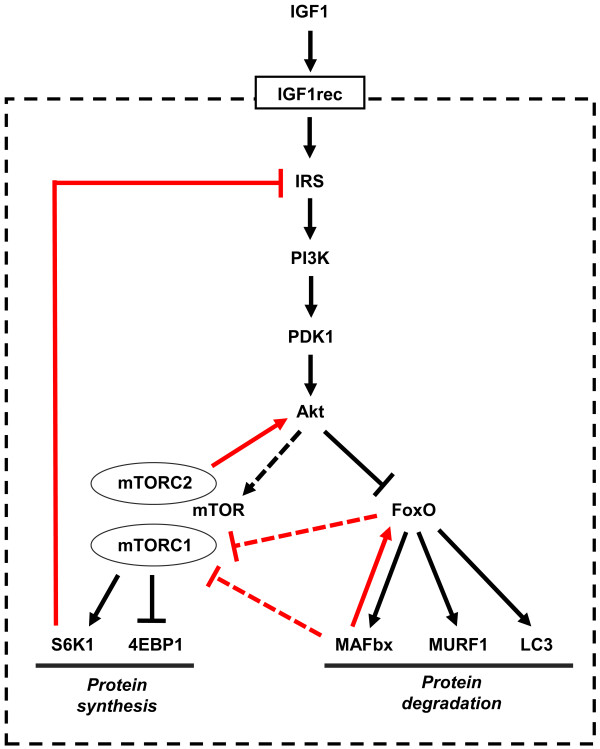
“(AMPK), an energy status sensor activated by ATP depletion, inhibits mTORC1 by phosphorylating and inactivating Raptor [23], and by phosphorylating and activating TSC2 [24].”
BackgroundInfluentialIdentification of ubiquitin ligases required for skeletal muscle atrophy.
Bodine, S. C., Latres, E., Baumhueter, S., Lai, V. K.-M., Nunez, L., Clarke, B. A., Poueymirou, W. T., Panaro, F. J., Na, E., Dharmarajan, K., et al. (2001) · Science · doi:10.1126/science.1065874 · Cited by 3,521 · cited 5×
“Their role in muscle-mass regulation is supported by the finding that muscle atrophy induced by denervation is partially prevented in both MuRF1 null and atrogin1/MAFbx null mice [ 92 ].”
BackgroundInfluentialTSC2 is phosphorylated and inhibited by Akt and suppresses mTOR signalling
Inoki, K., Li, Y., Zhu, T., Wu, J., & Guan, K.-L. (2002) · Nature Cell Biology · doi:10.1038/ncb839 · Cited by 3,083 · cited 3×
“Rictor-dependent phosphorylation of Akt at serine 473 is required for Akt-mediated phosphorylation of FoxO but not of TSC2, thus it does not affect activation of S6K [5].”
BackgroundInfluentialSandri, M., Sandri, C., Gilbert, A., Skurk, C., Calabria, E., Picard, A., Walsh, K., Schiaffino, S., Lecker, S. H., & Goldberg, A. L. (2004) · Cell · doi:10.1016/s0092-8674(04)00400-3 · Cited by 3,012 · cited 5×
“Small interfering RNA to FoxO1 and FoxO3 Knockdown of FoxO1 and FoxO3 by RNA interference Prevention of atrogin-1 reporter upregulation induced by starvation [ 91 ]”
BackgroundInfluentialSelf-citationLiu, J.-P., Baker, J. C., Perkins, A. S., Robertson, E. J., & Efstratiadis, A. (1993) · Cell · doi:10.1016/s0092-8674(05)80084-4 · Cited by 2,854
The Rag GTPases Bind Raptor and Mediate Amino Acid Signaling to mTORC1
Sancak, Y., Peterson, T. R., Shaul, Y. D., Lindquist, R. A., Thoreen, C. C., Bar-Peled, L., & Sabatini, D. M. (2008) · Science · doi:10.1126/science.1157535 · Cited by 2,773 · cited 13×
“(AMPK), an energy status sensor activated by ATP depletion, inhibits mTORC1 by phosphorylating and inactivating Raptor [23], and by phosphorylating and activating TSC2 [24].”
BackgroundInfluentialBodine, S. C., Stitt, T. N., Gonzalez, M., Kline, W. O., Stover, G. L., Bauerlein, R., Zlotchenko, E., Scrimgeour, A., Lawrence, J. C., Glass, D. J., et al. (2001) · Nature Cell Biology · doi:10.1038/ncb1101-1014 · Cited by 2,752 · cited 4×
“Activation of PI3K Muscle fiber hypertrophy [69, 78 ]”
BackgroundInfluentialRole of insulin-like growth factors in embryonic and postnatal growth
Baker, J., Liu, J.-P., Robertson, E. J., & Efstratiadis, A. (1993) · Cell · doi:10.1016/s0092-8674(05)80085-6 · Cited by 2,463 · cited 1×
“Igf1 null mice exhibit severe growth retardation and most die soon after birth [37-39], whereas targeted ablation experiments showed that liver-derived IGF1, although it is the principal source of IGF1 in the serum, is not required for postnatal body growth [40,41].”
BackgroundStitt, T. N., Drujan, D., Clarke, B. A., Panaro, F., Timofeyva, Y., Kline, W. O., Gonzalez, M., Yancopoulos, G. D., & Glass, D. J. (2004) · Molecular Cell · doi:10.1016/s1097-2765(04)00211-4 · Cited by 1,964 · cited 1×
“Direct evidence for FoxO-dependent activation of the ubiquitin-proteasome pathway was obtained with the demonstration that the muscle-specific ubiquitin ligases atrogin-1/MAFbx and MuRF1, which are induced in various models of muscle atrophy [92,93], are transcriptional targets of FoxO factors, and are upregulated by…”
BackgroundFoxO3 Controls Autophagy in Skeletal Muscle In Vivo
Mammucari, C., Milan, G., Romanello, V., Masiero, E., Rudolf, R., Del Piccolo, P., Burden, S. J., Di Lisi, R., Sandri, C., Zhao, J., et al. (2007) · Cell Metabolism · doi:10.1016/j.cmet.2007.11.001 · Cited by 1,953 · cited 4×
“An unexpected result was the finding that mTOR, which is considered as a major regulator of autophagy in different cell systems, does not appear to play a major role in muscle autophagy, as treatment with the mTOR inhibitor rapamycin has a limited effect on autophagy in skeletal muscle both in vitro and in vivo [ 81 ,…”
BackgroundInfluentialSelf-citationCho, H., Mu, J., Kim, J. K., Thorvaldsen, J. L., Chu, Q., Crenshaw, E. B., Kaestner, K. H., Bartolomei, M. S., Shulman, G. I., & Birnbaum, M. J. (2001) · Science · doi:10.1126/science.292.5522.1728 · Cited by 1,860 · cited 2×
“Disruption of the Akt1 gene causes growth retardation and apoptosis [74,75], whereas deletion of Akt2 causes defects in glucose metabolism but not altered growth [ 76 ].”
BackgroundDisruption of IRS-2 causes type 2 diabetes in mice
Withers, D. J., Gutierrez, J. S., Towery, H. H., Burks, D. J., Ren, J., Previs, S. F., Zhang, Y., Bernal, D., Pons, S., Shulman, G. I., et al. (1998) · Nature · doi:10.1038/36116 · Cited by 1,775 · cited 2×
“IRS2 null Viable Almost normal growth (birth weight 90% of control) [ 65 ]”
BackgroundAtrogin-1, a muscle-specific F-box protein highly expressed during muscle atrophy
Gomes, M. D., Lecker, S. H., Jagoe, R. T., Navon, A., & Goldberg, A. L. (2001) · Proceedings of the National Academy of Sciences · doi:10.1073/pnas.251541198 · Cited by 1,774 · cited 1×
“Direct evidence for FoxO-dependent activation of the ubiquitin-proteasome pathway was obtained with the demonstration that the muscle-specific ubiquitin ligases atrogin-1/MAFbx and MuRF1, which are induced in various models of muscle atrophy [92, 93 ], are transcriptional targets of FoxO factors, and are upregulated b…”
BackgroundRommel, C., Bodine, S. C., Clarke, B. A., Rossman, R., Nunez, L., Stitt, T. N., Yancopoulos, G. D., & Glass, D. J. (2001) · Nature Cell Biology · doi:10.1038/ncb1101-1009 · Cited by 1,583
Zhao, J., Brault, J. J., Schild, A., Cao, P., Sandri, M., Schiaffino, S., Lecker, S. H., & Goldberg, A. L. (2007) · Cell Metabolism · doi:10.1016/j.cmet.2007.11.004 · Cited by 1,489 · cited 2×
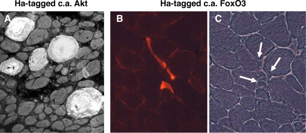
“The role of FoxO3 as an inducer of autophagy is supported by the finding that transfection of adult muscle fibers with constitutively active FoxO3 causes accumulation of autophagic vacuoles, whereas fasting-induced autophagy is blocked by dominant-negative FoxO3 and by RNA interference-mediated FoxO3 knockdown [81, 10…”
BackgroundSelf-citationGuertin, D. A., Stevens, D. M., Thoreen, C. C., Burds, A. A., Kalaany, N. Y., Moffat, J., Brown, M., Fitzgerald, K. J., & Sabatini, D. M. (2006) · Developmental Cell · doi:10.1016/j.devcel.2006.10.007 · Cited by 1,467 · cited 1×
“TORC2 is required for Akt phosphorylation and activation [6].”
BackgroundNormal growth and development in the absence of hepatic insulin-like growth factor I
Yakar, S., Liu, J., STANNARD, B. S., Butler, A. A., Accili, D., Sauer, B., & LeRoith, D. (1999) · Proceedings of the National Academy of Sciences · doi:10.1073/pnas.96.13.7324 · Cited by 1,418 · cited 2×
“This discrepancy was explained by the fact that a residual fraction of circulating IGF1 is detected in the liver-specific knockout [40,41] because of incomplete Igf1 gene excision [43].”
BackgroundFruman, D. A., Meyers, R. E., & Cantley, L. C. (1998) · Annual Review of Biochemistry · doi:10.1146/annurev.biochem.67.1.481 · Cited by 1,382
IKKβ/NF-κB Activation Causes Severe Muscle Wasting in Mice
Cai, D., Frantz, J. D., Tawa, N. E., Melendez, P. A., Oh, B.-C., Lidov, H. G. W., Hasselgren, P.-O., Frontera, W. R., Lee, J., Glass, D. J., et al. (2004) · Cell · doi:10.1016/j.cell.2004.09.027 · Cited by 1,367
Regulation of TORC1 by Rag GTPases in nutrient response
Kim, E., Goraksha-Hicks, P., Li, L., Neufeld, T. P., & Guan, K.-L. (2008) · Nature Cell Biology · doi:10.1038/ncb1753 · Cited by 1,349 · cited 13×
“(AMPK), an energy status sensor activated by ATP depletion, inhibits mTORC1 by phosphorylating and inactivating Raptor [23], and by phosphorylating and activating TSC2 [24].”
BackgroundInfluentialAutophagy is required to maintain muscle mass.
Masiero, E., Agatea, L., Mammucari, C., Blaauw, B., Loro, E., Komatsu, M., Metzger, D., Reggiani, C., Schiaffino, S., & Sandri, M. (2009) · Cell Metabolism · doi:10.1016/j.cmet.2009.10.008 · Cited by 1,282 · cited 2×
“Moreover, inhibition of autophagy exacerbated muscle loss during denervation and fasting, suggesting that the persistence of dysfunctional organelles affects the progression of muscle atrophy [ 102 ].”
BackgroundSelf-citationAlternative pathway of insulin signalling in mice with targeted disruption of the IRS-1 gene
Araki, E., Lipes, M. A., Patti, M.-E., Brüning, J. C., Haag III, B., Johnson, R. S., & Kahn, C. R. (1994) · Nature · doi:10.1038/372186a0 · Cited by 1,252 · cited 2×
“IRS1 null Viable Reduced growth (weight 30-60% of control) [ 64 ,122]”
BackgroundGrowth Hormone and the Insulin-Like Growth Factor System in Myogenesis*
Florini, J. R., Ewton, D. Z., & Coolican, S. A. (1996) · Endocrine Reviews · doi:10.1210/edrv-17-5-481 · Cited by 1,148 · cited 1×
“The role of the IGF1-Akt pathway in adult skeletal muscle There is no doubt that IGF1 is a major regulator of muscle mass during development, thanks to its effect on myogenic cell proliferation and differentiation [ 103 ].”
BackgroundMusarò, A., McCullagh, K., Paul, A., Houghton, L., Dobrowolny, G., Molinaro, M., Barton, E. R., L Sweeney, H., & Rosenthal, N. (2001) · Nature Genetics · doi:10.1038/84839 · Cited by 1,137 · cited 2×
“The effect of an IGF1 isoform (IGF1A) was investigated in a transgenic mouse model, in which expression of this isoform was driven by the myosin light chain 1 fast promoter [51].”
BackgroundThe TSC1-2 tumor suppressor controls insulin–PI3K signaling via regulation of IRS proteins
Harrington, L. S., Findlay, G. M., Gray, A., Толкачева, Т. А., Wigfield, S., Rebholz, H., Barnett, J., Leslie, N. R., Cheng, S., Shepherd, P. R., et al. (2004) · The Journal of Cell Biology · doi:10.1083/jcb.200403069 · Cited by 1,123 · cited 1×
“Negative feedback involves S6K, which inhibits IRS by phosphorylation at multiple sites, thus inducing its degradation and altered cell localization [7].”
BackgroundInsulin resistance and growth retardation in mice lacking insulin receptor substrate-1
Tamemoto, H., Kadowaki, T., Tobe, K., Yagi, T., Sakura, H., Hayakawa, T., Terauchi, Y., Ueki, K., Kaburagi, Y., Satoh, S., et al. (1994) · Nature · doi:10.1038/372182a0 · Cited by 1,081 · cited 1×
“IRS1 null Viable Reduced growth (weight 30-60% of control) [64, 122 ]”
BackgroundFoxOs are lineage-restricted redundant tumor suppressors and regulate endothelial cell homeostasis
Paik, J.-H., Kollipara, R., Chu, G., Ji, H., Xiao, Y., Ding, Z., Miao, L., Tothova, Z., Horner, J. W., Carrasco, D. R., et al. (2007) · Cell · doi:10.1016/j.cell.2006.12.029 · Cited by 1,032 · cited 2×
“FoxO3 null Viable but female sterility Normal growth [133,36] FoxO4 null Viable Normal growth [ 137 ]”
BackgroundSatellite cells as the source of nuclei in muscles of growing rats
Moss, F. P., & Leblond, C. P. (1971) · The Anatomical Record · doi:10.1002/ar.1091700405 · Cited by 1,025 · cited 1×
“Satellite-cell proliferation and fusion leading to increase in myonuclei occurs during postnatal muscle growth [ 110 ] and during compensatory hypertrophy induced by ablation of synergistic muscles [111], as demonstrated by electron microscope autoradiographs after 3 H-thymidine labeling.”
BackgroundSuppression of Ovarian Follicle Activation in Mice by the Transcription Factor Foxo3a
Castrillón, D. H., Miao, L., Kollipara, R., Horner, J. W., & DePinho, R. A. (2003) · Science · doi:10.1126/science.1086336 · Cited by 984
Sandri, M., Lin, J., Handschin, C., Yang, W., Arany, Z. P., Lecker, S. H., Goldberg, A. L., & Spiegelman, B. M. (2006) · Proceedings of the National Academy of Sciences · doi:10.1073/pnas.0607795103 · Cited by 974 · cited 1×
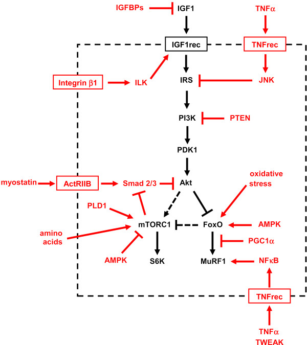
“PGC1a transfection protects adult muscles from FoxO-dependent atrophy, and the higher levels of PGC1a in oxidative muscle fibers can explain their greater resistance to muscle atrophy [31].”
BackgroundReversal of Cancer Cachexia and Muscle Wasting by ActRIIB Antagonism Leads to Prolonged Survival
Zhou, X., Wang, J. L., Lu, J., Song, Y., Kwak, K. S., Jiao, Q., Rosenfeld, R., Chen, Q., Boone, T. C., Simonet, W. S., et al. (2010) · Cell · doi:10.1016/j.cell.2010.07.011 · Cited by 960 · cited 1×
“In adult skeletal muscle, muscle hypertrophy can be induced and muscle wasting prevented by blocking myostatin either via postdevelopmental myostatin gene knockout, or with follistatin (a myostatin antagonist) or anti-myostatin antibodies or a soluble ActRIIB decoy receptor (see [19,20] and references therein).”
BackgroundCho, H., Thorvaldsen, J. L., Chu, Q., Feng, F., & Birnbaum, M. J. (2001) · Journal of Biological Chemistry · doi:10.1074/jbc.c100462200 · Cited by 959
Growth retardation and increased apoptosis in mice with homozygous disruption of the akt1 genePreprint
Chen, W. S., Xu, P.-Z., Gottlob, K., Chen, M.-L., Sokol, K. A., Shiyanova, T., Roninson, I. B., Weng, W., Suzuki, R., Tobe, K., et al. (2001) · Genes & Development · doi:10.1101/gad.913901 · Cited by 941 · cited 2×
“Akt1 null Viable but shorter life span Mild growth retardation (weight 80% of control) [74, 75 ]”
BackgroundSjögren, K., Liu, J., Blad, K., Skrtic, S., Vidal, O., Wallenius, V., LeRoith, D., Törnell, J., Isaksson, O. G. P., Jansson, J., et al. (1999) · Proceedings of the National Academy of Sciences · doi:10.1073/pnas.96.12.7088 · Cited by 940 · cited 2×
“This discrepancy was explained by the fact that a residual fraction of circulating IGF1 is detected in the liver-specific knockout [40,41] because of incomplete Igf1 gene excision [43].”
BackgroundSignaling in Muscle Atrophy and Hypertrophy
Sandri, M. (2008) · Physiology · doi:10.1152/physiol.00041.2007 · Cited by 866 · cited 1×
“Various aspects of the role of this pathway in skeletal muscle have been previously discussed [1-3].”
BackgroundBrunet, A., Park, J., Tran, H., Hu, L. S., Hemmings, B. A., & Greenberg, M. E. (2001) · Molecular and Cellular Biology · doi:10.1128/mcb.21.3.952-965.2001 · Cited by 860 · cited 1×
“SGK1 is activated by IGF1, PI3K and PDK1, and can induce phosphorylation of S6K and GSK3b in cardiomyocytes [54] and phosphorylation of FoxO3 in various cell types [55].”
BackgroundGreer, E. L., Oskoui, P. R., Banko, M. R., Maniar, J. M., Gygi, M. P., Gygi, S. P., & Brunet, A. (2007) · Journal of Biological Chemistry · doi:10.1074/jbc.m705325200 · Cited by 852 · cited 1×
“AMPK can activate FoxO factors by phosphorylation at several regulatory sites distinct from Akt phosphorylation sites [30].”
BackgroundPeng, X.-D., Xu, P.-Z., Chen, M.-L., Hahn-Windgassen, A., Skeen, J. E., Jacobs, J., Sundararajan, D., Chen, W. S., Crawford, S. E., Coleman, K. G., et al. (2003) · Genes & Development · doi:10.1101/gad.1089403 · Cited by 836 · cited 2×
“Akt1+Akt2 null Neonatal lethality Severe growth retardation (birth weight 50% of control), marked muscle atrophy [ 77 ]”
BackgroundInfluentialPDK1, the master regulator of AGC kinase signal transduction
Mora, A., Komander, D., van Aalten, D. M. F., & Alessi, D. R. (2004) · Seminars in Cell and Developmental Biology · doi:10.1016/j.semcdb.2003.12.022 · Cited by 818 · cited 1×
“PDK1 No isoforms [114]”
IGF-I is required for normal embryonic growth in mice.Preprint
Powell-Braxton, L., Hollingshead, P. G., Warburton, C., Dowd, M., Pitts-Meek, S., Dalton, D. K., Gillett, N. A., & Stewart, T. A. (1993) · Genes & Development · doi:10.1101/gad.7.12b.2609 · Cited by 795 · cited 1×
“Igf1 null mice exhibit severe growth retardation and most die soon after birth [37-39], whereas targeted ablation experiments showed that liver-derived IGF1, although it is the principal source of IGF1 in the serum, is not required for postnatal body growth [40,41].”
BackgroundMyostatin reduces Akt/TORC1/p70S6K signaling, inhibiting myoblast differentiation and myotube size
Trendelenburg, A. U., Meyer, A., Rohner, D., Boyle, J., Hatakeyama, S., & Glass, D. J. (2009) · American Journal of Physiology-Cell Physiology · doi:10.1152/ajpcell.00105.2009 · Cited by 792 · cited 2×
“Myostatin, together with activin A, another member of the TGFb family, acts via its receptor activin receptor IIB (ActRIIB) on Smad2 and Smad3, inhibitors of Akt; conversely, mTOR inhibits Smads [17,18].”
BackgroundPende, M., Um, S. H., Mieulet, V., Sticker, M., Goss, V. L., Mestan, J., Mueller, M., Fumagalli, S., Kozma, S. C., & Thomas, G. (2004) · Molecular and Cellular Biology · doi:10.1128/mcb.24.8.3112-3124.2004 · Cited by 766 · cited 2×
“S6K1+S6K2 null Perinatal lethality Reduced growth [ 130 ]”
BackgroundBarton-Davis, E. R., Shoturma, D. I., Musarò, A., Rosenthal, N. A., & Sweeney, H. L. (1998) · Proceedings of the National Academy of Sciences · doi:10.1073/pnas.95.26.15603 · Cited by 700 · cited 2×
“Igf1 (via virus) Overexpression of Igf1 Muscle fiber hypertrophy (and muscle regeneration) [50, 106 ]”
BackgroundHosaka, T., Biggs, W., Tieu, D. D., Boyer, A. D., Varki, N. M., Cavenee, W. K., & Arden, K. C. (2004) · Proceedings of the National Academy of Sciences · doi:10.1073/pnas.0400093101 · Cited by 684 · cited 2×
“FoxO3 null Viable but female sterility Normal growth [ 133 ,36]”
BackgroundThe E3 Ligase MuRF1 Degrades Myosin Heavy Chain Protein in Dexamethasone-Treated Skeletal Muscle
Clarke, B. A., Drujan, D., Willis, M. S., Murphy, L. O., Corpina, R. A., Burova, E., Rakhilin, S. V., Stitt, T. N., Patterson, C., Latres, E., et al. (2007) · Cell Metabolism · doi:10.1016/j.cmet.2007.09.009 · Cited by 683 · cited 1×
“MuRF1 is involved in the degradation of myosin heavy chains and other thick filament proteins, such as myosin light chains and myosin-binding protein C [ 95 ,96].”
BackgroundKwiatkowski, D. J. (2002) · Human Molecular Genetics · doi:10.1093/hmg/11.5.525 · Cited by 674 · cited 1×
“Akt1+Akt2 null Neonatal lethality Severe growth retardation (birth weight 50% of control), marked muscle atrophy [77] TSC1 null Embryonic lethality [ 126 ]”
BackgroundColeman, M. E., DeMayo, F., Yin, K. C., Lee, H. M., Geske, R., Montgomery, C., & Schwartz, R. J. (1995) · Journal of Biological Chemistry · doi:10.1074/jbc.270.20.12109 · Cited by 642 · cited 3×
“ASA-hIGF1 Viable Muscle hypertrophy [45,47]”
BackgroundBentzinger, C. F., Romanino, K., Cloëtta, D., Lin, S., Mascarenhas, J. B., Oliveri, F., Xia, J., Casanova, E., Costa, C. F., Brink, M., et al. (2008) · Cell Metabolism · doi:10.1016/j.cmet.2008.10.002 · Cited by 640 · cited 4×
“Rictor null in muscle (HSA-Cre) Viable Reduced postnatal growth with severe myopathy and premature death [ 87 ]”
BackgroundMethodInfluentialDisruption of the p70s6k/p85s6k gene reveals a small mouse phenotype and a new functional S6 kinase
Shima, H., Pende, M., Chen, Y., Fumagalli, S., THOMAS, G. V., & Kozma, S. C. (1998) · The EMBO Journal · doi:10.1093/emboj/17.22.6649 · Cited by 636 · cited 1×
“S6K1 null Viable Reduced growth (birth weight 80% of control), reduced muscle growth (fiber size 80% of control in adult mice) [89, 117 ]”
BackgroundmTOR Is Essential for Growth and Proliferation in Early Mouse Embryos and Embryonic Stem Cells
Murakami, M., Ichisaka, T., Maeda, M., Oshiro, N., Hara, K., Edenhofer, F., Kiyama, H., Yonezawa, K., & Yamanaka, S. (2004) · Molecular and Cellular Biology · doi:10.1128/mcb.24.15.6710-6718.2004 · Cited by 620 · cited 1×
“TSC2 null Embryonic lethality [127] mTOR null Embryonic lethality [128, 129 ]”
BackgroundCohen, S., Brault, J. J., Gygi, S. P., Glass, D. J., Valenzuela, D. M., Gartner, C. A., Latres, E., & Goldberg, A. L. (2009) · The Journal of Cell Biology · doi:10.1083/jcb.200901052 · Cited by 610
Kamei, Y., Miura, S., SUZUKI, M. T., Kai, Y., Mizukami, J., Taniguchi, T., Mochida, K., Tomoko, H. A. T. A., Matsuda, J., Aburatani, H., et al. (2004) · Journal of Biological Chemistry · doi:10.1074/jbc.m400674200 · Cited by 588 · cited 2×
“Mice overexpressing FoxO1 under a musclespecific promoter have muscle atrophy and increased levels of the lysosomal protease cathepsin L [ 90 ].”
BackgroundRegulation of Myocardial Contractility and Cell Size by Distinct PI3K-PTEN Signaling Pathways
Crackower, M. A., Oudit, G. Y., Kozieradzki, I., Sarao, R., Sun, H., Sasaki, T., Hirsch, E., Suzuki, A., Shioi, T., Irie-Sasaki, J., et al. (2002) · Cell · doi:10.1016/s0092-8674(02)00969-8 · Cited by 576 · cited 1×
“PTEN null in heart & muscle (MCK-Cre) Viable Cardiac hypertrophy but normal skeletal muscle growth; unaffected overload-induced muscle hypertrophy; improved muscle regeneration [70,71, 123 ]”
BackgroundSmad2 and 3 transcription factors control muscle mass in adulthood
Sartori, R., Milan, G., Patron, M., Mammucari, C., Blaauw, B., Abraham, R., & Sandri, M. (2009) · American Journal of Physiology-Cell Physiology · doi:10.1152/ajpcell.00104.2009 · Cited by 517 · cited 1×
“Myostatin, together with activin A, another member of the TGFb family, acts via its receptor activin receptor IIB (ActRIIB) on Smad2 and Smad3, inhibitors of Akt; conversely, mTOR inhibits Smads [17,18].”
BackgroundSelf-citationGangloff, Y.-G., Mueller, M., Dann, S. G., Svoboda, P., Sticker, M., Spetz, J.-F., Um, S. H., Brown, E. J., Cereghini, S., Thomas, G., et al. (2004) · Molecular and Cellular Biology · doi:10.1128/mcb.24.21.9508-9516.2004 · Cited by 485 · cited 1×
“TSC2 null Embryonic lethality [127] mTOR null Embryonic lethality [ 128 ,129]”
BackgroundMuscle-specific expression of insulin-like growth factor I counters muscle decline in mdx mice
Barton, E. R., Morris, L. D., Musarò, A., Rosenthal, N. A., & Sweeney, H. L. (2002) · The Journal of Cell Biology · doi:10.1083/jcb.200108071 · Cited by 450 · cited 1×
“Surprisingly, Akt activation was not detected in these mice [52], suggesting that IGF1 may signal via alternative pathways.”
BackgroundConditional Activation of Akt in Adult Skeletal Muscle Induces Rapid Hypertrophy
Lai, K.-M. V., Gonzalez, M., Poueymirou, W. T., Kline, W. O., Na, E., Zlotchenko, E., Stitt, T. N., Economides, A. N., Yancopoulos, G. D., & Glass, D. J. (2004) · Molecular and Cellular Biology · doi:10.1128/mcb.24.21.9295-9304.2004 · Cited by 442 · cited 1×
“HSA-Akt1, inducible Viable Muscle hypertrophy [79]”
Regulation of PI3-kinase/Akt signaling by muscle-enriched microRNA-486
Small, E. M., O'Rourke, J. R., Moresi, V., Sutherland, L. B., McAnally, J., Gerard, R. D., Richardson, J. A., & Olson, E. N. (2010) · Proceedings of the National Academy of Sciences · doi:10.1073/pnas.1000300107 · Cited by 417 · cited 1×
“A skeletal muscle- and cardiac-enriched microRNA, miR-486, has been shown to target and inhibit the PI3K inhibitor PTEN, thus activating the Akt pathway [ 72 ].”
BackgroundKOBAYASHI, T., DEAK, M., MORRICE, N., & COHEN, P. (1999) · Biochemical Journal · doi:10.1042/bj3440189 · Cited by 405 · cited 1×
“A potential alternative pathway could be the serum- and glucocorticoid-responsive kinase 1 (SGK1), a PI3K-dependent kinase with structural homology to Akt, which is strongly expressed in many tissues, including skeletal muscle and heart [53].”
BackgroundDraznin, B. (2006) · Diabetes · doi:10.2337/db06-0391 · Cited by 404 · cited 1×
“Activation of IRS types is inhibited by phosphorylation of serine residues induced by inflammatory cytokines such as tumor necrosis factor a (TNFa) via Jun N-terminal kinase (JNK) [15].”
BackgroundPallafacchina, G., Calabria, E., Serrano, A. L., Kalhovde, J. M., & Schiaffino, S. (2002) · Proceedings of the National Academy of Sciences · doi:10.1073/pnas.142166599 · Cited by 402 · cited 7×
“Note the striking hypertrophy of labeled myofibers compared with untransfected neighbouring fibers. Modified from [ 69 ].”
BackgroundInfluentialSelf-citationFast/Glycolytic Muscle Fiber Growth Reduces Fat Mass and Improves Metabolic Parameters in Obese Mice
Izumiya, Y., Hopkins, T., Morris, C., Sato, K., Zeng, L., Viereck, J., Hamilton, J. A., Ouchi, N., LeBrasseur, N. K., & Walsh, K. (2008) · Cell Metabolism · doi:10.1016/j.cmet.2007.11.003 · Cited by 395 · cited 1×
“MCK-myrAkt1, inducible Viable Muscle hypertrophy [80]”
Adipose tissue reduction in mice lacking the translational inhibitor 4E-BP1
Tsukiyama–Kohara, K., Poulin, F., Kohara, M., DeMaria, C. T., Cheng, A., Wu, Z., Gingras, A., Katsume, A., Elchebly, M., Spiegelman, B. M., et al. (2001) · Nature Medicine · doi:10.1038/nm1001-1128 · Cited by 392 · cited 1×
“4EBP3 is not expressed in muscle [120]”
Ohanna, M., Sobering, A. K., Lapointe, T., Lorenzo, L., Praud, C., Petroulakis, E., Sonenberg, N., Kelly, P. A., Sotiropoulos, A., & Pende, M. (2005) · Nature Cell Biology · doi:10.1038/ncb1231 · Cited by 392 · cited 2×
“S6K1 and S6K2 doubleknockout mice have reduced muscle fiber size with unchanged number of myonuclei [ 89 ].”
BackgroundProtein Kinase Bα/Akt1 Regulates Placental Development and Fetal Growth
Yang, Z.-Z., Tschopp, O., Hemmings-Mieszczak, M., Feng, J., Brodbeck, D., Perentes, E., & Hemmings, B. A. (2003) · Journal of Biological Chemistry · doi:10.1074/jbc.m302847200 · Cited by 384 · cited 1×
“Akt3 is not expressed in muscle [4,116]”
Muscle inactivation of mTOR causes metabolic and dystrophin defects leading to severe myopathy
Risson, V., Mazelin, L., Roceri, M., Sanchez, H., Moncollin, V., Corneloup, C., Richard-Bulteau, H., Vignaud, A., Baas, D., Defour, A., et al. (2009) · Journal of Cell Biology · doi:10.1083/jcb.200903131 · Cited by 381
Localized infusion of IGF-I results in skeletal muscle hypertrophy in rats
Adams, G. R., & McCue, S. A. (1998) · Journal of Applied Physiology · doi:10.1152/jappl.1998.84.5.1716 · Cited by 377
FoxOs Inhibit mTORC1 and Activate Akt by Inducing the Expression of Sestrin3 and Rictor
Chen, C.-C., Jeon, S., Bhaskar, P. T., Nogueira, V., Sundararajan, D., Tonic, I., Park, Y., & Hay, N. (2010) · Developmental Cell · doi:10.1016/j.devcel.2010.03.008 · Cited by 374 · cited 1×
“FoxO factors have been shown to inhibit mTORC1 and activate Akt by inducing the expression of sestrin3 and Rictor in cultured mammalian cells [12], but this pathway has not been characterized in skeletal muscle in vivo.”
BackgroundFunctional inactivation of the IGF-I and insulin receptors in skeletal muscle causes type 2 diabetes
Fernández, A. M., Kim, J. K., Yakar, S., Dupont, J., Hernandez-Sanchez, C., Castle, A. L., Filmore, J., Shulman, G. I., & Le Roith, D. (2001) · Genes & Development · doi:10.1101/gad.908001 · Cited by 369 · cited 1×
“IGF1 receptor(2) Viable Transient delay of postnatal muscle growth; unaffected overload-induced muscle hypertrophy; impaired muscle regeneration [59-62]”
BackgroundWaddell, D. S., Baehr, L. M., van den Brandt, J., Johnsen, S. A., Reichardt, H. M., Furlow, J. D., & Bodine, S. C. (2008) · American Journal of Physiology-Endocrinology and Metabolism · doi:10.1152/ajpendo.00646.2007 · Cited by 368
Onda, H., Lueck, A., Marks, P. W., Warren, H. B., & Kwiatkowski, D. J. (1999) · Journal of Clinical Investigation · doi:10.1172/jci7319 · Cited by 349 · cited 1×
“TSC1 null Embryonic lethality [126] TSC2 null Embryonic lethality [ 127 ]”
BackgroundBacquer, O. L., Petroulakis, E., Paglialunga, S., Poulin, F., Richard, D., Cianflone, K., & Sonenberg, N. (2007) · Journal of Clinical Investigation · doi:10.1172/jci29528 · Cited by 348
Abnormal Angiogenesis in Foxo1 (Fkhr)-deficient Mice
Furuyama, T., Kitayama, K., Shimoda, Y., Ogawa, M., Sone, K., Yoshida-Araki, K., Hisatsune, H., Nishikawa, S.-I., Nakayama, K., Nakayama, K., et al. (2004) · Journal of Biological Chemistry · doi:10.1074/jbc.m314214200 · Cited by 345 · cited 1×
“4EBP1+4EBP2 null Viable Normal growth [131] FoxO1 null Embryonic lethality [ 132 ,133]”
BackgroundLagirand-Cantaloube, J., Offner, N., Csibi, A., Leibovitch, M.-P., Batonnet‐Pichon, S., Tintignac, L. A. J., Segura, C. T., & Leibovitch, S. A. (2008) · The EMBO Journal · doi:10.1038/emboj.2008.52 · Cited by 344 · cited 1×
“MAFbx also appears to control protein synthesis by ubiquitylating and thus promoting the degradation of the eukariotic translation initiation factor 3 subunit F (eIF3F), thus suppressing S6K1 activation by mTOR [10,11].”
BackgroundPI3 Kinase Regulation of Skeletal Muscle Hypertrophy and Atrophy
Glass, D. J. (2010) · Current topics in microbiology and immunology · doi:10.1007/82_2010_78 · Cited by 343 · cited 1×
“Various aspects of the role of this pathway in skeletal muscle have been previously discussed [1-3].”
BackgroundEssential role of PDK1 in regulating cell size and development in mice
Lawlor, M. A., Mora, A., Ashby, P. R., Williams, M. R., Murray-Tait, V., Malone, L., Prescott, A. R., Lucocq, J. M., & Alessi, D. R. (2002) · The EMBO Journal · doi:10.1093/emboj/cdf387 · Cited by 320 · cited 1×
“PTEN null in heart & muscle (MCK-Cre) Viable Cardiac hypertrophy but normal skeletal muscle growth; unaffected overload-induced muscle hypertrophy; improved muscle regeneration [70,71,123] PDK1 null Embryonic lethality [ 124 ]”
BackgroundHornberger, T. A., Chu, W. K., Mak, Y. W., Hsiung, J. W., Huang, S. A., & Chien, S. (2006) · Proceedings of the National Academy of Sciences · doi:10.1073/pnas.0600678103 · Cited by 312 · cited 1×
“mTOR can also be directly activated independently of PI3K/Akt signaling, by phosphatidic acid, generated by phospholipase D (PLD) in response to mechanical signals induced by eccentric muscle contractions [27-29].”
BackgroundFruman, D. A., Mauvais‐Jarvis, F., Pollard, D. A., Yballe, C. M., Brazil, D. P., Bronson, R. T., Kahn, C. R., & Cantley, L. C. (2000) · Nature Genetics · doi:10.1038/81715 · Cited by 287
A Foxo/Notch pathway controls myogenic differentiation and fiber type specification
Kitamura, T., Kitamura, Y. I., Funahashi, Y., Shawber, C. J., Castrillon, D. H., Kollipara, R., DePinho, R. A., Kitajewski, J., & Accili, D. (2007) · Journal of Clinical Investigation · doi:10.1172/jci32054 · Cited by 285 · cited 1×
“FoxO1 null in muscle (HSA-Cre) Viable Normal growth, slow to fast switch in muscle [ 134 ,135]”
BackgroundContribution of satellite cells to IGF‐I induced hypertrophy of skeletal muscle
Barton‐Davis, Shoturma, & Sweeney (1999) · Acta Physiologica Scandinavica · doi:10.1046/j.1365-201x.1999.00618.x · Cited by 283 · cited 1×
“Satellite cells were reported to play a crucial role in the hypertrophic response induced by viral-mediated gene transfer of IGF1 in adult mouse muscles, as shown by the fact that gamma-irradiation, used to block satellite-cell proliferation, was found to reduce the hypertrophic effect of IGF1 overexpression [ 107 ].”
BackgroundMyosin accumulation and striated muscle myopathy result from the loss of muscle RING finger 1 and 3
Fielitz, J., Kim, M.-S., Shelton, J. M., Latif, S., Spencer, J. A., Glass, D. J., Richardson, J. A., Bassel‐Duby, R., & Olson, E. N. (2007) · Journal of Clinical Investigation · doi:10.1172/jci32827 · Cited by 280 · cited 1×
“By contrast, MuRF1 and 3 double-knockout mice develop skeletal and cardiac muscle myopathy with myosin accumulation [ 99 ], and MuRF1 and 2 doubleknockout mice have mild skeletal muscle hypertrophy and have reduced muscle loss during aging [100].”
BackgroundBAILYES, E. M., NAVÉ, B. T., Soos, M. A., ORR, S. R., Hayward, A. C., & Siddle, K. (1997) · Biochemical Journal · doi:10.1042/bj3270209 · Cited by 278 · cited 1×
“No isoforms, but hybrids with insulin receptor are present in skeletal muscle [112]”
BackgroundRegulation of Muscle Mass by Follistatin and Activins
Lee, S.-J., Lee, Y.-S., Zimmers, T. A., Soleimani, A., Matzuk, M. M., Tsuchida, K., Cohn, R. D., & Barton, E. R. (2010) · Molecular Endocrinology · doi:10.1210/me.2010-0127 · Cited by 273 · cited 1×
“In adult skeletal muscle, muscle hypertrophy can be induced and muscle wasting prevented by blocking myostatin either via postdevelopmental myostatin gene knockout, or with follistatin (a myostatin antagonist) or anti-myostatin antibodies or a soluble ActRIIB decoy receptor (see [19,20] and references therein).”
BackgroundHornberger, T. A., Stuppard, R., Conley, K. E., Fedele, M. J., Fiorotto, M. L., Chin, E. R., & Esser, K. A. (2004) · Biochemical Journal · doi:10.1042/bj20040274 · Cited by 266 · cited 1×
“mTOR can also be directly activated independently of PI3K/Akt signaling, by phosphatidic acid, generated by phospholipase D (PLD) in response to mechanical signals induced by eccentric muscle contractions [ 27-29 ].”
BackgroundIGF-I restores satellite cell proliferative potential in immobilized old skeletal muscle
Chakravarthy, M. V., Davis, B. S., & Booth, F. W. (2000) · Journal of Applied Physiology · doi:10.1152/jappl.2000.89.4.1365 · Cited by 262
Structure/function relationships underlying regulation of FOXO transcription factors
Obšil, T., & Obšilová, V. (2008) · Oncogene · doi:10.1038/onc.2008.20 · Cited by 261
Spangenburg, E. E., Roith, D. L., Ward, C. W., & Bodine, S. C. (2007) · The Journal of Physiology · doi:10.1113/jphysiol.2007.141507 · Cited by 259
Li, H., Willis, M. S., Lockyer, P. P., Miller, N., McDonough, H., Glass, D. J., & Patterson, C. (2007) · Journal of Clinical Investigation · doi:10.1172/jci31757 · Cited by 254 · cited 1×
“In another feed-forward mechanism, MAFbx, which is activated by FoxO, acts in turn as coactivator of FoxO [9].”
BackgroundTNF‐related weak inducer of apoptosis (TWEAK) is a potent skeletal muscle‐wasting cytokine
Dogra, C., Changoua, H., Wedhas, N., Qin, X., Wergedal, J. E., & Kumar, A. (2007) · The FASEB Journal · doi:10.1096/fj.06-7537com · Cited by 248 · cited 1×
“Circulating cytokines, such as TNFa and TNF-related weak inducer of apoptosis (TWEAK), can activate MuRF1 via the transcription factor nuclear factor B( NFB) [32, 33 ].”
BackgroundBlaauw, B., Canato, M., Agatea, L., Toniolo, L., Mammucari, C., Masiero, E., Abraham, R., Sandri, M., Schiaffino, S., & Reggiani, C. (2009) · The FASEB Journal · doi:10.1096/fj.09-131870 · Cited by 244 · cited 1×
“MLC1f-myrAkt1, inducible Viable Muscle hypertrophy [82]”
Self-citationBailey, J. L., Zheng, B., Hu, Z., Price, S. R., & Mitch, W. E. (2006) · Journal of the American Society of Nephrology · doi:10.1681/asn.2004100842 · Cited by 238 · cited 1×
“IRS IRS1 and IRS2 [65,113]”
The hormonal action of IGF1 in postnatal mouse growth
Στρατικόπουλος, Η., Szabolcs, M. J., Dragatsis, I., Klinakis, A., & Efstratiadis, A. (2008) · Proceedings of the National Academy of Sciences · doi:10.1073/pnas.0809223105 · Cited by 232 · cited 1×
“However, this interpretation was challenged by the demonstration that conditional expression of IGF1 in the liver of an Igf1 null background contributes to about 30% of the adult body size [42].”
BackgroundO’Neil, T. K., Duffy, L. R., Frey, J. W., & Hornberger, T. A. (2009) · The Journal of Physiology · doi:10.1113/jphysiol.2009.173609 · Cited by 230
Increased insulin sensitivity in mice lacking p85β subunit of phosphoinositide 3-kinase
Ueki, K., Yballe, C. M., Brachmann, S. M., Vicent, D., Watt, J. M., Kahn, C. R., & Cantley, L. C. (2001) · Proceedings of the National Academy of Sciences · doi:10.1073/pnas.012581799 · Cited by 229 · cited 2×
“PI3K p85a null mice have perinatal lethality, apparently due to liver necrosis [66], whereas PI3K p85b null mice are viable and have normal growth [ 67 ].”
BackgroundCellular and molecular responses to increased skeletal muscle loading after irradiation.
Adams, G., Caiozzo, V., Haddad, F., & Baldwin, K. (2002) · American Journal of Physiology - Cell Physiology · doi:10.1152/ajpcell.00173.2002 · Cited by 227
Ras is involved in nerve-activity-dependent regulation of muscle genes
Murgia, M., Serrano, A. L., Calabria, E., Pallafacchina, G., Lømo, T., & Schiaffino, S. (2000) · Nature Cell Biology · doi:10.1038/35004013 · Cited by 218 · cited 1×
“IGF1-dependent signaling via the mitogen-activated protein kinase/extracellular signal-regulated receptor kinase (MAPK/ERK) pathway has also been implicated in IGF1-dependent muscle growth regulation [56,57], however a constitutively active Ras double mutant that selectively activates the ERK pathway did not induce hy…”
BackgroundSelf-citationMavalli, M. D., DiGirolamo, D. J., Fan, Y., Riddle, R. C., Campbell, K. S., van Groen, T., Frank, S. J., Sperling, M. A., Esser, K. A., Bamman, M. M., et al. (2010) · Journal of Clinical Investigation · doi:10.1172/jci42447 · Cited by 217
Cellular mechanisms regulating protein synthesis and skeletal muscle hypertrophy in animals
Miyazaki, M., & Esser, K. A. (2008) · Journal of Applied Physiology · doi:10.1152/japplphysiol.91355.2008 · Cited by 204
Class I A Phosphoinositide 3-Kinase Regulates Heart Size and Physiological Cardiac Hypertrophy
Luo, J., McMullen, J. R., Sobkiw, C. L., Zhang, L., Dorfman, A. L., Sherwood, M. C., Logsdon, M. N., Horner, J. W., DePinho, R. A., Izumo, S., et al. (2005) · Molecular and Cellular Biology · doi:10.1128/mcb.25.21.9491-9502.2005 · Cited by 198
Deficiency of PDK1 in cardiac muscle results in heart failure and increased sensitivity to hypoxia
Mora, A., Davies, A. M., Bertrand, L., Sharif, I., Budas, G. R., Jovanović, A., Mouton, V., Kahn, C. R., Lucocq, J. M., Gray, G. A., et al. (2003) · The EMBO Journal · doi:10.1093/emboj/cdg469 · Cited by 194 · cited 2×
“Conditional deletion of PDK1 in heart and muscle leads to dilated cardiomyopathy but no apparent change in muscle [ 73 ].”
BackgroundCooperative control of striated muscle mass and metabolism by MuRF1 and MuRF2
Witt, C. C., Witt, S. H., Lerche, S., Labeit, D., Back, W. E., & Labeit, S. (2007) · The EMBO Journal · doi:10.1038/sj.emboj.7601952 · Cited by 186 · cited 1×
“By contrast, MuRF1 and 3 double-knockout mice develop skeletal and cardiac muscle myopathy with myosin accumulation [99], and MuRF1 and 2 doubleknockout mice have mild skeletal muscle hypertrophy and have reduced muscle loss during aging [ 100 ].”
BackgroundMuscle Ring Finger 1, but not Muscle Ring Finger 2, Regulates Cardiac Hypertrophy In Vivo
Willis, M. S., Ike, C., Li, L., Wang, D.-Z., Glass, D. J., & Patterson, C. (2007) · Circulation Research · doi:10.1161/01.res.0000259559.48597.32 · Cited by 173 · cited 1×
“A redundant function of the different MuRFs is suggested by the finding that mice lacking a single MuRF gene do not have a striking skeletal muscle phenotype [92, 97 ,98].”
BackgroundMyogenic Akt Signaling Regulates Blood Vessel Recruitment during Myofiber Growth
Takahashi, A., Kureishi, Y., Jiang, Y., Luo, Z., Guo, K. Y., Mukhopadhyay, D., Ivashchenko, Y., Branellec, D., & Walsh, K. X. (2002) · Molecular and Cellular Biology · doi:10.1128/mcb.22.13.4803-4814.2002 · Cited by 157 · cited 1×
“c.a. Akt (via virus) Activation of Akt Muscle fiber hypertrophy [ 139 ]”
BackgroundGout, I. T., Minami, T., Hara, K., Tsujishita, Y., Filonenko, V. V., Waterfield, M. D., & Yonezawa, K. (1998) · Journal of Biological Chemistry · doi:10.1074/jbc.273.46.30061 · Cited by 148
PTEN Inhibition Improves Muscle Regeneration in Mice Fed a High-Fat Diet
Hu, Z., Wang, H., Lee, I. H., Modi, S., Wang, X., Du, J., & Mitch, W. E. (2010) · Diabetes · doi:10.2337/db09-1155 · Cited by 136 · cited 2×
“Overload-induced skeletal muscle hypertrophy is also unaffected by PTEN knockout [70], but maturation of regenerating muscle fibers is accelerated [ 71 ].”
BackgroundBayascas, J. R., Wullschleger, S., Sakamoto, K., García‐Martínez, J. M., Clacher, C., Komander, D., van Aalten, D. M. F., Boini, K. M., Lang, F. J. W., Lipina, C., et al. (2008) · Molecular and Cellular Biology · doi:10.1128/mcb.02032-07 · Cited by 136 · cited 1×
“PDK1 knock-in mutant unable to bind phosphoinositides Viable Reduced growth (weight 35% of control) [ 125 ]”
BackgroundImportant role for AMPKαl in limiting skeletal muscle cell hypertrophy
Mounier, R., Lantier, L., Leclerc, J., Sotiropoulos, A., Pende, M., Daegelen, D., Sakamoto, K., Foretz, M., & Viollet, B. (2009) · The FASEB Journal · doi:10.1096/fj.08-119057 · Cited by 133 · cited 1×
“The role of AMPK in the control of muscle fiber size through mTOR inhibition is demonstrated by the phenotype of AMPKa1 knockout and AMPKa1/a2 double knockout mice, which develop muscle hypertrophy [25,26].”
BackgroundThe fate of newly formed satellite cells during compensatory muscle hypertrophy
Schiaffino, S., Pierobon Bormioli, S., & Aloisi, M. (1976) · Virchows Archiv B Cell Pathology · doi:10.1007/bf02899148 · Cited by 132 · cited 1×
“Satellite-cell proliferation and fusion leading to increase in myonuclei occurs during postnatal muscle growth [110] and during compensatory hypertrophy induced by ablation of synergistic muscles [ 111 ], as demonstrated by electron microscope autoradiographs after 3 H-thymidine labeling.”
BackgroundSelf-citationFernandez, A. M., Dupont, J., Farrar, R. P., Lee, S., STANNARD, B. S., & Roith, D. L. (2002) · Journal of Clinical Investigation · doi:10.1172/jci13503 · Cited by 132 · cited 2×
“IGF1 receptor 2 Viable Transient delay of postnatal muscle growth; unaffected overload-induced muscle hypertrophy; impaired muscle regeneration [ 59-62 ]”
BackgroundMichael, A., Haq, S., Chen, X., Hsich, E., Cui, L., Walters, B. M., Shao, Z., Bhattacharya, K., Kilter, H., Huggins, G. S., et al. (2004) · Journal of Biological Chemistry · doi:10.1074/jbc.m401413200 · Cited by 132
Csibi, A., Cornille, K., Leibovitch, M.-P., Poupon, A., Tintignac, L. A. J., Sanchez, A. M. J., & Leibovitch, S. A. (2010) · PLoS ONE · doi:10.1371/journal.pone.0008994 · Cited by 130 · cited 1×
“MAFbx also appears to control protein synthesis by ubiquitylating and thus promoting the degradation of the eukariotic translation initiation factor 3 subunit F (eIF3F), thus suppressing S6K1 activation by mTOR [10,11].”
BackgroundAoyama, T., Matsui, T., Novikov, M., Park, J., Hemmings, B. A., & Rosenzweig, A. (2005) · Circulation · doi:10.1161/01.cir.0000160352.58142.06 · Cited by 128 · cited 1×
“SGK1 is activated by IGF1, PI3K and PDK1, and can induce phosphorylation of S6K and GSK3b in cardiomyocytes [54] and phosphorylation of FoxO3 in various cell types [55].”
BackgroundThe ABCs of IGF-I isoforms: impact on muscle hypertrophy and implications for repair
Barton, E. R. (2006) · Applied Physiology, Nutrition, and Metabolism · doi:10.1139/h06-054 · Cited by 127 · cited 2×
“Two isoforms, IGF1A and IGF1B, are found in most mammals, and an additional form, IGF1C, is present in primates and humans [49].”
BackgroundInhibition of MAP/ERK kinase prevents IGF-I-induced hypertrophy in rat muscles
Haddad, F., & Adams, G. R. (2004) · Journal of Applied Physiology · doi:10.1152/japplphysiol.00856.2003 · Cited by 123 · cited 1×
“IGF1-dependent signaling via the mitogen-activated protein kinase/extracellular signal-regulated receptor kinase (MAPK/ERK) pathway has also been implicated in IGF1-dependent muscle growth regulation [ 56 ,57], however a constitutively active Ras double mutant that selectively activates the ERK pathway did not induce…”
BackgroundGoodman, C. A., Miu, M. H., Frey, J. W., Mabrey, D. M., Lincoln, H. C., Ge, Y., Chen, J., & Hornberger, T. A. (2010) · Molecular Biology of the Cell · doi:10.1091/mbc.e10-05-0454 · Cited by 121
Wang, H.-V., Chang, L.-W., Brixius, K., Wickström, S. A., Montanez, E., Thievessen, I., Schwander, M., Müller, U., Bloch, W., Mayer, U., et al. (2008) · The Journal of Cell Biology · doi:10.1083/jcb.200707175 · Cited by 112 · cited 1×
“Mechanical signals via integrin b 1a nd integrin-linked kinase (ILK) lead to phosphorylation of the IGF1 receptor and activation of the PI3K-Akt pathway in muscle cells [ 13 ].”
BackgroundCoordinated maintenance of muscle cell size control by AMP‐activated protein kinase
Lander, L., Mourner, R., Leclerc, J., Pende, M., Foretz, M., & Viollet, B. (2010) · The FASEB Journal · doi:10.1096/fj.10-155994 · Cited by 111 · cited 1×
“The role of AMPK in the control of muscle fiber size through mTOR inhibition is demonstrated by the phenotype of AMPKa1 knockout and AMPKa1/a2 double knockout mice, which develop muscle hypertrophy [25,26].”
BackgroundMitogen-activated protein kinase signaling is necessary for the maintenance of skeletal muscle mass
Shi, H., Scheffler, J. M., Zeng, C., Pleitner, J. M., Hannon, K., Grant, A. L., & Gerrard, D. E. (2009) · American Journal of Physiology-Cell Physiology · doi:10.1152/ajpcell.00475.2008 · Cited by 102 · cited 1×
“IGF1-dependent signaling via the mitogen-activated protein kinase/extracellular signal-regulated receptor kinase (MAPK/ERK) pathway has also been implicated in IGF1-dependent muscle growth regulation [56,57], however a constitutively active Ras double mutant that selectively activates the ERK pathway did not induce hy…”
BackgroundCriswell, D. S., Booth, F. W., DeMayo, F. J., Schwartz, R. J., Gordon, S. E., & Fiorotto, M. L. (1998) · American Journal of Physiology-Endocrinology and Metabolism · doi:10.1152/ajpendo.1998.275.3.e373 · Cited by 101 · cited 2×
“IGF1 concentration in the serum was similar in wild-type and transgenic mice, but transgenic mice developed skeletal muscle hypertrophy [45-47].”
BackgroundFielitz, J., van Rooij, E., Spencer, J. A., Shelton, J. M., Latif, S., van der Nagel, R., Bezprozvannaya, S., Windt, L. J. D., Richardson, J. A., Bassel‐Duby, R., et al. (2007) · Proceedings of the National Academy of Sciences · doi:10.1073/pnas.0611726104 · Cited by 97 · cited 1×
“A redundant function of the different MuRFs is suggested by the finding that mice lacking a single MuRF gene do not have a striking skeletal muscle phenotype [92,97, 98 ].”
BackgroundA growth stimulus is needed for IGF-1 to induce skeletal muscle hypertrophy in vivo
Shavlakadze, T., Chai, R. J., Maley, K., Cozens, G. S., Grounds, G., Winn, N., Rosenthal, N. A., & Grounds, M. D. (2010) · Journal of Cell Science · doi:10.1242/jcs.061119 · Cited by 97 · cited 1×
“This interpretation would be consistent with the view that a growth stimulus is required for IGF1 to induce muscle hypertrophy in vivo [ 109 ].”
ResultAl‐Zghoul, M. B., Gerrard, D. E., Watkins, B. A., & Hannon, K. (2003) · The FASEB Journal · doi:10.1096/fj.03-0293fje · Cited by 96 · cited 1×
“Igf1 Overexpression of Igf1 Prevention of glucocorticoid-induced muscle atrophy [48, 138 ]”
BackgroundSchakman, O., Gilson, H., de Coninck, V., Lause, P., Verniers, J., Havaux, X., Ketelslegers, J., & Thissen, J. (2005) · Endocrinology · doi:10.1210/en.2004-1594 · Cited by 95 · cited 3×
“In adult rats, local infusion of recombinant IGF1 results in muscle hypertrophy [104,105] and plasmid-mediated IGF1 gene transfer prevents corticosteroid-induced muscle atrophy [ 48 ].”
BackgroundInfluentialNebulin and N-WASP Cooperate to Cause IGF-1–Induced Sarcomeric Actin Filament Formation
Takano, K., Watanabe‐Takano, H., Suetsugu, S., Kurita, S., Tsujita, K., Kimura, S., Karatsu, T., Takenawa, T., & Endo, T. (2010) · Science · doi:10.1126/science.1197767 · Cited by 90
Blaauw, B., Mammucari, C., Toniolo, L., Agatea, L., Abraham, R., Sandri, M., Reggiani, C., & Schiaffino, S. (2008) · Human Molecular Genetics · doi:10.1093/hmg/ddn264 · Cited by 84 · cited 1×
“Akt activation causes multiple changes in muscle gene expression, documented by microarray analyses [80,82, 83 ].”
BackgroundSelf-citationWu, Y., Sun, H., Yakar, S., & LeRoith, D. (2009) · Endocrinology · doi:10.1210/en.2009-0272 · Cited by 83 · cited 1×
“In a similar study, elevated serum concentrations of IGF1, induced by overexpressing a rat Igf1 transgene specifically in the liver of Igf1 null mice, were able to rescue the severe growth-retarded phenotype observed in these mice [44].”
BackgroundSchakman, O., Kalista, S., Bertrand, L., Lause, P., Verniers, J., Ketelslegers, J. M., & Thissen, J. (2008) · Endocrinology · doi:10.1210/en.2008-0439 · Cited by 83 · cited 1×
“Finally, corticosteroids inhibit the IGF1-Akt pathway by acting at multiple levels, such as inducing decreased production of IGF1 and increased production of myostatin [34].”
BackgroundBarton, E. R., DeMeo, J., & Lei, H. (2010) · Journal of Applied Physiology · doi:10.1152/japplphysiol.01308.2009 · Cited by 78 · cited 3×
“The E peptides appear to be essential for muscle growth regulation because viral delivery of IGF1A or IGF1B promoted functional hypertrophy in mouse muscles, whereas delivery of mature IGF1 devoid of E peptide failed to cause an increase in muscle mass [ 50 ].”
BackgroundInfluentialHamilton, D. L., Philp, A., MacKenzie, M. G., & Baar, K. (2010) · PLoS ONE · doi:10.1371/journal.pone.0011624 · Cited by 71
Persistent IGF‐I overexpression in skeletal muscle transiently enhances DNA accretion and growth 1
Fiorotto, M. L., Schwartz, R. J., & Delaughter, M. C. (2003) · The FASEB Journal · doi:10.1096/fj.02-0289fje · Cited by 69
Pilot‐Storck, F., Chopin, E., Rual, J.-F. M., Baudot, A., Dobrokhotov, P., Robinson‐Rechavi, M., Brun, C., Cusick, M. E., HILL, D. E., Schaeffer, L., et al. (2010) · Molecular & Cellular Proteomics · doi:10.1074/mcp.m900568-mcp200 · Cited by 63 · cited 1×
“Indeed, a recent interactome map based on a yeast two-hybrid screen for 33 components of the PI3K-mTOR pathway has identified 802 interactions, including 67 new validated interactions [36].”
BackgroundThe cathepsin L gene is a direct target of FOXO1 in skeletal muscle
Yamazaki, Y., Kamei, Y., Sugita, S., Akaike, F., Kanai, S., Miura, S., Hirata, Y., Troen, B. R., Kitamura, T., Nishino, I., et al. (2010) · Biochemical Journal · doi:10.1042/bj20091346 · Cited by 63 · cited 1×
“FoxO1 null in muscle (HSA-Cre) Viable Normal growth, slow to fast switch in muscle [134, 135 ]”
BackgroundDurieux, A., D’Antona, G., Dominique Desplanches, C., Freyssenet, D. G., Klossner, S., Bottinelli, R., & Flück, M. (2009) · The Journal of Physiology · doi:10.1113/jphysiol.2009.171355 · Cited by 62 · cited 1×
“Overexpression of FAK by electrotransfer was found to induce slight hypertrophy in adult rat muscles, but the effect on the IGF1-Akt pathway was not investigated [14].”
BackgroundCloning and Characterization of p70S6KβDefines a Novel Family of p70 S6 Kinases
Saitoh, M., ten Dijke, P., Miyazono, K., & Ichijo, H. (1998) · Biochemical and Biophysical Research Communications · doi:10.1006/bbrc.1998.9784 · Cited by 51 · cited 1×
“S6K S6K1 and S6K2 [117-119]”
Héron‐Milhavet, L., Mamaeva, D. V., LeRoith, D., Lamb, N. J. C., & Fernandez, A. (2010) · Journal of Cellular Physiology · doi:10.1002/jcp.22218 · Cited by 32
Tang, Z., Yu, R., Lu, Y., Parlow, A. F., & Liu, J. (2005) · American Journal of Physiology-Endocrinology and Metabolism · doi:10.1152/ajpendo.00494.2004 · Cited by 22 · cited 1×
“This discrepancy was explained by the fact that a residual fraction of circulating IGF1 is detected in the liver-specific knockout [40,41] because of incomplete Igf1 gene excision [ 43 ].”
BackgroundCohen, S., Brault, J. J., Gygi, S. P., Glass, D. J., Valenzuela, D. M., Gartner, C. A., Latres, E., & Goldberg, A. L. (2009) · The Journal of Experimental Medicine · doi:10.1084/jem2067oia13 · Cited by 20
Muscle inactivation of mTOR causes metabolic and dystrophin defects leading to severe myopathy
Risson, V., Mazelin, L., Roceri, M., Sanchez, H., Moncollin, V., Corneloup, C., Richard‐Bulteau, H., Vignaud, A., Baas, D., Defour, A., et al. (2009) · The Journal of Experimental Medicine · doi:10.1084/jem20613oia33 · Cited by 17
Blaauw, B., Canato, M., Agatea, L., Toniolo, L., Mammucari, C., Masiero, E., Abraham, R., Sandri, M., Schiaffino, S., & Reggiani, C. (2010) · Biophysical Journal · doi:10.1016/j.bpj.2009.12.823 · Cited by 17
Self-citationMiller, N., Willis, M. S., Li, H., McDonough, H., Glass, D. J., Lockyer, P. P., & Patterson, C. (2020) · Carolina Digital Repository (University of North Carolina at Chapel Hill) · doi:10.17615/e39n-kz48 · Cited by 12
Regulation of Muscle Mass by Follistatin and Activins
Lee, S., Lee, Y., Zimmers, T. A., Soleimani, A. A., Matzuk, M. M., Tsuchida, K., Cohn, R. D., & Barton, E. R. (2010) · Endocrine Reviews · doi:10.1210/edrv.31.5.9985 · Cited by 9
Fernandez, A. M., Dupont, J., Farrar, R. P., Lee, S., STANNARD, B. S., & Roith, D. L. (2002) · Journal of Clinical Investigation · doi:10.1172/jci200213503 · Cited by 7
Persistent IGF-I overexpression in skeletal muscle transiently enhances DNA accretion and growth
Fiorotto, M. L. (2002) · The FASEB Journal · doi:10.1096/fj.02-0189fje · Cited by 5 · cited 2×
“ASA-hIGF1 Viable Muscle hypertrophy [45,47]”
BackgroundA growth stimulus is needed for IGF-1 to induce skeletal muscle hypertrophy in vivo
Shavlakadze, T., Chai, J., Maley, K., Cozens, G. S., Grounds, G., Winn, N., Rosenthal, N. A., & Grounds, M. D. (2013) · Journal of Cell Science · doi:10.1242/jcs.141143 · Cited by 4
Cited by
Sorted by citations
Mechanisms regulating skeletal muscle growth and atrophy
Schiaffino, S., Dyar, K. A., Ciciliot, S., Blaauw, B., & Sandri, M. (2013) · The FEBS Journal · doi:10.1111/febs.12253 · Cited by 1,492
Self-citationCellular and molecular mechanisms of muscle atrophy
Bonaldo, P., & Sandri, M. (2013) · Disease Models & Mechanisms · doi:10.1242/dmm.010389 · Cited by 1,284
“Insulin-like growth factor 1 (IGF1), a circulating growth factor, is also produced locally by many tissues, including skeletal muscle (reviewed in Schiaffino and Mammucari, 2011).”
Muscle wasting in disease: molecular mechanisms and promising therapies
Cohen, S., Nathan, J. A., & Goldberg, A. L. (2015) · Nature Reviews Drug Discovery · doi:10.1038/nrd4467 · Cited by 1,089
A PGC-1α isoform induced by resistance training regulates skeletal muscle hypertrophy
Ruas, J. L., White, J. P., Rao, R. R., Kleiner, S., Brannan, K. T., Harrison, B. C., Greene, N. P., Wu, J., Estall, J. L., Irving, B. A., et al. (2012) · Cell · doi:10.1016/j.cell.2012.10.050 · Cited by 677
Skeletal muscle: A review of molecular structure and function, in health and disease
Mukund, K., & Subramaniam, S. (2019) · WIREs Systems Biology and Medicine · doi:10.1002/wsbm.1462 · Cited by 789
“The major signaling pathway that regulates muscle mass and protein synthesis is the IGF1-Akt/PKB-mTOR signaling pathway (Schiaffino & Mammucari, 2011; Figure 12).”
Background
Metadata
- Authors
- Schiaffino, S., & Mammucari, C.
- Affiliations
- Veneto Institute of Molecular Medicine (IT)
- University of Padua (IT)
- Corresponding author
- Schiaffino, Stefano
- Venue
- Skeletal Muscle
- Published
- 2011
- Volume
- 1
- Issue
- 1
- Pages
- 4
- ISSN
- 2044-5040 (ISSN-L)
- PMC
- PMC3143906
- OpenAlex
- W2150059080
- Cited by
- 816
- References
- 150
- Citations by year
- 2026: 42 · 2025: 70 · 2024: 68 · 2023: 63 · 2022: 55 · 2021: 59 · 2020: 56 · 2019: 69 · 2018: 68 · 2017: 57 · 2016: 42 · 2015: 46 · 2014: 48 · 2013: 44 · 2012: 23
- FWCI
- 15.73
- Type
- journal article
- Language
- en
- Open access
- gold
- doi.org
- Licence
- cc-by
- Locations
- Skeletal Muscle (publishedVersion, cc-by)
- Europe PMC (PubMed Central) (publishedVersion, cc-by)
- PubMed Central (publishedVersion, cc-by)
- PubMed (publishedVersion)
- Padua Research Archive (University of Padova) (submittedVersion, cc-by)
- DOAJ (DOAJ: Directory of Open Access Journals) (submittedVersion)
- Funders
- European Commission
- Università degli Studi di Padova
- Topics
- Life Sciences › Biochemistry, Genetics and Molecular Biology › Molecular Biology › Muscle Physiology and Disorders
- Health Sciences › Nursing › Nutrition and Dietetics › Muscle metabolism and nutrition
- Life Sciences › Biochemistry, Genetics and Molecular Biology › Molecular Biology › Metabolism, Diabetes, and Cancer
- Keywords
- skeletal muscle growth, IGF-1 signaling, mTOR, GSK3β, FOXO transcription factors, muscle regeneration, myogenesis, mechanical loading, muscle atrophy, muscle protein synthesis, protein degradation
About these checks
Registry, retraction, Cites and statistics results are computed, not judged, with one exception: a printed reference that no index lists is read once by a model before it counts as a citation error. Claims are extracted by a model and kept only when their quote appears verbatim in the text; each cited source is then checked, with a model’s help, for whether it says what the claim relies on. Those verdicts can be wrong — read the claim’s quote and the cited source before relying on one.
Statistics and arithmetic are recomputed, not judged.
Confidence bands are shown only for fields calibrated on our annotated test set. None is calibrated yet, so no band is shown.
What counts as a claim ›How we measure confidence ›How we check this ›
Are you an author, or do you see a problem with this page? Request a review
