

The DOI matches its Crossref record; 24 of the 25 references it cites were found in a registry or an index.
The nasal cavity microbiota of healthy adults
Open accessAbstract
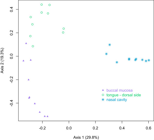
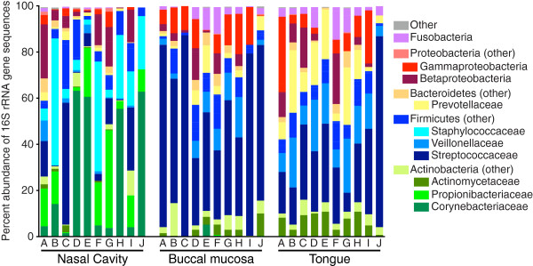
BACKGROUND: The microbiota of the nares has been widely studied. However, relatively few studies have investigated the microbiota of the nasal cavity posterior to the nares. This distinct environment has the potential to contain a distinct microbiota and play an important role in health. RESULTS: We obtained 35,142 high-quality bacterial 16S rRNA-encoding gene sequence reads from the nasal cavity and oral cavity (the dorsum of the tongue and the buccal mucosa) of 12 healthy adult humans and deposited these data in the Sequence Read Archive (SRA) of the National Center for Biotechnology Information (NCBI) (Bioproject: PRJNA248297). In our initial analysis, we compared the bacterial communities of the nasal cavity and the oral cavity from ten of these subjects. The nasal cavity bacterial communities were dominated by Actinobacteria, Firmicutes, and Proteobacteria and were statistically distinct from those on the tongue and buccal mucosa. For example, the same Staphylococcaceae operational taxonomic unit (OTU) was present in all of the nasal cavity samples, comprising up to 55% of the community, but Staphylococcaceae was comparatively uncommon in the oral cavity. CONCLUSIONS: There are clear differences between nasal cavity microbiota and oral cavity microbiota in healthy adults. This study expands our knowledge of the nasal cavity microbiota and the relationship between the microbiota of the nasal and oral cavities.
Overview
The claims and statistics of this paper haven’t been checked yet.
Run the check — free, no account needed. It reads the open-access text and checks the sources it cites.
Integrity signals
Registry
Cites
Statistics
Figures
Verification
Claims the paper makes
The claims and statistics of this paper haven’t been checked yet.
Run the check — free, no account needed. It reads the open-access text and checks the sources it cites.
Claims other papers make about this one
Each is checked against this paper’s own text: does it say what the citing paper says it does?
In the citing paper
“In healthy adults, bacteria present in the nasal cavity are typically those associated with skin, predominantly members of the Actinobacteria (e.g., Corynebacterium spp., Propionibacterium spp.), followed by Firmicutes (e.g., Staphylococcus spp.), and Proteobacteria”
From Respiratory viral infection-induced microbiome alterations and secondary bacterial pneumonia. (2018) · cites this paper as [69]
Self-check
Percentages, sums and reported differences re-computed from the paper’s own numbers.
The self-check needs the paper’s full text.
Citations
Cites
The SILVA ribosomal RNA gene database project: improved data processing and web-based tools
Quast, C., Pruesse, E., Yilmaz, P., Gerken, J., Schweer, T., Yarza, P., Peplies, J., & Glöckner, F. O. (2012) · Nucleic Acids Research · doi:10.1093/nar/gks1219 · Cited by 33,055 · cited 1×
“The sequences were aligned to the Silva reference alignment [17,18].”
MethodSchloss, P. D., Westcott, S. L., Ryabin, T., Hall, J. R., Hartmann, M., Hollister, E. B., Lesniewski, R. A., Oakley, B. B., Parks, D. H., Robinson, C. J., et al. (2009) · Applied and Environmental Microbiology · doi:10.1128/aem.01541-09 · Cited by 19,549 · cited 1×
“0 according to the Schloss SOP of November 27, 2012 [15,16].”
MethodUCHIME improves sensitivity and speed of chimera detection
Edgar, R. C., Haas, B. J., Clemente, J. C., Quince, C., & Knight, R. (2011) · Bioinformatics · doi:10.1093/bioinformatics/btr381 · Cited by 15,114
A new method for non-parametric multivariate analysis of variance
Anderson, M. J. (2001) · Austral Ecology · doi:10.1111/j.1442-9993.2001.01070.pp.x · Cited by 13,828 · cited 4×
“Principle coordinates analysis (PCoA) was used to visualize the θ distance matrix, and analysis of molecular variance (AMOVA) [22] was used to test the statistical significance of the differences between bacterial communities of different groups (i.e., nasal cavity communities versus buccal mucosa communities).”
MethodInfluentialStructure, function and diversity of the healthy human microbiome
The Human Microbiome Project Consortium (2012) · Nature · doi:10.1038/nature11234 · Cited by 11,726
Bacterial Community Variation in Human Body Habitats Across Space and Time
Costello, E. K., Lauber, C. L., Hamady, M., Fierer, N., Gordon, J. I., & Knight, R. (2009) · Science · doi:10.1126/science.1177486 · Cited by 3,231 · cited 1×
“However, many studies have characterized the microbiota of the healthy human nares [6-11], which are adjacent and anterior to the nasal cavity.”
BackgroundTopographical and Temporal Diversity of the Human Skin Microbiome
Grice, E. A., Kong, H. H., Conlan, S., Deming, C. B., Davis, J., Young, A. C., Program, N. C. S., Bouffard, G. G., Blakesley, R. W., Murray, P. R., et al. (2009) · Science · doi:10.1126/science.1171700 · Cited by 2,953 · cited 1×
“However, many studies have characterized the microbiota of the healthy human nares [6-11], which are adjacent and anterior to the nasal cavity.”
BackgroundThe role of the gut microbiota in nutrition and health
Flint, H. J., Scott, K. P., Louis, P., & Duncan, S. H. (2012) · Nature Reviews Gastroenterology & Hepatology · doi:10.1038/nrgastro.2012.156 · Cited by 2,283
Reducing the Effects of PCR Amplification and Sequencing Artifacts on 16S rRNA-Based Studies
Schloss, P. D., Gevers, D., & Westcott, S. L. (2011) · PLoS ONE · doi:10.1371/journal.pone.0027310 · Cited by 2,112 · cited 1×
“0 according to the Schloss SOP of November 27, 2012 [15,16].”
MethodSorokin, S. P. (1983) · doi:10.1007/978-1-349-06859-3_23 · Cited by 1,783
Cited by
Sorted by citations
Bassis, C. M., Erb-Downward, J. R., Dickson, R. P., Freeman, C. M., Schmidt, T. M., Young, V. B., Beck, J. M., Curtis, J. L., & Huffnagle, G. B. (2015) · mBio · doi:10.1128/mbio.00037-15 · Cited by 839
Self-citationHeintz‐Buschart, A., Pandey, U., Wicke, T., Sixel‐Döring, F., Janzen, A., Sittig‐Wiegand, E., Trenkwalder, C., Oertel, W. H., Mollenhauer, B., & Wilmes, P. (2018) · Movement Disorders · doi:10.1002/mds.27105 · Cited by 554
Microbiome and Human Health: Current Understanding, Engineering, and Enabling Technologies
Aggarwal, N., Kitano, S., Puah, G. R. Y., Kittelmann, S., Hwang, I. Y., & Chang, M. W. (2022) · Chemical Reviews · doi:10.1021/acs.chemrev.2c00431 · Cited by 571
Respiratory viral infection-induced microbiome alterations and secondary bacterial pneumonia.
Hanada, S., Pirzadeh, M., Carver, K. Y., & Deng, J. C. (2018) · Frontiers in Immunology · doi:10.3389/fimmu.2018.02640 · Cited by 495
“In healthy adults, bacteria present in the nasal cavity are typically those associated with skin, predominantly members of the Actinobacteria (e.g., Corynebacterium spp., Propionibacterium spp.), followed by Firmicutes (e.g., Staphylococcus spp.), and Proteobacteria (67) (68) (69) .”
BackgroundMicrobiota-mediated colonization resistance: mechanisms and regulation
Caballero-Flores, G., Pickard, J. M., & Núñez, G. (2023) · Nature Reviews Microbiology · doi:10.1038/s41579-022-00833-7 · Cited by 474
The microbiome of the upper respiratory tract in health and disease
Kumpitsch, C., Koskinen, K., Schöpf, V., & Moissl‐Eichinger, C. (2019) · BMC Biology · doi:10.1186/s12915-019-0703-z · Cited by 490
Pathogenesis of allergic diseases and implications for therapeutic interventions
Wang, J., Zhou, Y., Zhang, H., Hu, L., Liu, J., Wang, L., Wang, T., Zhang, H., Cong, L., & Wang, Q. (2023) · Signal Transduction and Targeted Therapy · doi:10.1038/s41392-023-01344-4 · Cited by 431
The human microbiome in disease and pathology
Manos, J. (2022) · Apmis · doi:10.1111/apm.13225 · Cited by 252
“A distinct difference in microbiome composition was detected between the nasal and oral cavities Streptococcaceae was the most abundant Firmicute genera in the oral cavity while Staphylococceae were not detected at all [91].”
BackgroundOral and nasal microbiota in Parkinson's disease
Pereira, P. A. B., Aho, V. T. E., Paulin, L. G., Pekkonen, E., Auvinen, P., & Scheperjans, F. (2017) · Parkinsonism & Related Disorders · doi:10.1016/j.parkreldis.2017.02.026 · Cited by 210
“These results are consistent with previous studies where oropharyngeal microbiota were found to be more diverse than nasal microbiota, and more similar in a specific anatomical site across individuals than within a specific individual [18].”
Bacterial microbiota of the upper respiratory tract and childhood asthma
Depner, M., Ege, M. J., Cox, M. J., Dwyer, S., Walker, A. W., Birzele, L. T., Genuneit, J., Horak, E., Braun‐Fahrländer, C., Danielewicz, H., et al. (2016) · Journal of Allergy and Clinical Immunology · doi:10.1016/j.jaci.2016.05.050 · Cited by 203
“In neonates, the colonization of the throat with M catarrhalis has been reported as a potential risk factor for the development of childhood asthma.”
Earl, J. P., Adappa, N. D., Król, J. E., Bhat, A. S., Balashov, S. V., Ehrlich, R. L., Palmer, J. N., Workman, A. D., Blasetti, M. T., Sen, B., et al. (2018) · Microbiome · doi:10.1186/s40168-018-0569-2 · Cited by 196
The Human Respiratory System and its Microbiome at a Glimpse
Santacroce, L., Charitos, I. A., Ballini, A., Inchingolo, F., Luperto, P., Nitto, E. D., & Topi, S. (2020) · Biology · doi:10.3390/biology9100318 · Cited by 203
“Age-related immune system changes contribute to the increased incidence of respiratory infections [28,30,31].”
BackgroundThe nasal microbiota in health and disease: variation within and between subjects
Biswas, K., Hoggard, M., Jain, R., Taylor, M. W., & Douglas, R. G. (2015) · Frontiers in Microbiology · doi:10.3389/fmicb.2015.00134 · Cited by 190
“Based on the findings of Yan et al. (2013) and Bassis et al. (2014) and the different microenvironments of the study sites, we might have expected the nasal microbiota of the chosen sites to differ.”
InfluentialBacterial community collapse: a meta‐analysis of the sinonasal microbiota in chronic rhinosinusitis
Mackenzie, B. W., Waite, D. W., Hoggard, M., Douglas, R. G., Taylor, M. W., & Biswas, K. (2016) · Environmental Microbiology · doi:10.1111/1462-2920.13632 · Cited by 187
Boeck, I. D., Wittouck, S., Wuyts, S., Oerlemans, E. F. M., van den Broek, M. F. L., Vandenheuvel, D., Vanderveken, O. M., & Lebeer, S. (2017) · Frontiers in Microbiology · doi:10.3389/fmicb.2017.02372 · Cited by 142
“…nose and the nasopharynx are low-complexity (in terms of observed richness or total number of bacterial genera present) and lowbiomass (in terms of total amount of bacterial cells) microbial niches (Biesbroek et al., 2012), an accurate discrimination between these biological variants is essential.”
BackgroundThe microbiota and autoimmunity: Their role in thyroid autoimmune diseases
Köhling, H.-L., Plummer, S. F., Marchesi, J. R., Davidge, K. S., & Ludgate, M. (2017) · Clinical Immunology · doi:10.1016/j.clim.2017.07.001 · Cited by 139
“Most of the studies compared the nasal microbiome of healthy persons [210, 224-227] with those of persons with chronic rhinosinusitis and other nasal inflammatory diseases [228-232].”
Bacteriophage and their potential roles in the human oral cavity
Edlund, A., Santiago-Rodríguez, T. M., Boehm, T. K., & Pride, D. T. (2015) · Journal of Oral Microbiology · doi:10.3402/jom.v7.27423 · Cited by 141
Environmental and mucosal microbiota and their role in childhood asthma
Birzele, L. T., Depner, M., Ege, M. J., Engel, M., Kublik, S., Bernau, C., Loss, G. J., Genuneit, J., Horak, E., Schloter, M., et al. (2017) · Allergy · doi:10.1111/all.13002 · Cited by 124
“Though this Pinaceae Chloroplast OTU was occasionally detected in nasal samples as well, the main nasal taxa were local bacteria such as Moraxella and Streptococcus, which are well known from clinical experience and previous studies (36-38), particularly in children (38).”
BackgroundMacrophage metabolic reprogramming during chronic lung disease
Ogger, P. P., & Byrne, A. J. (2020) · Mucosal Immunology · doi:10.1038/s41385-020-00356-5 · Cited by 123
Hoggard, M., Mackenzie, B. W., Jain, R., Taylor, M. W., Biswas, K., & Douglas, R. G. (2016) · Clinical Microbiology Reviews · doi:10.1128/cmr.00060-16 · Cited by 122
The oral cavity microbiota: between health, oral disease, and cancers of the aerodigestive tract
Bars, P. L., Matamoros, S., Montassier, E., Vacon, F. L., Potel, G., Soueidan, A., Jordana, F., & de la Cochetière, M.-F. (2017) · Canadian Journal of Microbiology · doi:10.1139/cjm-2016-0603 · Cited by 111
“At least 15 phyla have been detected , and the most predominant among these include Fusobacteria, Actinobacteria, Proteobacteria, Bacteroidetes and Spirochaetae (Rogers et al. 2013; Nadell et al, 2009).”
BackgroundInfluentialComposition and immunological significance of the upper respiratory tract microbiota
Schenck, L. P., Surette, M. G., & Bowdish, D. M. E. (2016) · FEBS Letters · doi:10.1002/1873-3468.12455 · Cited by 108
The gut microbiome in Parkinson's disease: A culprit or a bystander?
Keshavarzian, A., Engen, P. A., Bonvegna, S., & Cilia, R. (2020) · Progress in brain research · doi:10.1016/bs.pbr.2020.01.004 · Cited by 108
The microbiome: Composition and locations
Kennedy, M. S., & Chang, E. B. (2020) · Progress in molecular biology and translational science · doi:10.1016/bs.pmbts.2020.08.013 · Cited by 108
Kim, H. J., Jo, A., Jeon, Y. J., An, S., Lee, K., Yoon, S. S., & Choi, J. Y. (2019) · Microbiome · doi:10.1186/s40168-019-0691-9 · Cited by 92
Epithelial Barrier in the Nasal Mucosa, Related Risk Factors and Diseases
Zhang, R., Zhang, L., Li, P., Pang, K., Liu, H., & Tian, L. (2023) · International Archives of Allergy and Immunology · doi:10.1159/000528969 · Cited by 98
Mariani, J., Favero, C., Spinazzè, A., Cavallo, D. M., Carugno, M., Motta, V., Bonzini, M., Cattaneo, A., Pesatori, A. C., & Bollati, V. (2017) · Environmental Research · doi:10.1016/j.envres.2017.12.016 · Cited by 90
“The structure we observed is coherent with other studies (Bassis et al., 2014; Biswas et al., 2015; Costello et al., 2009) on the nasal bacterial community, where Actinobacteria was the most represented taxa.”
BackgroundResultCorrea‐Fiz, F., Fraile, L. J., & Aragón, V. (2016) · BMC Genomics · doi:10.1186/s12864-016-2700-8 · Cited by 88
“The diversity of the nasal microbiota was as low as was reported before in different animal and in human studies [24–27].”
The microbiome: an integral player in immune homeostasis and inflammation in the respiratory tract
Perdijk, O., Azzoni, R., & Marsland, B. J. (2023) · Physiological Reviews · doi:10.1152/physrev.00020.2023 · Cited by 94
Anterior Nares Diversity and Pathobionts Represent Sinus Microbiome in Chronic Rhinosinusitis
Boeck, I. D., Wittouck, S., Martens, K., Claes, J., Jorissen, M., Steelant, B., van den Broek, M. F. L., Seys, S., Hellings, P. W., Vanderveken, O. M., et al. (2019) · mSphere · doi:10.1128/msphere.00532-19 · Cited by 79
“This bacterial genus is known to be an important member of the anterior nares (27, 28), so this should be kept in mind in the interpreFIG 5 Associations of numerical (A) and categorical (B) microbiome covariates with microbiome-based subject clusters.”
BackgroundCastro‐Nallar, E., Shen, Y., Freishtat, R. J., Pérez‐Losada, M., Manimaran, S., Liu, G., Johnson, W. E., & Crandall, K. A. (2015) · BMC Medical Genomics · doi:10.1186/s12920-015-0121-1 · Cited by 73
“have been detected in sinus nasal studies of healthy individuals as well as in cases of rhinitis and rhinosinusitis, where their prevalence nears 100 % and their abundance is relatively high [46–48].”
BackgroundArsenic and the gastrointestinal tract microbiome
McDermott, T. R., Stolz, J. F., & Oremland, R. S. (2019) · Environmental Microbiology Reports · doi:10.1111/1758-2229.12814 · Cited by 76
Shilts, M. H., Rosas‐Salazar, C., Tovchigrechko, A., Larkin, E. K., Torralba, M., Akopov, A., Halpin, R., Peebles, R. S., Moore, M. L., Anderson, L. J., et al. (2015) · Microbial Ecology · doi:10.1007/s00248-015-0663-y · Cited by 70
“At the phylum level, the taxonomic profiles in the present study with nasal sampling by filter paper largely corroborate previous research of the human nasopharyngeal microbiome [27-30], although differences in the population studied, sampling methods, DNA extraction protocols, amplification primers, and data processi…”
BackgroundResultInfluentialRhoades, N. S., Pinski, A. N., Monsibais, A. N., Jankeel, A., Doratt, B. M., Cinco, I. R., Ibraim, I. C., & Messaoudi, I. (2021) · Cell Reports · doi:10.1016/j.celrep.2021.109637 · Cited by 73
“The healthy nasal microbiome is dominated by Corynebacterium, Staphylococcus, Streptococcus, Dolosigranulum, and Moraxella (Whelan et al., 2014; Bassis et al., 2014; Stearns et al., 2015).”
Rawls, M., & Ellis, A. K. (2018) · Annals of Allergy Asthma & Immunology · doi:10.1016/j.anai.2018.05.009 · Cited by 71
Environmental and Human Microbiome for Health
Panthee, B., Gyawali, S., Panthee, P., & Techato, K. (2022) · Life · doi:10.3390/life12030456 · Cited by 68
“Actinobacteria, Bacteroidetes, Firmicutes, or Proteobacteria were the commonly found microorganisms throughout the human body [23–25].”
BackgroundDoes the maternal vaginal microbiota play a role in seeding the microbiota of neonatal gut and nose?
Sakwińska, O., Foata, F., Berger, B. W., Brüssow, H., Combremont, S., Mercenier, A., Dogra, S. K., Soh, S., Chan, J. K. Y., Heong, G. Y. S., et al. (2017) · Beneficial Microbes · doi:10.3920/bm2017.0064 · Cited by 62
“The considerable change in infant nasal microbiota from birth to week 3 resulted in marked resemblance to adult nasal microbiota, i.e. domination by Staphylococcus and Corynebacterium as observed previously (Bassis et al. , 2014).”
Cotton and Surgical Face Masks in Community Settings: Bacterial Contamination and Face Mask Hygiene
Delanghe, L., Cauwenberghs, E., Spacova, I., Boeck, I. D., Beeck, W. V., Pepermans, K., Claes, I. J. J., Vandenheuvel, D., Verhoeven, V., & Lebeer, S. (2021) · Frontiers in Medicine · doi:10.3389/fmed.2021.732047 · Cited by 64
Nasal Microbiota, Olfactory Health, Neurological Disorders and Aging—A Review
Thangaleela, S., Sivamaruthi, B. S., Kesika, P., Bharathi, M., & Chaiyasut, C. (2022) · Microorganisms · doi:10.3390/microorganisms10071405 · Cited by 65
“It is predominantly colonized by Bacteroidetes and Firmicutes [150], Actinobacteria, Cyanobacteria, Fusobacteria, Proteobacteria, and Verrucobacteria [151], which can directly or indirectly influence the host immune responses [152].”
BackgroundAssociation between Microbiota and Nasal Mucosal Diseases in terms of Immunity
Tai, J., Han, M. S., Kwak, J., & Kim, T. H. (2021) · International Journal of Molecular Sciences · doi:10.3390/ijms22094744 · Cited by 60
“The microbiota of healthy adults’ anterior nares is mainly composed of Actinobacteria, Firmicutes, and Proteobactera [28].”
BackgroundHyun, D., Min, H. J., Kim, M., Whon, T. W., Shin, N.-R., Kim, P. S., Kim, H. S., Lee, J., Kang, W., Choi, A. M. K., et al. (2018) · Infection and Immunity · doi:10.1128/iai.00934-17 · Cited by 56
“Unlike the microbial pattern of the inferior turbinate microbiota noted in healthy individuals, both previously (37, 38) and in this study, the Firmicutes phylum was dominant in the inferior turbinate microbiota of AR patients under high IgE conditions, a condition attributable to low diversity of the inferior turbina…”
ResultThe Microbiota/Host Immune System Interaction in the Nose to Protect from COVID-19
Stadio, A. D., Costantini, C., Renga, G., Pariano, M., Ricci, G., & Romani, L. R. (2020) · Life · doi:10.3390/life10120345 · Cited by 55
The emerging role of probiotics as a mitigation strategy against coronavirus disease 2019 (COVID-19)
Mirzaei, R., Attar, A., Papizadeh, S., Jeda, A. S., Hosseini‐Fard, S. R., Jamasbi, E., Kazemi, S., Amerkani, S., Talei, G. R., Moradi, P., et al. (2021) · Archives of Virology · doi:10.1007/s00705-021-05036-8 · Cited by 59
Size, Composition, and Source Profiles of Inhalable Bioaerosols from Colorado Dairies
Schaeffer, J. W., Reynolds, S. J., Magzamen, S. L., VanDyke, A., Gottel, N., Gilbert, J. A., Owens, S. M., Hampton‐Marcell, J., & Volckens, J. (2017) · Environmental Science & Technology · doi:10.1021/acs.est.7b00882 · Cited by 55
Bacterial microbiome of the nose of healthy dogs and dogs with nasal disease
Treß, B., Dorn, E. S., Suchodolski, J. S., Nisar, T., Ravindran, P., Weber, K., Hartmann, K., & Schulz, B. S. (2017) · PLoS ONE · doi:10.1371/journal.pone.0176736 · Cited by 51
Chen, C., Liou, M., Lee, C., Chang, M.-C., Kuo, H., & Chang, T. (2019) · Scientific Reports · doi:10.1038/s41598-019-42548-5 · Cited by 48
Alterations in Oral–Nasal–Pharyngeal Microbiota and Salivary Proteins in Mouth-Breathing Children
Fan, C., Guo, L., Gu, H., Huo, Y., & Lin, H. (2020) · Frontiers in Microbiology · doi:10.3389/fmicb.2020.575550 · Cited by 50
Shukla, S. K., Ye, Z., Sandberg, S., Reyes, I., Fritsche, T. R., & Keifer, M. (2017) · PLoS ONE · doi:10.1371/journal.pone.0183898 · Cited by 53
Friend or Foe: Interbacterial Competition in the Nasal Cavity
Hardy, B. L., & Merrell, D. S. (2020) · Journal of Bacteriology · doi:10.1128/jb.00480-20 · Cited by 42
“netically, competition for similar environmental niches leads to well-documented competition between various members of the Staphylococcus genus; this competition in turn contributes to the overall structure of the nasal microbiota (13, 31).”
BackgroundInfluentialEffects of nasopharyngeal microbiota in respiratory infections and allergies
Kang, H. M., & Kang, J. H. (2021) · Korean Journal of Pediatrics · doi:10.3345/cep.2020.01452 · Cited by 46
Exploring the bacterial assemblages along the human nasal passage
Wos‐Oxley, M. L., Chaves‐Moreno, D., Jáuregui, R., Oxley, A. P. A., Kaspar, U., Plumeier, I., Kahl, S., Rudack, C., Becker, K., & Pieper, D. H. (2016) · Environmental Microbiology · doi:10.1111/1462-2920.13378 · Cited by 40
“…has been the comparison between human anterior nares (or less commonly other nasal passage 2010; Lemon et al., 2010; Human Microbiome Project, 2012; Bassis et al., 2014), while comparisons between specific anatomical regions of the human nasal passage have only recently been attempted (Yan et al.,…”
BackgroundTwo sampling methods yield distinct microbial signatures in the nasopharynges of asthmatic children
Pérez‐Losada, M., Crandall, K. A., & Freishtat, R. J. (2016) · Microbiome · doi:10.1186/s40168-016-0170-5 · Cited by 42
“Consequently, several recent metataxonomic and metagenomic (see [7] for distinction) studies have investigated how nasopharyngeal microbial communities change during health and disease in relation to clinical variation [1, 3, 5, 8–16].”
BackgroundThe microbiome of pediatric patients with chronic rhinosinusitis
Stapleton, A. L., Shaffer, A. D., Morris, A. M., Li, K., Fitch, A. C., & Methé, B. A. (2020) · International Forum of Allergy & Rhinology · doi:10.1002/alr.22597 · Cited by 40
Peterson, S. W., Knox, N., Golding, G. R., Tyler, S. D., Tyler, A. D., Mabon, P., Embree, J. E., Fleming, F., Fanella, S. T., Domselaar, G. V., et al. (2016) · PLoS ONE · doi:10.1371/journal.pone.0152493 · Cited by 39
“In conjunction with increased risk for transmission of persistent/colonizing antimicrobial resistant (AMR) strains, asymptomatic nasal carriage is associated with increased risk for acute respiratory illnesses of both the upper and lower respiratory tracts, skin and soft tissue infections, and bacteremia [17,18].”
BackgroundDifferential flora in the microenvironment of lung tumor and paired adjacent normal tissues
Mao, Q., Ma, W., Wang, Z., Liang, Y., Zhang, T., Yang, Y., Xia, W., Jiang, F., Hu, J., & Xu, L. (2020) · Carcinogenesis · doi:10.1093/carcin/bgaa044 · Cited by 36
Possible Therapeutic Mechanisms and Future Perspectives of Vaginal Microbiota Transplantation
Tuniyazi, M., Li, S., Fu, Y., Hu, X., & Zhang, N. (2023) · Microorganisms · doi:10.3390/microorganisms11061427 · Cited by 34
The Upper Airway Microbiota, Environmental Exposures, Inflammation, and Disease
Elgamal, Z., Singh, P., & Geraghty, P. M. (2021) · Medicina · doi:10.3390/medicina57080823 · Cited by 43
“The nasal cavity is constantly interacting with the external environment [18,20].”
BackgroundGao, X., Xu, Y., Cai, Y., Shi, J., Chen, F., Lin, Z., Chen, T., Xia, Y., Shi, W., & Zhao, Z. (2019) · Environmental Research · doi:10.1016/j.envres.2019.108749 · Cited by 32
Ebrahimi, K. H. (2020) · FEBS Letters · doi:10.1002/1873-3468.13845 · Cited by 32
Huntley, K. S., Raber, J., Fine, L. M., & Bernstein, J. A. (2021) · Frontiers in Allergy · doi:10.3389/falgy.2021.737086 · Cited by 34
“evaluated the middle meatus in healthy adults via cultureindependent 16S rRNA gene analysis with 269 sequences per sample (22).”
BackgroundMethodLiving Therapeutics: The Next Frontier of Precision Medicine
Kelly, V. W., Liang, B. K., & Sirk, S. J. (2020) · ACS Synthetic Biology · doi:10.1021/acssynbio.0c00444 · Cited by 30
Do Aging Factors Influence the Clinical Presentation and Management of Chronic Rhinosinusitis?
Renteria, A. E., Endam, L. M., & Desrosiers, M. Y. (2017) · Otolaryngology · doi:10.1177/0194599817691258 · Cited by 31
Harrass, S., Yi, C., & Chen, H. (2021) · International Journal of Molecular Sciences · doi:10.3390/ijms222011207 · Cited by 31
“The healthy nasal cavity is colonised with Actinobacteria, Bacteroidetes, Firmicutes, and Proteobacteria on the phylum level, and Bifidobacterium, Corynebacterium, Staphylococcus, Streptococcus, Dolosigranulum and Moraxella on the genus level [23,24].”
BackgroundXie, J., Tian, S., Liu, J., Cao, R., Yue, P., Cai, X., Shang, Q., Yang, M. H., Han, L., & Zhang, D. (2022) · Pharmacological Research · doi:10.1016/j.phrs.2022.106189 · Cited by 30
Arca-Lafuente, S., Núñez-Corcuera, B., Ramis, R., Karakitsios, S., Sarigiannis, D. A., dos Santos, S. G., Fernández‐Rodríguez, A., & Briz, V. (2025) · Respiratory Research · doi:10.1186/s12931-025-03179-9 · Cited by 33
“…Actinobacteria and Firmicutes are the predominant phyla in the nasopharynx microbiome of healthy individuals [40, 41], whereas in the oral cavity , the prevalence of Actinobacteria is lower than that in the nasopharynx, and Firmicutes is dominant, followed by Bacteroidetes and Proteobacteria [42].”
Jeon, Y. J., Gil, C. H., Won, J., Jo, A., & Kim, H. J. (2020) · BMC Microbiology · doi:10.1186/s12866-020-01974-6 · Cited by 28
Rudder, C. D., García‐Timermans, C., Boeck, I. D., Lebeer, S., de Wiele, T. V., & Calatayud, M. (2020) · Scientific Reports · doi:10.1038/s41598-020-73857-9 · Cited by 27
Vendl, C., Slavich, E., Wemheuer, B., Nelson, T. M., Ferrari, B. C., Thomas, T., & Rogers, T. L. (2020) · Scientific Reports · doi:10.1038/s41598-020-69602-x · Cited by 27
Habibi, N., Mustafa, A. S., & Khan, M. W. (2021) · PLoS ONE · doi:10.1371/journal.pone.0260314 · Cited by 26
Factors Limiting the Translatability of Rodent Model–Based Intranasal Vaccine Research to Humans
Cai, L., Xu, H., & Cui, Z. (2022) · AAPS PharmSciTech · doi:10.1208/s12249-022-02330-9 · Cited by 26
“Like adults, the nasal microbiota of young children is dominated by Actinobacteria and Firmicutes, with more limited Proteobacteria [106].”
BackgroundInfluentialBender, M. E., Read, T. D., Edwards, T. S., Hargita, M. N., Cutler, A. J., Wissel, E. F., & Wise, S. K. (2020) · The Laryngoscope · doi:10.1002/lary.28599 · Cited by 24
“DISCUSSION Prior studies have shown that AR patients, in general, have increased bacterial diversity in their microbiomes.(5,7) In gastrointestinal disease, the dysbiosis theory is commonly invoked, postulating that bacterial diversity declines with infection and/or inflammation as the normal microbiome ecosystem is d…”
BackgroundRapid separation of bacteria from primary nasal samples using inertial microfluidics
Shrestha, J., Bazaz, S. R., Ding, L., Vasilescu, S. A., Idrees, S., Söderström, B., Hansbro, P. M., Ghadiri, M., & Warkiani, M. E. (2022) · Lab on a Chip · doi:10.1039/d2lc00794k · Cited by 24
Rudder, C. D., Calatayud, M., Lebeer, S., & de Wiele, T. V. (2020) · mSphere · doi:10.1128/msphere.00916-19 · Cited by 24
Lu, C., Wang, F., Liu, Z., Li, B., Yang, W., & Liao, H. (2023) · Chemosphere · doi:10.1016/j.chemosphere.2023.139296 · Cited by 23
Rudder, C. D., Calatayud, M., Lebeer, S., & de Wiele, T. V. (2018) · Microbiome · doi:10.1186/s40168-018-0462-z · Cited by 24
Pérez‐Losada, M., Crandall, K. A., & Freishtat, R. J. (2016) · AIMS Microbiology · doi:10.3934/microbiol.2016.2.108 · Cited by 21
“Numerous metagenomic and metataxonomic studies have identified commensal and pathogenic members of the nares and investigated how nasal microbiotas change during health and disease [18–30].”
Human nasal microbiota shifts in healthy and chronic respiratory disease conditions
Konovalovas, A., Armalytė, J., Klimkaitė, L., Liveikis, T., Jonaitytė, B., Danila, E., Bironaitė, D., Mieliauskaitė, D., Bagdonas, E., & Aldonytė, R. (2024) · BMC Microbiology · doi:10.1186/s12866-024-03294-5 · Cited by 21
Rouchka, E. C., Chariker, J. H., Alejandro, B. S., Adcock, R. S., Singhal, R. A., Ramírez, J. A., Palmer, K. E., Lasnik, A. B., Carrico, R., Arnold, F. W., et al. (2021) · Scientific Reports · doi:10.1038/s41598-021-95197-y · Cited by 20
Ortega‐Peña, S., Rodríguez–Martínez, S., Cancino-Diaz, M. E., & Cancino‐Díaz, J. C. (2022) · Life · doi:10.3390/life12030341 · Cited by 22
“Corynebacterium and Cutibacterium are the most abundant and prevalent species of the genus Actinobacteria in the nasal cavity [53,57,60].”
BackgroundInfluentialAs, L., Azizov, I., & Kozlov, R. S. (2021) · Russian Rhinology · doi:10.17116/rosrino20212901123 · Cited by 21
Casadei, E., Tacchi, L., Lickwar, C. R., Espenschied, S. T., Davison, J. M., Muñoz, P., Rawls, J. F., & Salinas, I. (2019) · Chemical Senses · doi:10.1093/chemse/bjz050 · Cited by 19
Ahmed, N., Mahmoud, N., Solyman, S. M., & Hanora, A. (2019) · OMICS A Journal of Integrative Biology · doi:10.1089/omi.2019.0144 · Cited by 19
“Bassis et al. (2014) also recorded predominance of Actinobacteria and Firmicutes in the nares of healthy individuals.”
Wu, C., Ko, W., Ho, M., Lin, H.-H., Yang, Y.-L., Lin, J., Huang, I., Wang, H., Lai, J., Shiau, Y., et al. (2017) · Journal of Oral Microbiology · doi:10.1080/20002297.2017.1322446 · Cited by 18
“could be a reservoir of CA-MRSA due to overcrowding and unsanitary conditions [20,23].”
BackgroundResultIslam, M. Z., Johannesen, T. B., Lilje, B., Urth, T. R., Larsen, A. R., Angen, Ø., & Larsen, J. (2020) · PLoS ONE · doi:10.1371/journal.pone.0232456 · Cited by 17
Renteria, A. E., Maniakas, A., Endam, L. M., Asmar, M., González, E., & Desrosiers, M. Y. (2020) · International Forum of Allergy & Rhinology · doi:10.1002/alr.22653 · Cited by 18
Vangrinsven, E., Fastrès, A., Taminiau, B., Billen, F., Daube, G., & Clercx, C. (2021) · BMC Veterinary Research · doi:10.1186/s12917-021-03055-w · Cited by 19
Upper respiratory microbial communities of healthy populations are shaped by niche and age
Zelasko, S. E., Swaney, M. H., Sandstrom, S., Davenport, T. C., Seroogy, C. M., Gern, J. E., Kalan, L., & Currie, C. R. (2024) · Microbiome · doi:10.1186/s40168-024-01940-8 · Cited by 17
“First, based on shotgun metagenomic sequencing, we find that the microbiota inhabiting nasal and oral cavities are unique among infants and adults, providing further evidence that healthy respiratory microbiota are niche dependent [13, 15].”
BackgroundMethodMicrobiota Profile of the Nasal Cavity According to Lifestyles in Healthy Adults in Santiago, Chile
Toro‐Ascuy, D., Cárdenas, J. P., Zorondo‐Rodríguez, F., González, D., Silva-Moreno, E., Puebla, C., Nunez-Parra, A., Reyes-Cerpa, S., & Fuenzalida, L. (2023) · Microorganisms · doi:10.3390/microorganisms11071635 · Cited by 18
Beyond Hygiene: Commensal Microbiota and Allergic Diseases
Hong, S., Kim, K. S., & Surh, C. D. (2017) · Immune Network · doi:10.4110/in.2017.17.1.48 · Cited by 17
“In normal skin, Firmicutes, Actinobacteria and Proteobacteria are the predominant phyla relative to other mucosal tissues; e.g., Bacteriodetes predominate in the gut, but is a minor component of skin microbiota (17).”
BackgroundInfluentialBowers, J. R., Valentine, M., Harrison, V., Fofanov, V. Y., Gillece, J. D., Delisle, J., Patton, B. K., Schupp, J. M., Sheridan, K. L., Lemmer, D., et al. (2019) · mBio · doi:10.1128/mbio.02262-18 · Cited by 17
Bartosik, T. J., Campion, N. J., Freisl, K., Liu, D. T., Gangl, K., Stanek, V., Tu, A., Pjevac, P., Hausmann, B., Eckl‐Dorna, J., et al. (2023) · Frontiers in Immunology · doi:10.3389/fimmu.2023.1112345 · Cited by 16
“The microbiome of healthy nasal sinuses is dominated by the genera Corynebacterium, Staphylococcus, Streptococcus, Haemophilus and Moraxella (8–10).”
BackgroundJo, A., Won, J., Gil, C. H., Kim, S. G., Lee, K., Yoon, S. S., & Kim, H. J. (2022) · npj Biofilms and Microbiomes · doi:10.1038/s41522-022-00290-3 · Cited by 16
The animal's microbiome and cancer: A translational perspective
Aluai‐Cunha, C., Pinto, C. A., Correia, A., Serra, C. R., & Santos, A. A. F. (2023) · Veterinary and Comparative Oncology · doi:10.1111/vco.12892 · Cited by 17
Multi-Omics Approaches: The Key to Improving Respiratory Health in People With Cystic Fibrosis?
Lee, A. J., Einarsson, G. G., Gilpin, D. F., & Tunney, M. M. (2020) · Frontiers in Pharmacology · doi:10.3389/fphar.2020.569821 · Cited by 14
Ceraudo, M., Prior, A., Balestrino, A., Anania, P., Camera, M., Fiaschi, P., Gatto, F., Riccardi, N., Zona, G., & Rossi, D. C. (2020) · Acta Neurochirurgica · doi:10.1007/s00701-020-04560-x · Cited by 15
“infections due to the passages through the nares [3].”
BackgroundNasal Microbiome in COVID-19: A Potential Role of Corynebacterium in Anosmia
Nardelli, C., Scaglione, G. L., Testa, D., Setaro, M., Russo, F., Domenico, C. D., Atripaldi, L., Zollo, M., Corrado, F., Salvatore, P., et al. (2022) · Current Microbiology · doi:10.1007/s00284-022-03106-x · Cited by 15
Is Nasal Dysbiosis a Required Component for Neuroinflammation in Major Depressive Disorder?
Vásquez-Pérez, J. M., González‐Guevara, E., Gutiérrez-Buenabad, D., Martínez‐Gopar, P. E., Martínez‐Lazcano, J. C., & Cárdenas, G. (2024) · Molecular Neurobiology · doi:10.1007/s12035-024-04375-2 · Cited by 16
“Commensal and pathogenic microbiota can communicate with the host epithelial, immune, and CNS by producing extracellular vesicles (EVs) or exosomes.”
InfluentialCurrent Issues and Perspectives in Antimicrobials use in Dental Practice
Santacroce, L., Spirito, F., Bottalico, L., Muzio, E. L., Charitos, I. A., Potenza, M. A., Montagnani, M., & Jirillo, E. (2022) · Current Pharmaceutical Design · doi:10.2174/1381612828666220920114723 · Cited by 15
Alharbi, N. S. H. A. (2019) · Saudi Journal of Biological Sciences · doi:10.1016/j.sjbs.2019.05.008 · Cited by 13
“The human nasal cavity is a section of the respiratory system and all the parts of the respiratory system receive the inhaled air through the nasal cavity (Bassis et al., 2014; Ramakrishnan et al., 2016; Bomar et al., 2018).”
BackgroundCrosstalk between gut microbiota and renal ischemia/reperfusion injury
Huang, P., Cao, J., Chen, J., Luo, Y., Gong, X., Wu, C., & Wang, Y. H. D. (2022) · Frontiers in Cellular and Infection Microbiology · doi:10.3389/fcimb.2022.1015825 · Cited by 13
“, 2021), nasal cavity (Bassis et al., 2014), oral cavity (Blaustein et al.”
Background
Metadata
- Authors
- Bassis, C. M., Tang, A. L., Young, V. B., & Pynnonen, M. A.
- Affiliations
- University of Michigan (US)
- University of Cincinnati (US)
- Corresponding author
- Bassis, Christine M
- Venue
- Microbiome
- Published
- 2014-08-11
- Volume
- 2
- Issue
- 1
- Pages
- 27-27
- ISSN
- 2049-2618 (ISSN-L)
- PMC
- PMC4138944
- OpenAlex
- W2161225919
- Cited by
- 209
- References
- 25
- Citations by year
- 2026: 17 · 2025: 17 · 2024: 25 · 2023: 23 · 2022: 22 · 2021: 15 · 2020: 30 · 2019: 14 · 2018: 11 · 2017: 15 · 2016: 13 · 2015: 6
- FWCI
- 15.10
- Type
- journal article
- Language
- en
- Open access
- gold
- doi.org
- Locations
- Microbiome (publishedVersion)
- Europe PMC (PubMed Central) (publishedVersion, cc-by)
- PubMed Central (publishedVersion, cc-by)
- PubMed (publishedVersion)
- Deep Blue (University of Michigan) (submittedVersion)
- Funders
- National Institutes of Health
- National Heart, Lung, and Blood Institute
- Topics
- Health Sciences › Dentistry › Periodontics › Oral microbiology and periodontitis research
- Life Sciences › Biochemistry, Genetics and Molecular Biology › Molecular Biology › Gut microbiota and health
- Physical Sciences › Environmental Science › Ecology › Microbial Community Ecology and Physiology
- Keywords
- tongue dorsum, buccal mucosa, Staphylococcaceae, bacterial community composition, healthy adults, 16S rRNA gene sequencing, actinobacteria, Firmicutes, Proteobacteria
About these checks
Registry, retraction, Cites and statistics results are computed, not judged, with one exception: a printed reference that no index lists is read once by a model before it counts as a citation error. Claims are extracted by a model and kept only when their quote appears verbatim in the text; each cited source is then checked, with a model’s help, for whether it says what the claim relies on. Those verdicts can be wrong — read the claim’s quote and the cited source before relying on one.
Statistics and arithmetic are recomputed, not judged.
Confidence bands are shown only for fields calibrated on our annotated test set. None is calibrated yet, so no band is shown.
What counts as a claim ›How we measure confidence ›How we check this ›
Are you an author, or do you see a problem with this page? Request a review
